. 1999 May;73(5):4360-71.
doi: 10.1128/JVI.73.5.4360-4371.1999.

Cyanovirin-N binds to gp120 to interfere with CD4-dependent human immunodeficiency virus type 1 virion binding, fusion, and infectivity but does not affect the CD4 binding site on gp120 or soluble CD4-induced conformational changes in gp120

Affiliations

Cyanovirin-N binds to gp120 to interfere with CD4-dependent human immunodeficiency virus type 1 virion binding, fusion, and infectivity but does not affect the CD4 binding site on gp120 or soluble CD4-induced conformational changes in gp120

M T Esser et al. J Virol. 1999 May.

Abstract

Cyanovirin-N (CV-N), an 11-kDa protein isolated from the cyanobacterium Nostoc ellipsosporum, potently inactivates diverse strains of human immunodeficiency virus type 1 (HIV-1), HIV-2, and simian immunodeficiency virus. While it has been well established that the viral surface envelope glycoprotein gp120 is a molecular target of CV-N, the detailed mechanism of action is of further interest. We compared matched native and CV-N-treated virus preparations in a panel of assays that measure viral replication, assessing successive stages of the viral life cycle. CV-N-treated virions failed to infect cells as detected by p24 production and quantitative PCR for HIV-1 reverse transcription products, whereas treatment of the target cells did not block infection, confirming that CV-N acts at the level of the virus, not the target cell, to abort the initial infection process. Compared to native HIV-1 preparations, CV-N-treated HIV-1 virions showed impaired CD4-dependent binding to CD4(+) T cells and did not mediate "fusion from without" of CD4(+) target cells. CV-N also blocked HIV envelope glycoprotein Env-induced, CD4-dependent cell-cell fusion. Mapping studies with monoclonal antibodies (MAbs) to defined epitopes on the HIV-1 envelope glycoprotein indicated that CV-N binds to gp120 in a manner that does not occlude or alter the CD4 binding site or V3 loop or other domains on gp120 recognized by defined MAbs and does not interfere with soluble CD4-induced conformational changes in gp120. Binding of CV-N to soluble gp120 or virions inhibited subsequent binding of the unique neutralizing MAb 2G12, which recognizes a glycosylation-dependent epitope. However, prior binding of 2G12 MAb to gp120 did not block subsequent binding by CV-N. These results help clarify the mechanism of action of CV-N and suggest that the compound may act in part by preventing essential interactions between the envelope glycoprotein and target cell receptors. This proposed mechanism is consistent with the extensive activity profile of CV-N against numerous isolates of HIV-1 and other lentiviruses and supports the potential broad utility of this protein as a microbicide to prevent the sexual transmission of HIV.


Figures
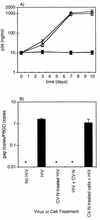
FIG. 1
FIG. 1
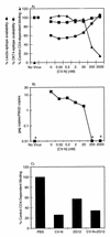
CV-N interacts with HIV virions but not host cells to inhibit HIV infection. HIV-1MN was either mock treated or pretreated with CV-N (200 nM) at 4°C for 90 min before performance of filtration dialysis twice through a 500-kDa-cutoff membrane to remove free CV-N. AA2 cells were either mock treated (—◊—) or pretreated with CV-N (—□—) (200 nM) at 37°C for 90 min and washed three times before being plated in duplicate at 0.5 × 10⁶ cells per well in a total volume of 3 ml and infected with HIV-1MN. CV-N-treated HIV-1MN (—●—) or HIV-1MN and CV-N (—▴—) were added to AA2 cells on day 0 and left in for the duration of the 10-day assay. (A) p24 accumulation. One hundred microliters of culture supernatant was sampled every 3 days to assay for virus production as measured by capsid p24 content. CV-N (200 nM) was nontoxic to the cells (by trypan blue exclusion [data not shown]). Nonincreasing amounts of p24 seen for CV-N-treated HIV or simultaneous addition of CV-N and HIV reflect residual virus inoculum. (B) Viral entry assay (PCR). The experiment in panel A was set up in duplicate, and the cells were harvested 24 h postinoculation and assayed for gag DNA by quantitative real-time PCR as described in Materials and Methods. ∗, no gag DNA detected (<30 copies per reaction). Data are representative of three independent experiments. PBGD, porphobilinogen deaminase.
FIG. 2
FIG. 2
CV-N blocks HIV-1-mediated fusion from without. (A) Untreated Sup-T1 T cells, highly susceptible to CD4-dependent, HIV-1 envelope-mediated cell fusion. (B) Following a 2-h incubation with concentrated native HIV-1 (6,400 ng of p24CA per ml), characteristic syncytia are seen, reflecting virion-mediated fusion from without. (C) CV-N pretreatment (200 nM) of virions blocks fusion. (D) Fusion mediated by native virions is inhibited by prior incubation of cells with anti-Leu3a MAb (10 μg/ml).
FIG. 3
FIG. 3
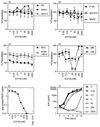
Effect of CV-N on Env-mediated cell fusion. The vaccinia virus-based reporter gene cell fusion assay was used (see Materials and Methods). The indicated CV-N concentrations represent those in the final fusion mixture. The background β-galactosidase activity value (0.5), obtained with the uncleavable nonfusogenic Env mutant, Unc, was subtracted from each value obtained with the active Envs. For each Env, 100% is defined as the β-galactosidase activity obtained in the absence of CV-N (optical density/minute × 1,000; LAV and 89.6, 36.8). Error bars indicate standard deviations of the mean values obtained from duplicate samples.
FIG. 4
FIG. 4
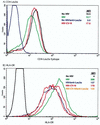
Binding of HIV and CV-N-treated virions to A3.01 cells. A3.01 (CD4-positive) cells were either mock treated or treated with 10 μg of anti-Leu3a MAb per ml for 30 min at 4°C and washed twice before addition of HIV-1MN, which was either mock treated or treated with CV-N (200 nM). The virions were added to A3.01 cells, and binding was assessed by quantitating virion-associated HLA-DR signal by flow cytometry, where HLA-DR acquisition indicates overall virion binding, and by Leu3a staining, where loss of availability of the Leu3a epitope is an indirect indication of CD4-dependent virion binding. Numbers in figure keys are MFI values. (A) CD4-Leu3a signal (gp120 binding epitope on CD4). A3.01 cells express the Leu3a epitope on CD4 (black trace), and saturating unlabeled anti-Leu3a MAb pretreatment blocks binding of fluorescein isothiocyanate-labeled anti-Leu3a MAb (compare blue and black traces). HIV binding blocks the Leu3a epitope (compare black and green traces). Cells with bound CV-N-treated virions have greater availability of Leu3a epitopes than do cells with bound untreated virions (compare red and green traces). (B) HLA-DR signal. A3.01 cells are HLA-DR negative (black trace). Addition of untreated HIV results in acquisition of HLA-DR staining (compare black and green traces). Approximately 56% of virion binding is CD4 dependent (compare green and blue traces). CV-N treatment of virions inhibits overall virion binding (compare green and red traces). CV-N inhibits CD4-dependent binding of virions; note the lack of increased inhibition of virion binding for CV-N-treated virions on unlabeled anti-Leu3a MAb-pretreated cells (compare blue and orange traces). At least 10,000 events were acquired for each sample.
FIG. 5
FIG. 5
CV-N concentration-dependent effects on virion binding (A) and infectivity (B) and effects of MAb 2G12 and CV-N on CD4-dependent binding (C). A3.01 (CD4-positive) cells were either mock treated with PBS or treated with 10 μg of unlabeled anti-Leu3a MAb per ml for 30 min at 4°C and washed twice before addition of HIV-1MN (17,000 ng of p24 per ml), which was either mock treated or treated with CV-N at various concentrations ranging from 0.02 to 2000 nM. (A) Percent CD4-dependent virion binding (anti-HLA-DR [—▴—]) was calculated by subtracting the MFI for CD4-independent binding (virion binding in the presence of the anti-Leu3a MAb) from total binding with 100% binding equal to binding in the absence of CV-N. MFI values for untreated virions: total binding to untreated cells, 286; binding to unlabeled anti-Leu3a MAb-pretreated cells, 127. Percent Leu3a epitope availability (—●—) and percent OKT4 epitope availability (—■—) were calculated by dividing the anti-Leu3a or anti-OKT4 signal in the presence of untreated virions by the anti-Leu3a MAb or anti-OKT4 MAb signal on untreated cells. MFI values for untreated cells: anti-Leu3a, 19.5; anti-OKT4, 33.1; for HIV-1 bound to cells, anti-Leu3a, 12.7; anti-OKT4, 30. (B) Parallel samples used for the binding assay shown in panel A were used in a quantitative PCR-based viral entry assay. ∗, no gag DNA detected (<30 copies per reaction). PBGD, porphobilinogen deaminase. (C) Effect of MAb 2G12 (50 μg/ml) alone or with CV-N (200 nM) on CD4-dependent HIV-1MN virion binding. Anti-HLA-DR MAb MFI values for virions (untreated cells and anti-Leu3a-treated cells, respectively): HIV-1-bound cells, 318 and 106 (67% CD4-dependent binding); CV-N-treated virions, (219 and 163; 2G12-treated virions, 222 and 102; CV-N–2G12-treated virions, 252 and 180. Percent CD4-dependent binding was calculated as for panel A. Each data point represents at least 10,000 acquired events.
FIG. 6
FIG. 6
Whole-virion immunoprecipitation of CV-N-treated virions. HIV-1MN (A, B, C, E, and F) or HIVIIIB (D) was either mock treated or treated with CV-N for 90 min at 4°C and precipitated with MAb or antisera to virion surface proteins. Values shown are means ± standard deviations for triplicate measurements in one experiment, representative of three independent experiments with similar results. (A) Polyclonal Ab to host cell-derived virion surface proteins, including MHC class I, MHC class II, and an antiserum raised against microvesicle preparations derived from H9 cells (7). (B) gp120 with IgG1b12 (CD4bs), W0/07 (V3), or F105 (CD4bs). (C) CD4i conformational epitope 48.d. After virions were pretreated with CV-N, sCD4 or 1% BSA was added, and the virions were precipitated with the 48.d MAb. (D) 2G12 MAb distinct neutralizing epitope on HIV-1MN and HIV-1IIIB. (E and F) Parallel samples from panels A to C (HIV-1MN) were used in a quantitative PCR-based viral entry assay (E) (∗, no gag DNA detected [<30 copies per reaction]) and a p24CA infectivity ELISA (F). Neg. Cont., negative control. PBGD, porphobilinogen deaminase.
FIG. 7
FIG. 7
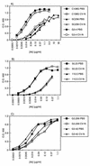
Effect of CV-N on binding of anti-gp120 MAbs to monomeric gp120: CV-N blocks 2G12 binding, but 2G12 does not block CV-N binding. HIVIIIB monomeric gp120 was applied as a coating to ELISA plates, and the ability of CV-N to compete with a panel of MAbs was examined. (A) V2 loop Abs; (B) V3 loop Abs; (C) C4 region Abs; (D) CD4bs Ab; (E) CD4i epitope Abs; (F) 2G12 MAb; (G and H) captured gp120 was pretreated with CV-N prior to incubation with 2G12 and detection of bound MAb (G) or pretreated with 2G12 prior to incubation with CV-N and detection of bound CV-N (H). O.D., optical density. The results for panels G and H were obtained by using the second assay protocol described under “ELISA studies” in Materials and Methods.
FIG. 7
FIG. 7
Effect of CV-N on binding of anti-gp120 MAbs to monomeric gp120: CV-N blocks 2G12 binding, but 2G12 does not block CV-N binding. HIVIIIB monomeric gp120 was applied as a coating to ELISA plates, and the ability of CV-N to compete with a panel of MAbs was examined. (A) V2 loop Abs; (B) V3 loop Abs; (C) C4 region Abs; (D) CD4bs Ab; (E) CD4i epitope Abs; (F) 2G12 MAb; (G and H) captured gp120 was pretreated with CV-N prior to incubation with 2G12 and detection of bound MAb (G) or pretreated with 2G12 prior to incubation with CV-N and detection of bound CV-N (H). O.D., optical density. The results for panels G and H were obtained by using the second assay protocol described under “ELISA studies” in Materials and Methods.

Similar articles

Cited by

References

    1. Alkhatib G, Broder C C, Berger E A. Cell type-specific fusion cofactors determine human immunodeficiency virus type 1 tropism for T-cell lines versus primary macrophages. J Virol. 1996;70:5487–5494. - PMC - PubMed
    1. Alkhatib, G., and E. A. Berger. Unpublished data.
    1. Arthos J, Deen K C, Chaikin M A, Fornwald J A, Sathe G, Sattentau Q J, Clapham P R, Weiss R A, McDougal J S, Pietropaolo C, et al. Identification of the residues in human CD4 critical for the binding of HIV. Cell. 1989;57:469–481. - PubMed
    1. Arthur L O, Bess J W, Jr, Sowder R C D, Benveniste R E, Mann D L, Chermann J C, Henderson L E. Cellular proteins bound to immunodeficiency viruses: implications for pathogenesis and vaccines. Science. 1992;258:1935–1938. - PubMed
    1. Attanasio R, Diley D, Buck D, Maino V C, Lohman K L, Kanda P, Kennedy R C. Structural characterization of a cross-reactive idiotype shared by monoclonal antibodies specific for the human CD4 molecule. J Biol Chem. 1991;266:14611–14619. - PubMed

Publication types