

Effects of interleukin-1 receptor antagonist overexpression on infection by Listeria monocytogenes
- PMID: 10085034
- PMCID: PMC96544
- DOI: 10.1128/IAI.67.4.1901-1909.1999
Effects of interleukin-1 receptor antagonist overexpression on infection by Listeria monocytogenes
Abstract
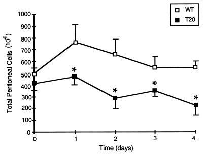
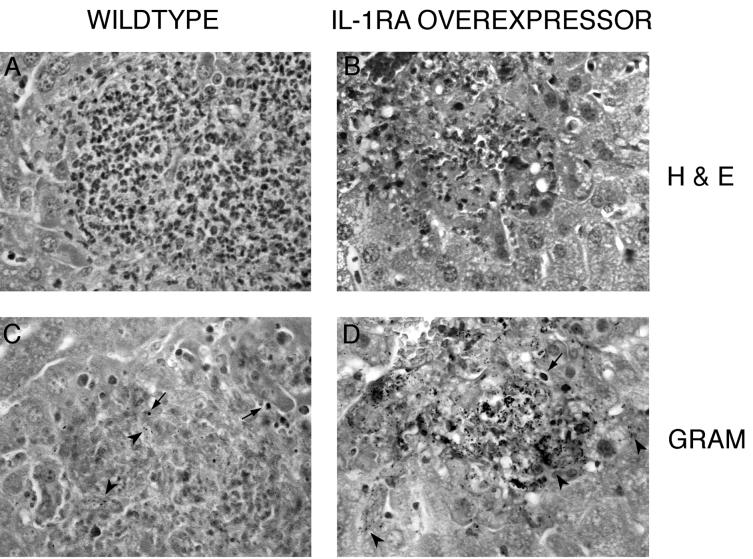
Interleukin-1 receptor antagonist (IL-1ra) is a naturally occurring cytokine whose only known function is the inhibition of interleukin-1 (IL-1). Using a reverse genetic approach in mice, we previously showed that increasing IL-1ra gene dosage leads to reduced survival of a primary listerial infection. In this study, we characterize further the role of endogenously produced IL-1ra and, by inference, IL-1 in murine listeriosis. IL-1ra overexpression inhibits, but does not eliminate, primary immune responses, reducing survival and increasing bacterial loads in the target organs. We demonstrate that IL-1ra functions in the innate immune response to regulate the peak leukocyte levels in the blood, the accumulation of leukocytes at sites of infection, and the activation of macrophages during a primary infection. Reduced macrophage class II major histocompatibility complex expression was observed despite increased gamma interferon (IFN-gamma) levels, suggesting that IL-1 activity is essential along with IFN-gamma for macrophage activation in vivo. We also show that IL-1ra plays a more limited role during secondary listeriosis, blunting the strength of the delayed-type hypersensitivity response to listerial antigen while not significantly altering cellular immunity to a second infectious challenge. When these results are compared to those for other mutant mice, IL-1ra appears to be unique among the cytokines studied to date in its regulation of leukocyte migration during primary listeriosis.
Figures


Similar articles
-
Functions of interleukin 1 receptor antagonist in gene knockout and overproducing mice.Proc Natl Acad Sci U S A. 1996 Oct 1;93(20):11008-13. doi: 10.1073/pnas.93.20.11008. Proc Natl Acad Sci U S A. 1996. PMID: 8855299 Free PMC article.
-
Differential regulation of cytokine and cytokine receptor mRNA expression upon infection of bone marrow-derived macrophages with Listeria monocytogenes.Infect Immun. 1996 Sep;64(9):3475-83. doi: 10.1128/iai.64.9.3475-3483.1996. Infect Immun. 1996. PMID: 8751887 Free PMC article.
-
The epistatic interrelationships of IL-1, IL-1 receptor antagonist, and the type I IL-1 receptor.J Immunol. 2002 Jul 1;169(1):393-8. doi: 10.4049/jimmunol.169.1.393. J Immunol. 2002. PMID: 12077269
-
Endogenous IL-1 is required for neutrophil recruitment and macrophage activation during murine listeriosis.J Immunol. 1994 Sep 1;153(5):2093-101. J Immunol. 1994. PMID: 8051414
-
IL-1 receptor antagonist (IL-1ra) expression, function, and cytokine-mediated regulation during mycobacterial and schistosomal antigen-elicited granuloma formation.J Immunol. 1996 Apr 1;156(7):2503-9. J Immunol. 1996. PMID: 8786311
Cited by
-
T cell-extrinsic IL-1 signaling controls long-term gammaherpesvirus infection by suppressing viral reactivation.Virology. 2022 Nov;576:134-140. doi: 10.1016/j.virol.2022.09.006. Epub 2022 Oct 7. Virology. 2022. PMID: 36244319 Free PMC article.
-
Interleukin-1 receptor type 1 is essential for control of cerebral but not systemic listeriosis.Am J Pathol. 2007 Mar;170(3):990-1002. doi: 10.2353/ajpath.2007.060507. Am J Pathol. 2007. PMID: 17322383 Free PMC article.
-
Sequelae of Fetal Infection in a Non-human Primate Model of Listeriosis.Front Microbiol. 2019 Sep 11;10:2021. doi: 10.3389/fmicb.2019.02021. eCollection 2019. Front Microbiol. 2019. PMID: 31572310 Free PMC article.
-
Interferon gamma signaling alters the function of T helper type 1 cells.J Exp Med. 2000 Oct 2;192(7):977-86. doi: 10.1084/jem.192.7.977. J Exp Med. 2000. PMID: 11015439 Free PMC article.
-
Alignment of practices for data harmonization across multi-center cell therapy trials: a report from the Consortium for Pediatric Cellular Immunotherapy.Cytotherapy. 2022 Feb;24(2):193-204. doi: 10.1016/j.jcyt.2021.08.007. Epub 2021 Oct 26. Cytotherapy. 2022. PMID: 34711500 Free PMC article.
References
-
- Czuprynski C J, Brown J F, Young K M, Cooley A J, Kurtz R S. Effects of murine recombinant interleukin 1α on the host response to bacterial infection. J Immunol. 1988;140:962–968. - PubMed
-
- Dai W J, Bartens W, Köhler G, Hufnagel M, Kopf M, Brombacher F. Impaired macrophage listericidal and cytokine activities are responsible for the rapid death of Listeria monocytogenes-infected IFN-γ receptor-deficient mice. J Immunol. 1997;158:5297–5304. - PubMed
-
- Dai W J, Köhler G, Brombacher F. Both innate and acquired immunity to Listeria monocytogenes infection are increased in IL-10-deficient mice. J Immunol. 1997;158:2259–2267. - PubMed
-
- Dinarello C A. Biologic basis for interleukin-1 in disease. Blood. 1996;87:2095–2147. - PubMed
Publication types
MeSH terms
Substances
Grants and funding
LinkOut - more resources
Full Text Sources
Medical
