. 1999 Mar;73(3):2509-16.
doi: 10.1128/JVI.73.3.2509-2516.1999.

Moloney murine leukemia virus infects cells of the developing hair follicle after neonatal subcutaneous inoculation in mice

Affiliations

Moloney murine leukemia virus infects cells of the developing hair follicle after neonatal subcutaneous inoculation in mice

M A Okimoto et al. J Virol. 1999 Mar.

Abstract

The nature of Moloney murine leukemia virus (M-MuLV) infection after a subcutaneous (s.c.) inoculation was studied. We have previously shown that an enhancer variant of M-MuLV, Mo+PyF101 M-MuLV, is poorly leukemogenic when used to inoculate mice s.c., but not when inoculated intraperitoneally. This attenuation of leukemogenesis correlated with an inability of Mo+PyF101 M-MuLV to establish infection in the bone marrow of mice at early times postinfection. These results suggested that a cell type(s) is infected in the skin by wild-type but not Mo+PyF101 M-MuLV after s.c. inoculation and that this infection is important for the delivery of infection to the bone marrow, as well as for efficient leukemogenesis. To determine the nature of the cell types infected by M-MuLV and Mo+PyF101 M-MuLV in the skin after a s.c. inoculation, immunohistochemistry with an anti-M-MuLV CA antibody was performed. Cells of developing hair follicles, specifically cells of the outer root sheath (ORS), were extensively infected by M-MuLV after s.c. inoculation. The Mo+PyF101 M-MuLV variant also infected cells of the ORS but the level of infection was lower. By Western blot analysis, the level of infection in skin by Mo+PyF101 M-MuLV was approximately 4- to 10-fold less than that of wild-type M-MuLV. Similar results were seen when a mouse keratinocyte line was infected in vitro with both viruses. Cells of the ORS are a primary target of infection in vivo, since a replication defective M-MuLV-based vector expressing beta-galactosidase also infected these cells after a s.c. inoculation.


Figures
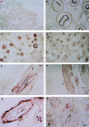
FIG. 1
FIG. 1
M-MuLV infection in the skin. Neonatal NIH Swiss mice were inoculated s.c. with ca. 106 fluorescence immunoassay PFU (see Materials and Methods). Animals were sacrificed at various times postinfection, and skin from around the site of inoculation was harvested, paraffin embedded, and sectioned. Immunohistochemistry was then performed on these sections to identify infected cells. The sections were first incubated with an antibody to the M-MuLV CA protein. This was followed by incubation with a horseradish peroxidase-conjugated goat anti-rabbit secondary antibody. The tissue was then incubated with a horseradish peroxidase substrate (that gives a purple color after reaction), and infected cells were visualized by light microscopy. Examples of this are shown here. Panels A and B show skin samples from control uninoculated mice that were stained with the anti-CA antibody. Panels C, E, and G show skin samples from mice that were infected with wild-type M-MuLV and sacrificed 4 weeks postinfection. Longitudinal (E) and transverse (C and G) cross-sections of hair follicles are shown; panel G is at higher magnification. Note the intense signal from cells that make up the outer region of the hair follicles (the ORS), indicating extensive infection. Panels D, F, and H show skin samples from mice that were infected with Mo+PyF101 M-MuLV which were also sacrificed 4 weeks postinfection. The regions that are infected with Mo+PyF101 M-MuLV are the same as those infected with wild-type M-MuLV (cells of the ORS), but the intensity of the signal is far less. Small cells that were not associated with hair follicles were also found to be infected (arrow in panel G) with both M-MuLV and Mo+PyF101 M-MuLV, but these cells were not common. Bar, 10 μm.
FIG. 2
FIG. 2
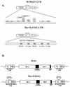
The M-MuLV and Mo+PyF101 LTRs. (A) M-MuLV LTR. The enhancer sequences of M-MuLV lie within two 75-bp direct repeats in the U3 region of the LTR. Each direct repeat contains consensus binding sites for a number of well-characterized transcription factors. The Mo+PyF101 LTR is shown in the lower half of panel A. The enhancer region of the polyomavirus strain F101 has been inserted directly downstream of the M-MuLV direct repeats. Binding motifs in the PyF101 enhancers include two copies of a BPV-like enhancer core (B core) and three copies of a polyoma enhancer core (C1 and C2) (7). None of the viral structural proteins are altered in Mo+PyF101 M-MuLV. (B) BAG and Mo+PyBAG vectors. The BAG vector has been previously described (19). It contains the wild-type M-MuLV LTR driving the transcription of the bacterial lacZ gene. It also contains the Neor gene as a selectable marker. The Mo+PyBAG vector is similar to the BAG vector except that it has the LTR from Mo+PyF101 M-MuLV regulating lacZ expression. Both vectors were obtained from transfected Psi-2 cells as described in Materials and Methods.
FIG. 3
FIG. 3
M-MuLV and Mo+PyF101 M-MuLV infection in the skin. Protein extracts were made from the skin of mice that had been infected s.c. with wild-type M-MuLV and Mo+PyF101 M-MuLV and sacrificed at various times postinfection. Western blot analysis was then performed on these extracts as described in Materials and Methods. Equal amounts of skin extract (10 μg of protein) were analyzed in each lane. With the anti-CA antibody, both the viral p30 and the Pr65 gag proteins were detected. As shown here, protein extracts from mice that had been infected with wild-type M-MuLV had significantly higher levels of both viral proteins than equivalent amounts of protein extract from age-matched mice infected with Mo+PyF101 M-MuLV.
FIG. 4
FIG. 4
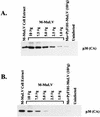
Quantification of Mo+PyF101 M-MuLV infection in the skin. To quantify the differences in the amount of virus present in the skin, serial dilutions of a wild-type M-MuLV-infected skin extract were compared to an extract from Mo+PyF101 M-MuLV-infected skin on the same Western blot. Panel A shows protein extract from mice sacrificed 4 weeks postinfection. A serial dilution of 10, 7.5, 5.0, 2.5, and 1.0 μg of M-MuLV-infected skin extract was compared to 10 μg of protein extract from an age-matched Mo+PyF101 M-MuLV-infected mouse. The viral p30 protein is shown. The intensity of the signal from 10 μg of the Mo+PyF101 M-MuLV skin extract was comparable to the signal seen from 1.0 μg of M-MuLV skin extract, indicating that there was approximately 10-fold less viral protein. A similar analysis was performed in panel B with mice that had been sacrificed 6 weeks postinfection. In this case, the Mo+PyF101 M-MuLV skin extract contained about fourfold less viral protein.
FIG. 5
FIG. 5
Infection by BAG vector in the skin. Neonatal NIH Swiss mice were inoculated with ca. 106 BAG vector particles. Mice were sacrificed 1 week postinfection, and skin was removed from around the site of inoculation and prepared as described above. Infected cells were visualized by immunohistochemistry with an anti-β-galactosidase antibody as described in Materials and Methods. In this protocol, a specific antibody reaction results in deposition of a brown stain. Panels A and B show skin samples from age-matched uninoculated mice stained with the anti-β-galactosidase antibody. Panels C and D show skin samples from BAG-infected mice. Note that in panel C cells in the ORS of the hair follicle are infected by the BAG vector and in panel D cells of the sebaceous gland are infected. Since the BAG vector is replication defective, these cells must be primary targets for M-MuLV infection. Bar, 10 μm.

Similar articles

Cited by

References

    1. Belli B, Fan H. The leukemogenic potential of an enhancer variant of Moloney murine leukemia virus varies with the route of inoculation. J Virol. 1994;68:6883–6889. - PMC - PubMed
    1. Brightman B K, Davis B R, Fan H. Preleukemic hematopoietic hyperplasia induced by Moloney murine leukemia virus is an indirect consequence of viral infection. J Virol. 1990;64:4582–4584. - PMC - PubMed
    1. Brightman B K, Okimoto M, Kulkarni V, Lander J K, Fan H. Differential behavior of the Mo+PyF101 enhancer variant of Moloney murine leukemia virus in rats and mice. Virology. 1998;242:60–67. - PubMed
    1. Chesebro B, Britt W, Evans L, Wehrly K, Nishio J, Cloyd M. Characterization of monoclonal antibodies reactive with murine leukemia viruses: use in analysis of strains of friend MCF and Friend ecotropic murine leukemia virus. Virology. 1983;127:134–148. - PubMed
    1. Coffin J, Hughes S H, Varmus H, editors. Retroviruses. Plainview, N.Y: Cold Spring Harbor Press; 1997. - PubMed

Publication types

LinkOut - more resources