. 1999 Jan;19(1):251-60.
doi: 10.1128/MCB.19.1.251.

hnRNP A1 recruited to an exon in vivo can function as an exon splicing silencer

Affiliations

hnRNP A1 recruited to an exon in vivo can function as an exon splicing silencer

F Del Gatto-Konczak et al. Mol Cell Biol. 1999 Jan.

Abstract

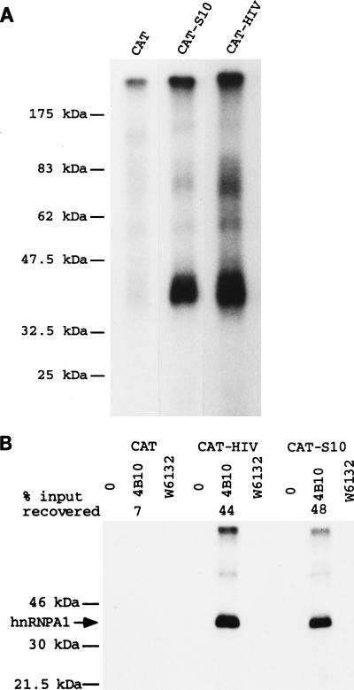
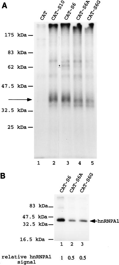
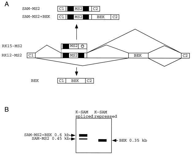
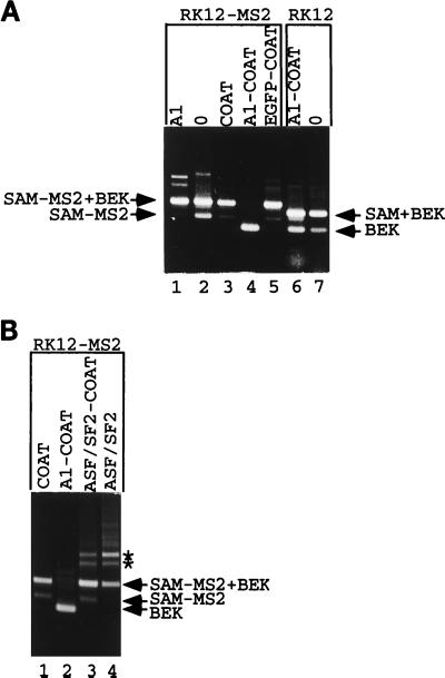
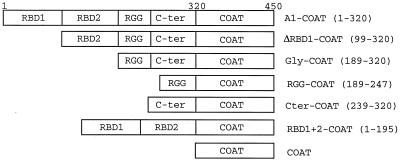
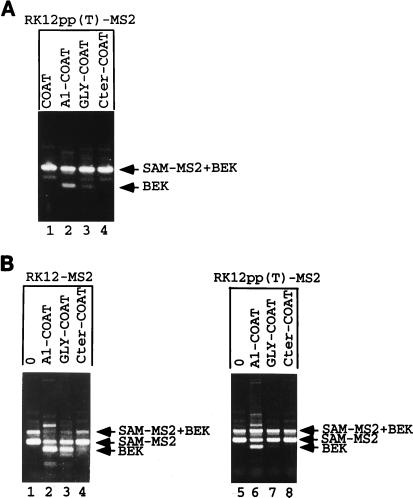
Some exons contain exon splicing silencers. Their activity is frequently balanced by that of splicing enhancers, and this is important to ensure correct relative levels of alternatively spliced mRNAs. Using an immunoprecipitation and UV-cross-linking assay, we show that RNA molecules containing splicing silencers from the human immunodeficiency virus type 1 tat exon 2 or the human fibroblast growth factor receptor 2 K-SAM exon bind to hnRNP A1 in HeLa cell nuclear extracts better than the corresponding RNA molecule without a silencer. Two different point mutations which abolish the K-SAM exon splicing silencer's activity reduce hnRNP A1 binding twofold. Recruitment of hnRNP A1 in the form of a fusion with bacteriophage MS2 coat protein to a K-SAM exon whose exon splicing silencer has been replaced by a coat binding site efficiently represses splicing of the exon in vivo. Recruitment of only the glycine-rich C-terminal domain of hnRNP A1, which is capable of interactions with other proteins, is sufficient to repress exon splicing. Our results show that hnRNP A1 can function to repress splicing, and they suggest that at least some exon splicing silencers could work by recruiting hnRNP A1.


Figures

FIG. 1
FIG. 1
Schematic representations of various minigenes. (A) The parent RK3 minigene with the Rous sarcoma virus long terminal repeat promoter (RSV) and the bovine growth hormone polyadenylation signal (BGH). Between the two are the constitutive exons C1 and C2 and the alternative exons K-SAM and BEK. Positions of primers used for RT-PCR are shown. Possible splicing patterns are shown, together with corresponding RNAs. (B) Structures of modified K-SAM exons found in other minigenes in the RK3 framework. ESS are stippled. Part of the CAT sequence is shown, and ESS sequences used to replace CAT sequences are underlined. Point mutations within the K-SAM exon ESS in RK12-S6A and -S6G are marked by asterisks. EcoRI and SalI sites used to remove fragments for in vitro transcription are marked.
FIG. 2
FIG. 2
RNA with either the K-SAM ESS or the HIV tat exon 2 ESS cross-links to hnRNP A1 in HeLa extracts. (A) The EcoRI-SalI fragments of RK12, RK12-S10, and RK12-HIV (Fig. 1B) were transcribed in vitro to yield 32P-labeled CAT, CAT-S10 (containing the K-SAM exon’s S10 ESS), or CAT-HIV (containing the HIV tat exon 2 ESS) RNAs, respectively. These RNAs were incubated in HeLa extract before UV cross-linking and analysis by SDS-PAGE. (B) CAT, CAT-HIV, and CAT-S10 RNAs as described above were added to a HeLa cell nuclear extract and immunoprecipitated with no antibody (0), anti-hnRNP A1 monoclonal antibody 4B10, or the irrelevant antibody W6132. Washed immunoprecipitates were exposed to UV light, treated with RNase T1, and subjected to SDS-PAGE. The percentage of input RNA recovered is shown only for the 4B10 series (average of five determinations); for all other series the percentage of RNA recovered was less than 2%. The expected migration of hnRNP A1 (35 kDa) is shown (arrow).
FIG. 3
FIG. 3
Effects of mutating the K-SAM exon’s ESS on UV-cross-linking results. (A) 32P-labeled CAT (lane 1), CAT-S10 (lane 2), CAT-S6 (lane 3), CAT-S6A (lane 4), and CAT-S6G (lane 5) RNAs as described in the text were obtained by in vitro transcription and incubated in HeLa extract before UV cross-linking and analysis by SDS-PAGE. Cross-linking to a 35-kDa protein is indicated by an arrow, and an asterisk marks a band discussed in the text for CAT-S6 RNA. (B) 32P-labeled CAT-S6 (lane 1), CAT-S6A (lane 2), and CAT-S6G (lane 3) RNAs were incubated in HeLa cell extract before immunoprecipitation with anti-hnRNP A1 antibody 4B10, UV cross-linking, and analysis by SDS-PAGE. Cross-linking to hnRNP A1 protein is indicated by an arrow. Relative quantification of the hnRNP A1 signals was by PhosphorImager analysis.
FIG. 4
FIG. 4
Possible splicing products of RNAs from minigenes with an MS2 operator within the K-SAM exon. (A) Schematic representation of fragments of minigenes RK12-MS2 and RK15-MS2. MS2, MS2 operator; —, K-SAM exon ESS. CAT sequences are in black. A partial structure of possible spliced RNAs is shown for RK12-MS2. (B) Representation of expected RT-PCR results following transfection of RK12-MS2 into 293 cells, depending on whether the K-SAM exon is spliced or repressed.
FIG. 5
FIG. 5
Recruitment of hnRNP A1 represses splicing. (A) RK12-MS2 was cotransfected into 293 cells with an expression vector coding for hnRNP A1 (lane 1), with the empty expression vector ΔCOAT (lane 2), or with expression vectors coding for coat (lane 3), an hnRNP A1-coat fusion (lane 4), or an EGFP-coat fusion (lane 5). RK12 was cotransfected into 293 cells with an expression vector coding for an hnRNP A1-coat fusion (lane 6) or the empty expression vector ΔCOAT (lane 7). Harvested RNA was subjected to RT-PCR with P1 and P2, and products were separated by gel electrophoresis. The origins of various fragments obtained are shown. The structures of named fragments are shown in Fig. 1A (for SAM+BEK [0.5 kb] and BEK [0.35 kb]) or Fig. 4 (for SAM-MS2 [0.45 kb] and SAM-MS2+BEK [0.6 kb]). (B) RK12-MS2 was cotransfected into 293 cells with the coat expression vector (lane 1) or with expression vectors coding for the hnRNP A1-coat fusion (lane 2), an ASF/SF2-coat fusion (lane 3), or ASF/SF2 devoid of any coat sequences (lane 4). Harvested RNA was analyzed as described for panel A. Asterisks mark two RT-PCR products discussed in the text which appear after overexpression of ASF/SF2 activity.
FIG. 6
FIG. 6
Schematic representations of hnRNP A1-coat fusions used, showing the various domains (RBD1, RBD2, RGG, and C-ter) making up the 320-amino-acid hnRNP A1. Numbers in parentheses indicate the amino acids of hnRNP A1 which have been fused to the 130-aa coat protein to make the different fusions. The full-length hnRNP A1 fusion (A1-COAT) is thus composed of 450 aa.
FIG. 7
FIG. 7
Recruitment of the glycine-rich C-terminal domain is sufficient to repress splicing. (A) Western analysis. 293 cells were transfected with FLAG-tagged expression vectors as marked (see Fig. 6 for the structures of hnRNP A1-derived fusions), and proteins were harvested and subjected to Western blotting with an anti-FLAG epitope antibody. A composite of two gels is shown. Sizes of fusion proteins, in kilodaltons: A1-COAT, 52; ΔRBD1-COAT, 41; GLY-COAT, 31; Cter-COAT, 25; RBD1+2-COAT, 40; COAT, 16.5; RGG-COAT, 23; and EGFP-COAT, 46. (B) 293 cells were transfected with RK15 (lane 1) or cotransfected with RK15-MS2 and expression vectors coding for the indicated coat fusion proteins (lanes 3 to 10) (see Fig. 6 for the structures of the hnRNP A1-derived fusions). 0, ΔCOAT (the empty expression vector) (lane 2). Harvested RNA was subjected to RT-PCR with P1 and P2, and products were separated by gel electrophoresis. The structures of the SAM-MS2+BEK and BEK fragments are shown in Fig. 4. (C) 293 cells were cotransfected with RK12-MS2 and expression vectors coding for the indicated coat fusion proteins (see Fig. 6 for their structures). Harvested RNA was subjected to RT-PCR with P1 and P2, and products were separated by gel electrophoresis. The structures of the SAM-MS2+BEK and BEK fragments are shown in Fig. 4.
FIG. 8
FIG. 8
hnRNP A1 repression can be relieved by reinforcing the exon’s polypyrimidine tract. (A) 293 cells were cotransfected with RK12pp(T)-MS2 and expression vectors coding for the indicated coat fusion proteins (see Fig. 6 for their structures). Harvested RNA was subjected to RT-PCR with P1 and P2, and products were separated by gel electrophoresis. The structures of the SAM-MS2+BEK and BEK fragments are shown in Fig. 4. (B) SVK14 cells were cotransfected with RK12-MS2 (lanes 1 to 4) or RK12pp(T)-MS2 (lanes 5 to 8) and expression vectors coding for the indicated coat fusion proteins (see Fig. 6 for their structures). Harvested RNA was subjected to RT-PCR with P1 and P2, and products were separated by gel electrophoresis. The structures of the SAM-MS2+BEK, SAM-MS2, and BEK fragments are shown in Fig. 4.

Similar articles

Cited by

References

    1. Adams M D, Tarng R S, Rio D C. The alternative splicing factor PSI regulates P-element third intron splicing in vivo. Genes Dev. 1997;11:129–138. - PubMed
    1. Amendt B A, Hesslein D, Chang L J, Stoltzfus C M. Presence of negative and positive cis-acting RNA splicing elements within and flanking the first Tat-coding exon of human immunodeficiency virus type 1. Mol Cell Biol. 1994;14:3960–3970. - PMC - PubMed
    1. Amendt B A, Si Z H, Stoltzfus C M. Presence of exon splicing silencers within human immunodeficiency virus type 1 Tat exon 2 and Tat-Rev exon 3: evidence for inhibition mediated by cellular factors. Mol Cell Biol. 1995;15:4606–4615. - PMC - PubMed
    1. Baba-Aissa F, Van Den Bosch L, Wuytack F, Raeymaekers L, Casteels R. Regulation of the sarco/endoplasmic reticulum Ca(2+)-ATPase (SERCA) 2 gene transcript in neuronal cells. Brain Res Mol Brain Res. 1998;55:92–100. - PubMed
    1. Black A C, Luo J, Watanabe C, Chun S, Bakker A, Fraser J K, Morgan J P, Rosenblatt J D. Polypyrimidine tract-binding protein and heterogeneous nuclear ribonucleoprotein A1 bind to human T-cell leukemia virus type 2 RNA regulatory elements. J Virol. 1995;69:6852–6858. - PMC - PubMed

Publication types

Substances

LinkOut - more resources