

Bradykinin stimulates type II alveolar cells to release neutrophil and monocyte chemotactic activity and inflammatory cytokines
- PMID: 9846978
- PMCID: PMC1866317
- DOI: 10.1016/S0002-9440(10)65702-1
Bradykinin stimulates type II alveolar cells to release neutrophil and monocyte chemotactic activity and inflammatory cytokines
Abstract
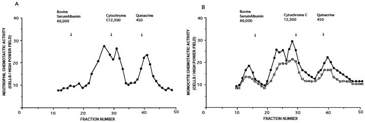
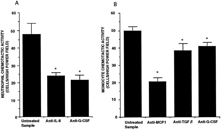
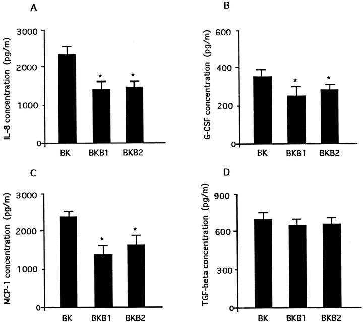
In the present study, we evaluated the potential of bradykinin (BK) to induce the release of neutrophil and monocyte chemotactic activity (NCA and MCA) and cytokines from an alveolar type II epithelial cell line, A549 cells. BK stimulated A549 cells to release NCA and MCA in a dose- and time-dependent manner (P < 0.001). Checkerboard analysis revealed that both NCA and MCA involved chemotactic and chemokinetic activity. Molecular sieve column chromatography showed three molecular weight masses (near 19 kd, 8 kd, and 400 d) for NCA and several molecular weight peaks (near 66 kd, 25 kd, 19 kd, 16 kd, and 400 d) for MCA. The release of NCA and MCA was inhibited by cycloheximide and lipoxygenase inhibitors (P < 0.01). The NCA and MCA were inhibited by leukotriene B4 (LTB4) receptor antagonist (P < 0.01), and the concentration of LTB4 was high enough for NCA and MCA. Antibodies to interleukin (IL)-8 and granulocyte colony-stimulating factor (G-CSF) attenuated NCA (P < 0.01), and antibodies to monocyte chemotactic protein-1 (MCP-1), G-CSF, and transforming growth factor (TGF)-beta attenuated MCA (P < 0.01). The levels of IL-8, G-CSF, MCP-1, and TGF-beta increased time dependently (P < 0.01). BK also stimulated the release of ILeukin-6 from A549 cells (P < 0.001). The receptors responsible for the release of NCA, MCA, and individual chemokines involved both BKB1 and BKB2 receptors. These data suggest that BK may stimulate alveolar type II pneumocytes to release inflammatory cytokines, which then may modulate the lung inflammation.
Figures


Similar articles
-
Bradykinin stimulates alveolar macrophages to release neutrophil, monocyte, and eosinophil chemotactic activity.J Immunol. 1996 Oct 1;157(7):3122-9. J Immunol. 1996. PMID: 8816423
-
Smoke extract stimulates lung fibroblasts to release neutrophil and monocyte chemotactic activities.Am J Physiol. 1999 Dec;277(6):L1149-57. doi: 10.1152/ajplung.1999.277.6.L1149. Am J Physiol. 1999. PMID: 10600885
-
Bradykinin stimulates lung fibroblasts to release neutrophil and monocyte chemotactic activity.Am J Respir Cell Mol Biol. 2000 Jan;22(1):75-84. doi: 10.1165/ajrcmb.22.1.3752. Am J Respir Cell Mol Biol. 2000. PMID: 10615068
-
Interleukin-8: An evolving chemokine.Cytokine. 2022 May;153:155828. doi: 10.1016/j.cyto.2022.155828. Epub 2022 Mar 2. Cytokine. 2022. PMID: 35247648 Review.
-
[The regulation of mast cell migration. Part 2: mast cell chemoattractants].Postepy Hig Med Dosw (Online). 2007 Sep 28;61:493-9. Postepy Hig Med Dosw (Online). 2007. PMID: 17909517 Review. Polish.
Cited by
-
Pulmonary epithelium, cigarette smoke, and chronic obstructive pulmonary disease.Int J Chron Obstruct Pulmon Dis. 2007;2(4):409-28. Int J Chron Obstruct Pulmon Dis. 2007. PMID: 18268916 Free PMC article. Review.
-
Effects of sivelestat, a new elastase inhibitor, on IL-8 and MCP-1 production from stimulated human alveolar epithelial type II cells.J Anesth. 2006;20(3):159-65. doi: 10.1007/s00540-006-0391-z. J Anesth. 2006. PMID: 16897233
-
Carbon black nanoparticles induce type II epithelial cells to release chemotaxins for alveolar macrophages.Part Fibre Toxicol. 2005 Dec 6;2:11. doi: 10.1186/1743-8977-2-11. Part Fibre Toxicol. 2005. PMID: 16332254 Free PMC article.
-
SARS-CoV-2 and ACE2: The biology and clinical data settling the ARB and ACEI controversy.EBioMedicine. 2020 Aug;58:102907. doi: 10.1016/j.ebiom.2020.102907. Epub 2020 Aug 6. EBioMedicine. 2020. PMID: 32771682 Free PMC article. Review.
-
Kinin B2 receptor regulates chemokines CCL2 and CCL5 expression and modulates leukocyte recruitment and pathology in experimental autoimmune encephalomyelitis (EAE) in mice.J Neuroinflammation. 2008 Nov 5;5:49. doi: 10.1186/1742-2094-5-49. J Neuroinflammation. 2008. PMID: 18986535 Free PMC article.
References
-
- Weiland JE, Davis WB, Holter JF, Mohammed JR, Dorinsky PM, Gadek JE: Lung neutrophils in the adult respiratory distress syndrome: clinical and pathophysiological significance. Am Rev Respir Dis 1986, 133:218-225 - PubMed
-
- Hunninghake GW, Garrett KC, Richerson HB, Fantone JC, Ward PA, Rennard SI, Bitterman PB, Crystal RG: Pathogenesis of the granulomatous lung disease. Am Rev Respir Dis 1984, 130:476-496 - PubMed
-
- Blusse van Oud Alblas A, van der Linden-Schrever B, van Furth R: Origin and kinetics of pulmonary macrophages during an inflammatory reaction induced by intra-alveolar administration of aerosolized heat-killed BCG. Am Rev Respir Dis 1983, 128:276-281 - PubMed
-
- Adanson IYR, Bowden JH: Role of monocytes and interstitial cell cells in the generation of alveolar macrophages. II. Kinetic studies after carbon loading. Lab Invest 1980, 42:518-531 - PubMed
MeSH terms
Substances
LinkOut - more resources
Full Text Sources
Research Materials
Miscellaneous
