

doi: 10.1073/pnas.95.25.14950.
The akt kinase: molecular determinants of oncogenicity
Affiliations
- PMID: 9843996
- PMCID: PMC24556
- DOI: 10.1073/pnas.95.25.14950
Item in Clipboard
The akt kinase: molecular determinants of oncogenicity
Proc Natl Acad Sci U S A.
.
Abstract
The serine-threonine kinase Akt is a downstream target of phosphoinositide 3-kinase (PI 3-kinase); it is activated by the phosphoinositide 3-phosphate-dependent kinases PDK1 and PDK2. Certain mutated forms of Akt induce oncogenic transformation in chicken embryo fibroblast cultures and hemangiosarcomas in young chickens. This ability to transform cells depends on localization of Akt at the plasma membrane and on the kinase activity of Akt. A transdominant negative form of Akt interferes with oncogenic transformation induced by the p3k oncogene, which codes for an activated form of PI 3-kinase. Akt is therefore an essential mediator of p3k-induced oncogenicity.
Figures
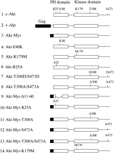
Schematic structures of Akt constructs. Group-specific antigen (Gag) sequence in v-Akt or myristylation signals are highlighted as black boxes. Functionally important amino acid residues in the PH and kinase domains are noted. The HA epitope tag is on the amino terminus of constructs 1 and 4–8; on all others, it is on the carboxyl terminus.
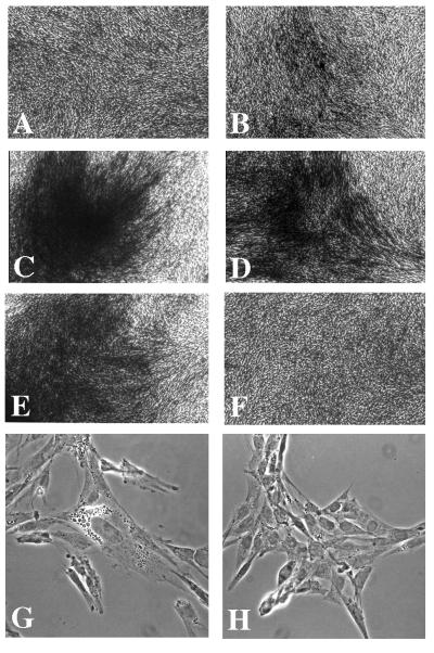
Morphology of CEF transfected with Akt constructs. (A) RCAS vector control. (B) c-Akt. (C) v-Akt. (D) Akt-E40K. (E) Akt-Myr. (F) Akt-K179M. (G and H) Phase-contrast micrographs of CEFs transfected with Akt-Myr (G) or RCAS vector (H).
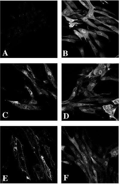
Subcellular localization of the HA-tagged Akt proteins in CEF. (A) RCAS vector control. (B) c-Akt. (C) v-Akt. (D) Akt-E40K. (E) Akt-Myr. (F) Akt-K179M.
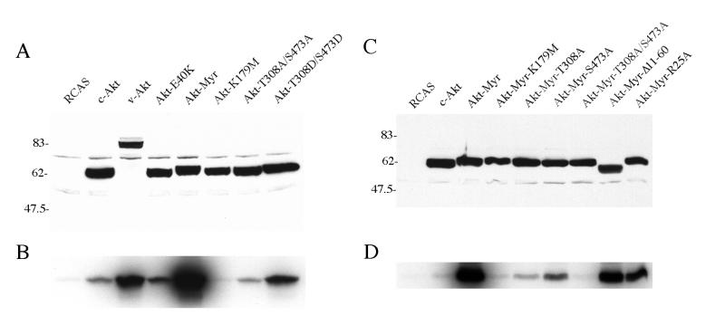
Expression and in vitro kinase activity of Akt proteins. (A and C) Lysates of CEF stably transfected with HA-tagged Akt were probed with anti-HA antibody in Western blots. (B and D) In vitro immune complex kinase assay of the HA-tagged Akt constructs with histone H2B as substrate.
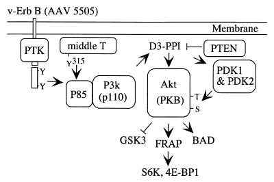
Akt and cellular signaling. See text for details.
Similar articles
-
Transformation of chicken cells by the gene encoding the catalytic subunit of PI 3-kinase.Science. 1997 Jun 20;276(5320):1848-50. doi: 10.1126/science.276.5320.1848. Science. 1997. PMID: 9188528
-
A role of the kinase mTOR in cellular transformation induced by the oncoproteins P3k and Akt.Proc Natl Acad Sci U S A. 2001 Jan 2;98(1):136-41. doi: 10.1073/pnas.98.1.136. Proc Natl Acad Sci U S A. 2001. PMID: 11134523 Free PMC article.
-
Inhibition of protein synthesis by Y box-binding protein 1 blocks oncogenic cell transformation.Mol Cell Biol. 2005 Mar;25(6):2095-106. doi: 10.1128/MCB.25.6.2095-2106.2005. Mol Cell Biol. 2005. PMID: 15743808 Free PMC article.
-
Molecular targeting: PI3 kinase pathway.Ann Oncol. 2004;15 Suppl 4:iv233-9. doi: 10.1093/annonc/mdh932. Ann Oncol. 2004. PMID: 15477314 Review. No abstract available.
-
Kinase phosphorylation: Keeping it all in the family.Curr Biol. 1999 Jul 15;9(14):R521-4. doi: 10.1016/s0960-9822(99)80326-1. Curr Biol. 1999. PMID: 10421571 Review.
Cited by
-
Evi1 regulates Notch activation to induce zebrafish hematopoietic stem cell emergence.EMBO J. 2016 Nov 2;35(21):2315-2331. doi: 10.15252/embj.201593454. Epub 2016 Sep 16. EMBO J. 2016. PMID: 27638855 Free PMC article.
-
Restricted 12p amplification and RAS mutation in human germ cell tumors of the adult testis.Am J Pathol. 2000 Oct;157(4):1155-66. doi: 10.1016/S0002-9440(10)64631-7. Am J Pathol. 2000. PMID: 11021820 Free PMC article.
-
Akt3 and mutant V600E B-Raf cooperate to promote early melanoma development.Cancer Res. 2008 May 1;68(9):3429-39. doi: 10.1158/0008-5472.CAN-07-5867. Cancer Res. 2008. PMID: 18451171 Free PMC article.
-
Phosphatidylinositol 3-kinase-dependent activation of mammalian protein kinase B/Akt in Saccharomyces cerevisiae, an in vivo model for the functional study of Akt mutations.J Biol Chem. 2009 May 15;284(20):13373-13383. doi: 10.1074/jbc.M807867200. Epub 2009 Mar 23. J Biol Chem. 2009. PMID: 19307184 Free PMC article.
-
Liver clock protein BMAL1 promotes de novo lipogenesis through insulin-mTORC2-AKT signaling.J Biol Chem. 2014 Sep 12;289(37):25925-35. doi: 10.1074/jbc.M114.567628. Epub 2014 Jul 25. J Biol Chem. 2014. PMID: 25063808 Free PMC article.
References
-
- Chang H W, Aoki M, Fruman D, Auger K R, Bellacosa A, Tsichlis P N, Cantley L C, Roberts T M, Vogt P K. Science. 1997;276:1848–1850. - PubMed
-
- Toker A, Cantley L C. Nature (London) 1997;387:673–676. - PubMed
-
- Rodriguez-Viciana P, Warne P H, Dhand R, Vanhaesebroeck B, Gout I, Fry M J, Waterfield M D, Downward J. Nature (London) 1994;370:527–532. - PubMed
Publication types
MeSH terms
Substances
Grants and funding
LinkOut - more resources
Full Text Sources
Other Literature Sources
Research Materials
Miscellaneous
