. 1998 Dec;9(12):3561-78.
doi: 10.1091/mbc.9.12.3561.

Retrograde transport from the pre-Golgi intermediate compartment and the Golgi complex is affected by the vacuolar H+-ATPase inhibitor bafilomycin A1

Affiliations
Free PMC article

Retrograde transport from the pre-Golgi intermediate compartment and the Golgi complex is affected by the vacuolar H+-ATPase inhibitor bafilomycin A1

H Palokangas et al. Mol Biol Cell. 1998 Dec.
Free PMC article

Abstract

The effect of the vacuolar H+-ATPase inhibitor bafilomycin A1 (Baf A1) on the localization of pre-Golgi intermediate compartment (IC) and Golgi marker proteins was used to study the role of acidification in the function of early secretory compartments. Baf A1 inhibited both brefeldin A- and nocodazole-induced retrograde transport of Golgi proteins to the endoplasmic reticulum (ER), whereas anterograde ER-to-Golgi transport remained largely unaffected. Furthermore, p58/ERGIC-53, which normally cycles between the ER, IC, and cis-Golgi, was arrested in pre-Golgi tubules and vacuoles, and the number of p58-positive approximately 80-nm Golgi (coatomer protein I) vesicles was reduced, suggesting that the drug inhibits the retrieval of the protein from post-ER compartments. In parallel, redistribution of beta-coatomer protein from the Golgi to peripheral pre-Golgi structures took place. The small GTPase rab1p was detected in short pre-Golgi tubules in control cells and was efficiently recruited to the tubules accumulating in the presence of Baf A1. In contrast, these tubules showed no enrichment of newly synthesized, anterogradely transported proteins, indicating that they participate in retrograde transport. These results suggest that the pre-Golgi structures contain an active H+-ATPase that regulates retrograde transport at the ER-Golgi boundary. Interestingly, although Baf A1 had distinct effects on peripheral pre-Golgi structures, only more central, p58-containing elements accumulated detectable amounts of 3-(2, 4-dinitroanilino)-3'-amino-N-methyldipropylamine (DAMP), a marker for acidic compartments, raising the possibility that the lumenal pH of the pre-Golgi structures gradually changes in parallel with their translocation to the Golgi region.


Figures

Figure 1
Figure 1
Baf A1 inhibits the BFA-induced redistribution of Golgi mannosidase II to the ER. Control NRK cells (A and B) and cells pretreated for 3 h in medium containing 10−6 M Baf A1 (C and D) were either fixed directly (A and C) or after a further 10 min incubation in the presence of BFA (1 μg/ml) (B and D), followed by staining with antibodies against mannosidase II. In control cells, BFA causes an efficient relocation of mannosidase II to the ER (B), whereas in Baf A1-treated cells the redistribution of the protein is considerably slowed down (D). Accordingly, tubular intermediates of the transfer process can be observed (D and inset; arrows). The cells in D also display weak ER-like staining, indicating that the retrograde transport of mannosidase II is not completely blocked by Baf A1. Bars, 10 μm.
Figure 2
Figure 2
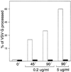
Baf A1 inhibits the BFA-induced redistribution of Golgi glycosyltransferases as measured by processing of the VSV tsO45 mutant G protein retained in the ER at the restrictive temperature (39.5°C). Chinese hamster ovary cells were infected with VSV tsO45 and, after virus adsorption, cultured for 3 h at 39.5°C in the presence or absence of Baf A1. The cells were pulse-labeled at 39.5°C with 35S-methionine/cysteine and then either transferred directly on ice or chased for 45–90 min at 39.5°C in the presence of different concentrations of BFA (0.2 or 5 μg/ml). The pulse–chase of Baf A1-pretreated cells was performed in the continuous presence of the drug. The tsO45 G protein was immunoprecipitated from cell lysates and analyzed, either undigested or after digestion with endo H, by SDS-PAGE. The amount of conversion of the G protein to the endo H-resistant form was quantitated as described in MATERIALS AND METHODS. White columns, Control cells; black columns, Baf A1-treated cells.
Figure 3
Figure 3
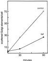
Baf A1 inhibits the nocodazole-induced redistribution of Golgi enzymes. Control and Baf A1-treated NRK cells were either fixed directly or after a further 30 or 90 min incubation in the presence of the microtubule-depolymerizing drug nocodazole (5 μM). The cells were stained for immunofluorescence microscopy using antibodies against Golgi mannosidase II, and the number of scattered, peripheral Golgi structures was determined as described in MATERIALS AND METHODS. Figure 1, A and C, also illustrates the typical distribution of mannosidase II in control and Baf A1-pretreated cells, respectively, before the addition of nocodazole (0 min time point).
Figure 4
Figure 4
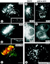
Accumulation of ERGIC-53 in globular and tubular pre-Golgi structures in Baf A1-treated cells. Human Hep-2 and HeLa cells were incubated for 3 h in the presence or absence of Baf A1 and immunostained for ERGIC-53 using monoclonal (G1/93) antibodies. (A and B) Confocal immunofluorescence images from the middle plane of control and Baf A1-treated Hep-2 cells, respectively. (C) Quantitation showing that Baf A1 significantly increases the number of detectable, ERGIC-53-positive pre-Golgi structures in both Hep-2 and HeLa cells. (D and inset) Optical section from the top of the nuclei, which best illustrates the accumulation of ERGIC-53 in both centrally and peripherally located pre-Golgi tubules (arrowheads) in Baf A1-treated Hep-2 cells. Bars, 10 μm (A, B, and D) and 2.5 μm (inset in D).
Figure 5
Figure 5
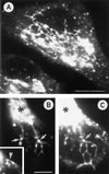
Visualization of the pre-Golgi tubules in Baf A1-treated PC12 cells using antibodies against rab1p. (A) Confocal immunofluorescence image demonstrating tubulation of the pre-Golgi structures in the drug-treated cells and the formation of an extensive, partly continuous reticulum. (B and C) Two partly overlapping confocal sections from the same cell showing details of a rab1p-positive reticulum that extends from the Golgi region (asterisks) towards the cell periphery. Note the localization of the globular IC domains to the branchpoints of the reticulum (arrows). The arrowheads indicate rab1p-positive tubules extending from and connecting the peripheral, globular pre-Golgi structures. Bars, 10 μm (A) and 5 μm (B).
Figure 6
Figure 6
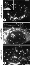
Pre-Golgi tubules can also be detected in control and 15°C-treated cells. Control NRK (A) and PC12 cells (A, inset) and NRK cells incubated for 2 h at 15°C (B and inset) were stained with antibodies against rab1p. Note the tubular extensions and connections of the pre-Golgi structures (arrowheads) that become more pronounced in response to the low-temperature treatment. The asterisk in B denotes the Golgi region. (C) The pre-Golgi tubules accumulating in NRK cells at 15°C also contain p58. Bars, 5 μm (A, B, and insets) and 2.5 μm (C).
Figure 7
Figure 7
Newly synthesized, anterogradely transported proteins are not concentrated in the pre-Golgi tubules accumulating in Baf A1-treated cells. (A–C) BHK-21 cells were infected with an SFV vector encoding the human TfR, treated with Baf-A1, and double-stained for rab1p (A) and TfR (B). C shows the merged confocal image. Partial colocalization of rab1p and TfR is observed in the punctate pre-Golgi structures (arrows), whereas both central and peripheral (see insets in A and B) rab1p-positive tubules do not contain detectable amounts of TfR. (D–G) Double-localization of rab1p (D and F) and the VSV-G protein (E and G) in tsO45 mutant-infected, Baf A1-treated cells incubated for 3 h at 39.5°C (D and E) or shifted for an additional 5 min to 32°C in the continuous presence of the drug (F and G), to synchronize the export of the G protein from the ER. At 39.5°C, the bulk of the G protein remains arrested in the ER and is not detected in the rab1p-positive tubules (arrowheads in D and E). In cells shifted to 32°C, the G protein and rab1p partially colocalize in the Golgi region (asterisk) and many of the punctate pre-Golgi structures (arrows), whereas the rab1p-positive tubules remain mostly devoid of the G protein. (H) To study the effect of Baf A1 on endocytic compartments, BHK-21 cells expressing human TfR were incubated in medium containing mouse anti-TfR antibodies. After Baf A1-treatment the cells were fixed, and the internalized TfR-antibody complexes were visualized using fluorochrome-coupled secondary antibodies. Note the tubular connections between peripheral and central endocytic structures (arrowheads). Bars, 5 μm (A–C, H) and 10 μm (D–G).
Figure 8
Figure 8
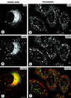
The lumenal pH of centrally located pre-Golgi structures is acidic. (A–C) NRK cells were incubated for 30 min at 37°C in medium containing the weak base DAMP (50 μM), followed by fixation and double-staining for p58 (A) and DAMP (B). C is the merged image, showing apparent colocalization of the two markers in the perinuclear Golgi region (asterisks). Some colocalization is also seen in a few more peripherally located pre-Golgi structures that can be distinguished as individual elements (arrows). (D–F) To disperse both endocytic and pre-Golgi structures, cells were treated for 2 h with 5 μM nocodazole, incubated in the presence of DAMP, and processed for confocal immunofluorescence microscopy as above. A number of the p58-positive structures colocalize with DAMP in these cells (arrows). The arrowheads indicate pre-Golgi structures that contain very weak DAMP labeling, giving rise to a light green color (instead of yellow) in the merged image. Bar, 10 μm.
Figure 9
Figure 9
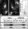
β-COP is redistributed in response to Baf A1 treatment, but its membrane-bound pool is not significantly affected. (A and B) Confocal immunofluorescence images from control (A) and Baf A1-treated NRK cells (B), stained with antibodies against β-COP, demonstrating an increased association of β-COP with peripheral pre-Golgi structures in response to Baf A1. Bar, 10 μm. The quantitation in C shows that Baf A1 causes an approximately twofold increase in the number of detectable pre-Golgi structures. (D) Association of β-COP with membranes. Postnuclear supernatant (pns), total membrane (m), and cytosol (cyto) fractions were prepared from control and Baf A1-treated PC12 cells. Equal amounts of protein from each fraction were run in SDS-PAGE, transferred to nitrocellulose, and analyzed for their content of β-COP, p58, and rab1p by quantitative immunoblotting.
Figure 10
Figure 10
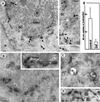
Baf A1 inhibits the formation of p58-containing 80 nm (COPI) vesicles. Control and Baf A1-treated PC12 cells were stained with antibodies against p58 and processed for immunoperoxidase electron microscopy. (A and inset) Control cells showing the presence of p58 in both cisternal and pleiomorphic elements (arrowheads) and a number of ∼80 nm vesicles or buds (arrows) at the cis face of the Golgi complex (GC). (B–D) In Baf A1-treated cells the cisternal organization and polarity of the Golgi are maintained, but both trans-elements (asterisks) and p58-positive cis-elements appear dilated. In spite of the more intensive staining of the cis-Golgi cisternae (B and inset), the p58-positive 80 nm vesicles are largely absent. C and D show p58-positive vacuoles (v) and tubules (arrows) observed in the drug-treated cells. (E) Quantitation showing that Baf A1 considerably reduces the number of p58-positive 80 nm vesicles and buds. A (inset) and B (inset)–D correspond to ∼50 nm and ∼200 nm sections, respectively. N, Nucleus; M, mitochondria. Bars, 0.2 μm.

Similar articles

Cited by

References

    1. Anderson RGW, Falck JR, Goldstein JL, Brown MS. Visualization of acidic organelles in intact cells by electron microscopy. Proc Natl Acad Sci USA. 1984;81:4838–4842. - PMC - PubMed
    1. Anderson RGW, Pathak RK. Vesicles and cisternae in the trans Golgi apparatus of human fibroblasts are acidic compartments. Cell. 1985;40:635–643. - PubMed
    1. Aniento F, Gu F, Parton RG, Gruenberg J. An endosomal β-COP is involved in the pH-dependent formation of transport vesicles destined for late endosomes. J Cell Biol. 1996;133:29–41. - PMC - PubMed
    1. Arar C, Carpentier V, Le Caer J-P, Monsigny M, Legrand A, Roche A-A. ERGIC-53, a membrane protein of the endoplasmic reticulum-Golgi intermediate compartment, is identical to MR60, an intracellular mannose-specific lectin of myelomonocytic cells. J Biol Chem. 1995;270:3551–3553. - PubMed
    1. Aridor M, Balch WE. Principles of selective transport: coat complexes hold the key. Trends Cell Biol. 1996;96:315–320. - PubMed

Publication types

MeSH terms