. 1998 Dec;9(12):3455-73.
doi: 10.1091/mbc.9.12.3455.

Sec61p serves multiple roles in secretory precursor binding and translocation into the endoplasmic reticulum membrane

Affiliations
Free PMC article

Sec61p serves multiple roles in secretory precursor binding and translocation into the endoplasmic reticulum membrane

M Pilon et al. Mol Biol Cell. 1998 Dec.
Free PMC article

Abstract

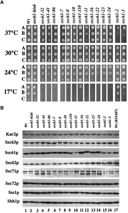
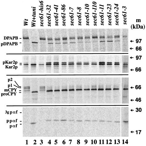
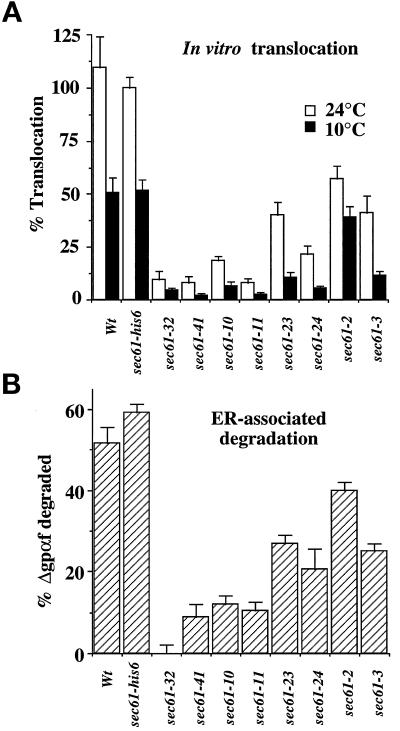
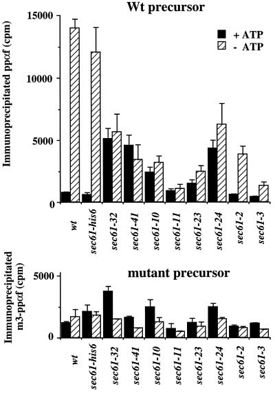
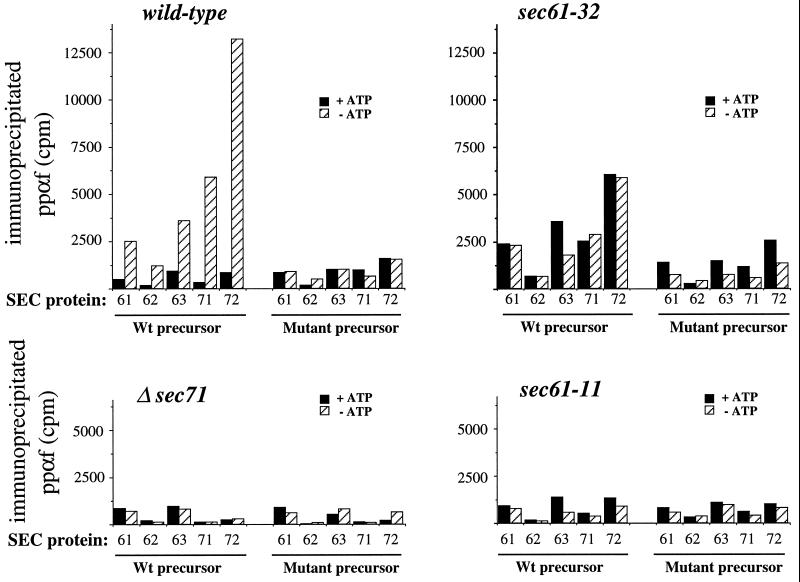
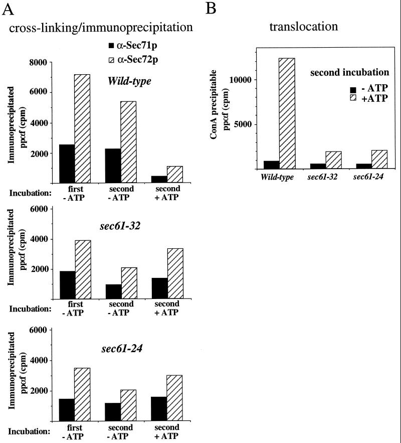
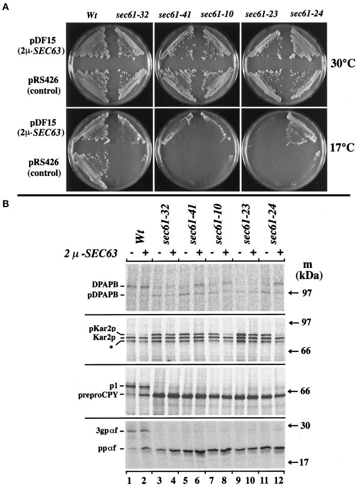
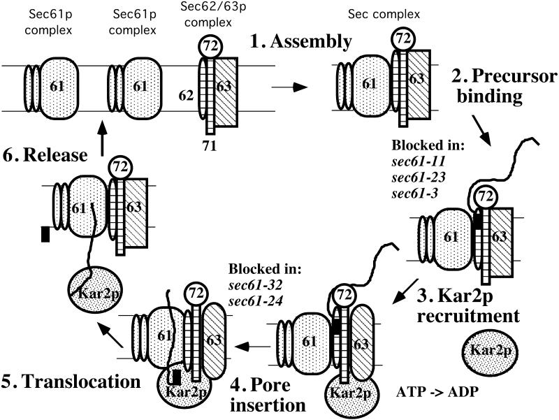
The evolutionarily conserved Sec61 protein complex mediates the translocation of secretory proteins into the endoplasmic reticulum. To investigate the role of Sec61p, which is the main subunit of this complex, we generated recessive, cold-sensitive alleles of sec61 that encode stably expressed proteins with strong defects in translocation. The stage at which posttranslational translocation was blocked was probed by chemical crosslinking of radiolabeled secretory precursors added to membranes isolated from wild-type and mutant strains. Two classes of sec61 mutants were distinguished. The first class of mutants was defective in preprotein docking onto a receptor site of the translocon that included Sec61p itself. The second class of mutants allowed docking of precursors onto the translocon but was defective in the ATP-dependent release of precursors from this site that in wild-type membranes leads to pore insertion and full translocation. Only mutants of the second class were partially suppressed by overexpression of SEC63, which encodes a subunit of the Sec61 holoenzyme complex responsible for positioning Kar2p (yeast BiP) at the translocation channel. These mutants thus define two early stages of translocation that require SEC61 function before precursor protein transfer across the endoplasmic reticulum membrane.


Figures

Figure 1
Figure 1
(A) Cold sensitivity of sec61 mutant strains. Cells carrying plasmids with the indicated SEC61 alleles were grown to an OD600 of 1. Aliquots (10 μl) of each culture (A) and 10-fold (B) and 100-fold (C) dilutions were plated on YPD plates, which were incubated at the indicated temperatures for 3 d (37, 30, and 24°C) or 5 d (17°C). Strain sec61-his6 has the wild-type SEC61 sequence with an N-terminal 6-histidine repeat; the presence of the 6-histidine repeat had no influence on growth, expression, or translocation. The sec61-2 and sec61-3 strains are previously published chromosomal sec61 mutants; sec61-32, sec61-41, and sec61-86 were obtained by hydroxylamine mutagenesis; the other Cs alleles were obtained by PCR mutagenesis. (B) Sec complex protein levels in SEC61 mutant membranes. Microsomal membranes were isolated from the indicated strains and grown at the permissive temperature (30°C), and equal amounts of protein were separated by SDS-PAGE on 7.5–17.5% gradient gels. Proteins were analyzed by immunoblotting using specific antibodies for the indicated Sec proteins. The shift in electrophoretic mobility of Sec61p caused by the presence of the 6-histidine repeat can be seen. RSY607 carries a wild-type chromosomal copy of SEC61.
Figure 2
Figure 2
Cs SEC61 mutant cells are deficient for protein translocation into the ER in vivo. Wild-type and mutant cells were pulse labeled with [35S]methionine/cysteine at 30°C for 15 min as described in MATERIALS AND METHODS, and secretory proteins were immunoprecipitated. The glycosylation inhibitor tunicamycin was present at 10 μg/ml in one culture of wild-type cells. The positions of precursor forms (pDPAPB, pKar2p, and ppαf), signal-cleaved, unglycosylated proteins (Kar2p, proCPY, and pαf), and signal-cleaved, glycosylated forms (DPAPB, p1, p2, mCPY, and 3 gpαf) are indicated.
Figure 3
Figure 3
Sec61 mutant strains are defective both for ppαf translocation into the ER and export for degradation (ERAD) of an unglycosylated pαf form. (A) In vitro translocation. ppαf was incubated with membranes in the presence of ATP at the indicated temperature for 40 min. Translocation was measured by the formation of fully glycosylated, protease-protected pαf as described in MATERIALS AND METHODS. Each bar is the average of five experiments with SE. Translocation in sec61-his6 at 24°C was set at 100%. Approximately one-third of the added precursor was translocated in these membranes. (B) ERAD. The glycosylation site mutant pΔgpαf was translocated at 24°C for 50 min, and then the membranes were washed and reincubated with 6 mg/ml cytosol in the presence of ATP. The decrease in the amount of signal-cleaved, unglycosylated pro-α-factor (Δgpαf) over 30 min was quantified as described in MATERIALS AND METHODS. Each bar is the average of three experiments with SE.
Figure 4
Figure 4
SEC61 mutants display defects in the interaction of ppαf with Sec72p in vitro. Radiolabeled ppαf wild-type or m3 (signal sequence)-mutant precursor were incubated with the indicated membranes (300,000 cpm precursor and 200 μg microsomal protein for each immunoprecipitation) either in the presence (filled bars) or absence (hatched bars) of ATP, followed by crosslinking with DSP and immunoprecipitation with Sec72p antibodies. The crosslinking and coimmunoprecipitation efficiency of ppαf was determined by liquid scintillation counting. Each bar is the average of three experiments with SE.
Figure 5
Figure 5
Sec61p, Sec62p, Sec63p, Sec71p, and Sec72p all interact with precursor proteins in the absence of ATP; these interactions are dependent on an intact signal sequence and are altered in sec61 mutant membranes. Radiolabeled ppαf wild-type or m3-mutant precursor (300,000 cpm and 200 μg microsomal protein per immunoprecipitation) were incubated with the indicated membranes either in the presence (filled bars) or absence (hatched bars) of ATP followed by crosslinking with DSP and immunoprecipitation using antibodies against the indicated Sec proteins. Each bar is the average of two separate experiments.
Figure 6
Figure 6
The Sec complex is intact in Cs sec61 strains. ER membrane proteins were fractionated after solubilization in digitonin. Total, total microsomal protein; Sol., digitonin-soluble protein recovered from the 100,000 × g supernatant; Free, fraction of digitonin-soluble proteins not binding to Con-A; Con-A, proteins binding to concanavalin A. Equal aliquots of each fraction were analyzed by SDS-PAGE and immunoblotting using the indicated antibodies in the first incubation. For Sec63p and Sec61p, [35S]protein A was used in the secondary incubation, and blots were exposed to a PhosphorImager. Sec72p was visualized using the ECL procedure. The band visible below Sec63p in the Sec63p immunoblots is unrelated.
Figure 7
Figure 7
Secretory precursor interaction with the receptor component Sec72p in sec61-11 membranes can be partly restored by adding wild-type Sec61p from an sec71 or sec72 deletion strain. Detergent (octylglucoside) extracts of the indicated wild-type (Wt) or mutant membranes were prepared and mixed in equal amounts before reconstitution of proteoliposomes by dialysis as described in MATERIALS AND METHODS. Binding of wild-type and m3-mutant ppαf precursor to Sec72p in the proteoliposomes reconstituted from 200 μg of solubilized membranes was assayed as in Figure 4. Each bar is the average of two experiments.
Figure 8
Figure 8
Sec61-32 and sec61-24 membranes are defective in the ATP-mediated precursor release from the Sec complex and translocation through the Sec61p pore. ppαf precursor (300,000 cpm for each precipitation) was incubated with membranes at 20°C in the absence of ATP to allow interaction with the receptor complex. Incubations were then cooled on ice for 2 min, followed by centrifugation for 5 min at 15,000 × g to collect the microsomes. Membranes were washed in buffer and reincubated either in the presence or absence of ATP for 20 min at 20°C. After 20 min each reaction was divided into three aliquots, and receptor interaction (A) was assessed by analyzing crosslinking to Sec71p and Sec72p as outlined in Figure 4; translocation (B) was assessed by trypsin treatment followed by Con-A precipitation to determine the amount of protease-protected glycosylated pαf in the incubations. Note that because only a fraction of ppαf can be crosslinked and immunoprecipitated, the amounts of precursor in A represent only a portion of the molecules bound to the translocation machinery.
Figure 9
Figure 9
Overexpression of SEC63 partially suppresses the cold sensitivity of a subset of sec61 mutant strains by enhancing translocation. (A) Strains with the indicated plasmids were streaked onto SC plates minus uracil and incubated at 30°C for 3 d or 17°C for 5 d. pDF15 is a vector with the full-length SEC63 sequence. pRS426 is a multicopy vector used as a control. (B) Indicated strains carrying either pRS426 control plasmid (−) or the 2μ-SEC63 plasmid pDF15 (+) were pulse labeled at 17°C followed by immunoprecipitation as in Figure 2. The positions of precursor forms (pDPAPB, pKar2p, preproCPY, and ppαf), signal-cleaved, unglycosylated proteins (Kar2p and pαf), and signal-cleaved, glycosylated forms (DPAPB, p1, and 3 gpαf) are indicated. The band marked with an asterisk is unrelated.
Figure 10
Figure 10
Cs sec61 mutants define two early stages of posttranslational translocation. The trimeric Sec61p complex consists of Sec61p and two smaller proteins, Sbh1p and Sss1p. The Sec61p complex and Sec62/Sec63p complex assemble to form the heptameric Sec complex (1), which binds precursor at a cytoplasmic docking site (2). After Kar2p recruitment and ATP hydrolysis (3), precursor is released from the docking site and inserts into the pore (4). Kar2p directly promotes the translocation (5) and release of precursors into the lumen (6). The drawing does not represent all the protein–protein interactions known to occur.

Similar articles

Cited by

References

    1. Allison DS, Young ET. Mutations in the signal sequence of prepro-α-factor inhibit both translocation into the endoplasmic reticulum and processing by signal peptidase in yeast cells. Mol Cell Biol. 1989;9:4977–4985. - PMC - PubMed
    1. Baldwin RL. Temperature dependence of the hydrophobic interaction in protein folding. Proc Natl Acad Sci USA. 1986;83:8069–8072. - PMC - PubMed
    1. Beckmann R, Bubeck D, Grassuci R, Penczek P, Verschoor A, Blobel G, Frank J. Alignment of conduits for the nascent polypeptide chain in the ribosome-Sec61 complex. Science. 1997;278:2123–2126. - PubMed
    1. Biederer T, Volkwein C, Sommer T. Degradation of subunits of the Sec61p complex, an integral component of the ER membrane, by the ubiquitin-proteasome pathway. EMBO J. 1996;15:2069–2076. - PMC - PubMed
    1. Brodsky JL, Goeckeler J, Schekman R. BiP and Sec63p are required for both co- and posttranslational protein translocation into the yeast endoplasmic reticulum. Proc Natl Acad Sci USA. 1995;92:9643–9646. - PMC - PubMed

Publication types

MeSH terms