. 1998 Nov 15;12(22):3579-90.
doi: 10.1101/gad.12.22.3579.

GATA6 regulates HNF4 and is required for differentiation of visceral endoderm in the mouse embryo

Affiliations

GATA6 regulates HNF4 and is required for differentiation of visceral endoderm in the mouse embryo

E E Morrisey et al. Genes Dev. .

Abstract

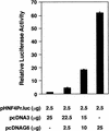
GATA6 belongs to a family of zinc finger transcription factors that play important roles in transducing nuclear events that regulate cellular differentiation and embryonic morphogenesis in vertebrate species. To examine the function of GATA6 during embryonic development, gene targeting was used to generate GATA6-deficient (GATA6(-/-)) ES cells and mice harboring a null mutation in GATA6. Differentiated embryoid bodies derived from GATA6(-/-) ES cells lack a covering layer of visceral endoderm and severely attenuate, or fail to express, genes encoding early and late endodermal markers, including HNF4, GATA4, alpha-fetoprotein (AFP), and HNF3beta. Homozygous GATA6(-/-) mice died between embryonic day (E) 6.5 and E7. 5 and exhibited a specific defect in endoderm differentiation including severely down-regulated expression of GATA4 and absence of HNF4 gene expression. Moreover, widespread programmed cell death was observed within the embryonic ectoderm of GATA6-deficient embryos, a finding also observed in HNF4-deficient embryos. Consistent with these data, forced expression of GATA6 activated the HNF4 promoter in nonendodermal cells. Finally, to examine the function of GATA6 during later embryonic development, GATA6(-/-)-C57BL/6 chimeric mice were generated. lacZ-tagged GATA6(-/-) ES cells contributed to all embryonic tissues with the exception of the endodermally derived bronchial epithelium. Taken together, these data suggest a model in which GATA6 lies upstream of HNF4 in a transcriptional cascade that regulates differentiation of the visceral endoderm. In addition, these data demonstrate that GATA6 is required for establishment of the endodermally derived bronchial epithelium.


Figures

Figure 1
Figure 1
Targeted disruption of the GATA6 gene. (A) Schematic representation of the GATA6 targeting strategy. (Top) Partial restriction endonuclease map of the murine GATA6 genomic locus. (•) Exon 3; (○) exons 4 and 5. Restriction enzymes: (Not) NotI; (Nco) NcoI; (B) BamHI. (Middle) GATA6 targeting vector containing the neomycin-resistance (neo) and herpes simplex virus thymidine kinase (tk) genes under the control of the PGK promoter. (Bottom) Structure of the targeted GATA6 mutant allele. (B) (Left) Southern blot analysis of DNA prepared from the offspring of a GATA6+/− × GATA6+/− mating. DNA was digested with BamHI and hybridized to the radiolabeled GATA6 genomic probe shown in Fig. 1A. The positions of the wild-type (13 kb) and targeted (8 kb) allele are indicated with arrows at left. (Right) Southern blot analysis of DNA prepared from wild-type (+/+), heterozygous (+/−), and homozygous mutant (−/−) ES cells demonstrating the wild-type (wt) and targeted (t) alleles. (C) (Top) Northern blot analysis of GATA6 gene expression in wild-type (+/+), heterozygous (+/−), and homozygous mutant (−/−) ES cells. (Bottom) Equal loading and integrity of RNA was assessed by ethidium bromide staining of the 28S RNA in the gel prior to transfer. (D) Western blot analysis of protein lysates prepared from wild-type (+/+) ES cells and homozygous mutant GATA6−/− (−/−) ES cells following 12 days of differentiation in vitro. Whole cell lysates prepared from differentiated wild-type and GATA6−/− ES cells were fractionated by SDS-PAGE, transferred to an Immobilon-P membrane, which was incubated with rabbit α-mouse GATA6 polyclonal antiserum, and visualized with goat anti-mouse horseradish peroxidase-coupled secondary antibody. The 56-kD marker is shown at right and the 48-kD band corresponding to GATA6 is indicated with an arrow at left.
Figure 1
Figure 1
Targeted disruption of the GATA6 gene. (A) Schematic representation of the GATA6 targeting strategy. (Top) Partial restriction endonuclease map of the murine GATA6 genomic locus. (•) Exon 3; (○) exons 4 and 5. Restriction enzymes: (Not) NotI; (Nco) NcoI; (B) BamHI. (Middle) GATA6 targeting vector containing the neomycin-resistance (neo) and herpes simplex virus thymidine kinase (tk) genes under the control of the PGK promoter. (Bottom) Structure of the targeted GATA6 mutant allele. (B) (Left) Southern blot analysis of DNA prepared from the offspring of a GATA6+/− × GATA6+/− mating. DNA was digested with BamHI and hybridized to the radiolabeled GATA6 genomic probe shown in Fig. 1A. The positions of the wild-type (13 kb) and targeted (8 kb) allele are indicated with arrows at left. (Right) Southern blot analysis of DNA prepared from wild-type (+/+), heterozygous (+/−), and homozygous mutant (−/−) ES cells demonstrating the wild-type (wt) and targeted (t) alleles. (C) (Top) Northern blot analysis of GATA6 gene expression in wild-type (+/+), heterozygous (+/−), and homozygous mutant (−/−) ES cells. (Bottom) Equal loading and integrity of RNA was assessed by ethidium bromide staining of the 28S RNA in the gel prior to transfer. (D) Western blot analysis of protein lysates prepared from wild-type (+/+) ES cells and homozygous mutant GATA6−/− (−/−) ES cells following 12 days of differentiation in vitro. Whole cell lysates prepared from differentiated wild-type and GATA6−/− ES cells were fractionated by SDS-PAGE, transferred to an Immobilon-P membrane, which was incubated with rabbit α-mouse GATA6 polyclonal antiserum, and visualized with goat anti-mouse horseradish peroxidase-coupled secondary antibody. The 56-kD marker is shown at right and the 48-kD band corresponding to GATA6 is indicated with an arrow at left.
Figure 2
Figure 2
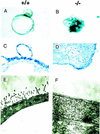
Morphology and ultrastructure of wild-type and GATA6−/− embryoid bodies following 12 days of differentiation in vitro. (A,B) Gross appearance of wild-type (+/+) (A) and homozygous mutant (−/−) (B) embryoid bodies. Magnification, 40×. (C,D) Toluidine blue-stained 1 μm cross-sections of differentiated wild-type (C) and GATA6−/− mutant (D) embryoid bodies. Magnification, 100×. (E,F) Representative electron microscopic analysis of the surface of differentiated embryoid bodies derived from wild-type ES cells (E) and GATA6−/− ES cells (F). Magnification, 10,000×.
Figure 3
Figure 3
Northern blot analysis of endodermal markers in wild-type and GATA6 null embryoid bodies. Northern blots were prepared with RNA harvested from wild-type (+/+) and GATA6 null (−/−) ES cells over a 12-day course of differentiation and were hybridized with radiolabeled α-fetoprotein (AFP), HNF3β, HNF4, or GATA4 probes. (Bottom) The 28S RNA band on the ethidium-stained gel prior to membrane transfer of RNA.
Figure 4
Figure 4
Expression of the GATA4 and GATA6 genes in the postimplantation mouse embryo. Histological and in situ hybridization analyses performed on E5.5 (A–C) and E6.5 (D–F) murine embryos. (A) A hematoxylin and eosin-stained section through the E5.5 embryo. (B) Hybridization of the GATA4 antisense riboprobe to yolk sac visceral endoderm (VE) and parietal endoderm (PE) of E5.5 embryos. (C) Hybridization of the antisense GATA6 riboprobe to the E5.5 embryo. (D) A hemotoxylin and eosin-stained section through the E6.5 embryo. (VE) Visceral endoderm, (PE) parietal endoderm, (EC) embryonic ectoderm. (E) Hybridization of the GATA4 (E) and GATA6 (F) antisense riboprobe to E6.5 embryos.
Figure 5
Figure 5
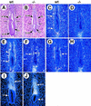
Expression of endodermal markers in wild-type and GATA6−/− mutant E6.5 embryos. Histological and in situ hybridization analyses performed on wild-type (wt) and GATA6−/− (−/−) mutant E6.5 embryos. (A,B) A hematoxylin and eosin-stained section of E6.5 wild-type (A) and GATA6−/− (B) embryos. (X-VE) Visceral endoderm at the extra-embryonic pole, (E-VE) visceral endoderm at the embryonic pole of the embryo, (PE) parietal endoderm, embryonic ectoderm (EC). (C,D) In situ hybridization performed with a radiolabeled GATA6 antisense riboprobe on a wild-type (C) and GATA6−/− (D) embryo. (E,F) In situ hybridization performed with a radiolabeled GATA4 antisense riboprobe on a wild-type (E) and GATA6−/− (D) embryo. (G,H) In situ hybridization performed with a radiolabeled HNF4 antisense riboprobe on a wild-type (G) and GATA6−/− (H) embryo. (Arrowheads) Extraembryonic VE. (I,J) In situ hybridization performed with a radiolabeled Pem antisense riboprobe in wild-type (I) and GATA6−/− (J) embryos. (EPC) Ectoplacental cone, (VE) proximal and distal visceral endoderm.
Figure 6
Figure 6
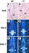
Expression of ectodermal markers in wild-type and GATA6−/− mutant E6.5 embryos. Histological and in situ hybridization analysis performed on wild-type (wt) and GATA6−/− (−/−) mutant E6.5 embryos. (A,B) Hematoxylin and eosin-stained section of an E6.5 wild-type (A) and GATA6−/− (B) embryo. (VE) Visceral endoderm, (EC) embryonic ectoderm. (C,D) In situ hybridization performed with an Otx2 antisense riboprobe on the wild-type (C) and GATA6−/− (D) embryos. (Arrowheads) Hybridization signal. (E,F) In situ hybridization was performed with an ENC1 antisense riboprobe on the wild-type (E) and GATA6−/− (F) mutant embryos. (Arrowheads) Hybridization. Of note, epifluorescence is also observed in red blood cells within the yolk sac cavity. Magnification, 40×.
Figure 7
Figure 7
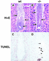
Cell death is observed in E7.0 GATA6−/− embryos. (A,B) Histological sections of E7.0 wild-type (wt) (A) and GATA6−/− (−/−) (B) embryos. (VE) Visceral endoderm, (PE) parietal endoderm. (C,D) E7.0 wild-type (C) and GATA6−/− mutant (D) embryos were subjected to the TUNEL reaction. Brown-stained nuclei indicate end incorporation in DNA. Rare TUNEL-positive nuclei were observed in red blood cells within the yolk sac cavity in wild-type embryos (C). In contrast, multiple TUNEL-positive nuclei (black arrowheads) were observed within the embryonic ectoderm of E7.0 GATA6−/− mutant embryos (D). In addition, an occasional TUNEL-positive cell (blue arrowhead) was observed within the parietal endoderm of GATA6−/− mutant embryos.
Figure 8
Figure 8
GATA6-modulated transactivation of the HNF4 promoter in NIH-3T3 cells. NIH-3T3 cells were transfected with 2.5 μg of the luciferase reporter plasmid (pcHNF4Pr.luc) containing the murine HNF4 promoter and varying amounts (μg) of an expression plasmid encoding GATA6 (pcDNAG6) or the control plasmid (pcDNA3). All transfection mixtures also contained 1 μg of the pMSVβgal reference plasmid. Forty-eight hours after transfection, luciferase and β-galactosidase activities were determined. Luciferase activities corrected for differences in transfection efficiencies were normalized to luciferase activity obtained following transfection of the pHNF4Pr.luc plasmid with the pcDNA3 control plasmid. The data are presented as relative luciferase activities ± s.e.m. The mean absolute light units obtained following transfection of the pHNF4Pr.luc reporter plasmid and the control plasmid pcDNA3 was 353.
Figure 9
Figure 9
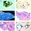
β-gal+GATA6−/− ES cells fail to contribute to the embryonic bronchial epithelium of GATA6−/−–C57BL/6 chimeric embryos. (A) X-gal staining of an E10.5 GATA6−/−–C57BL/6 chimeric embryo. (B) Histological analysis of a section through the thoracic region of an E10.5 GATA6−/−–C57BL/6 chimera. (v) Ventricular and (a) atrial chambers of the primitive heart, (nt) neural tube, (s) somites, (g) primitive foregut. Magnification, 10×. (C) In situ hybridization of a section through the primitive lung bud of an E13.5 mouse embryo by use of a GATA6 antisense riboprobe. (Arrowheads) Bronchial epithelium. (D) Histological analysis of a section through the primitive lung bud of an E13.5 chimeric embryo. (Arrowheads) Bronchial epithelium. Magnification 10×. (E) Same as D but magnification, 20×. (F) Histological analysis of a section through the primitive lung bud of a control E13.5 GATA6+/−–C57BL/6 chimeric embryo. β-gal+GATA6+/− cells contribute to both the bronchial epithelium (arrowheads) and the lung mesenchyme. Magnification, 20×.
Figure 10
Figure 10
A transcriptional cascade controlling differentiation of the visceral endoderm. As described in Results and published previously (Duncan et al. 1997; Kuo et al. 1997), analysis of the phenotypes of GATA6−/−, GATA4−/−, and HNF4−/− mutant embryos has shown that GATA6 activates (directly or indirectly) the expression of the early endodermal marker HNF4, which in turn, activates the expression of late endodermal markers, including α-fetoprotein (AFP), transferrin (TFN), Apo-AI, ApoAIV, and ApoB. In addition, GATA4 gene expression is down-regulated in GATA6-deficient embryos, whereas GATA6 gene expression is up-regulated in GATA4-deficient embryos suggesting cross-regulation between GATA6 and GATA4.

Similar articles

Cited by

References

    1. Arceci RJ, King AA, Simon MC, Orkin SH, Wilson DB. Mouse GATA-4: A retinoic acid-inducible GATA-binding transcription factor expressed in endodermally derived tissues and heart. Mol Cell Biol. 1993;13:2235–2246. - PMC - PubMed
    1. Bradley A. Teratocarcinomas and embryonic stem cells, a practical approach (ed. E.J. Robertson), pp. 113–151. IRL Press, Oxford, UK. 1987. Production and analysis of chimeric mice.
    1. Chang MW, Barr E, Seltzer J, Jiang Y-Q, Nabel GJ, Nabel EG, Parmacek MS, Leiden JM. Cytostatic gene therapy for vascular proliferative disorders with a constitutively active form of the retinoblastoma gene product. Science. 1995;267:518–522. - PubMed
    1. Chen WS, Manova K, Weinstein DC, Duncan SA, Plump AS, Prezioso VR, Bachvarova RF, Darnell JE. Disruption of the HNF-4 gene, expressed in visceral endoderm, leads to cell death in embryonic ectoderm and impaired gastrulation of mouse embryos. Genes & Dev. 1994;8:2466–2477. - PubMed
    1. Doetschman TC, Eistetter H, Katz M, Schmidt W, Kemler R. The in vitro development of blastocyst-derived embryonic stem cell lines: Formation of visceral yolk sac, blood islands and myocardium. J Embryol Exp Morphol. 1985;87:27–45. - PubMed

Publication types

MeSH terms