. 1998 Dec 1;18(23):9733-50.
doi: 10.1523/JNEUROSCI.18-23-09733.1998.

The vesicular GABA transporter, VGAT, localizes to synaptic vesicles in sets of glycinergic as well as GABAergic neurons

Affiliations

The vesicular GABA transporter, VGAT, localizes to synaptic vesicles in sets of glycinergic as well as GABAergic neurons

F A Chaudhry et al. J Neurosci. .

Abstract

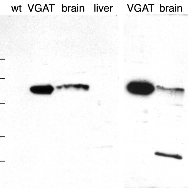
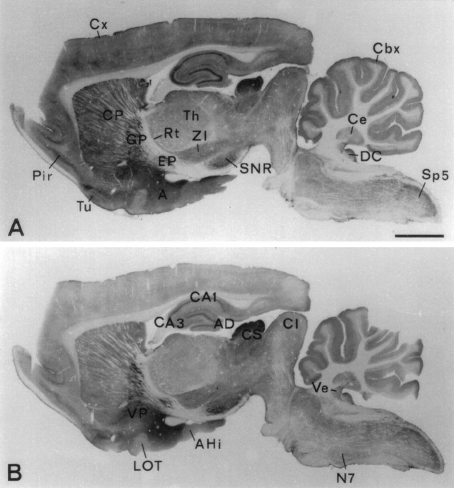
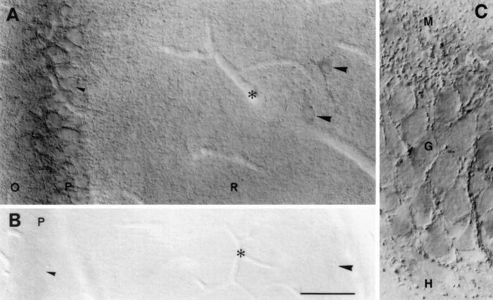
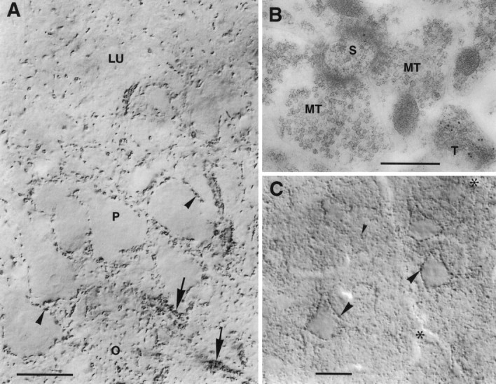
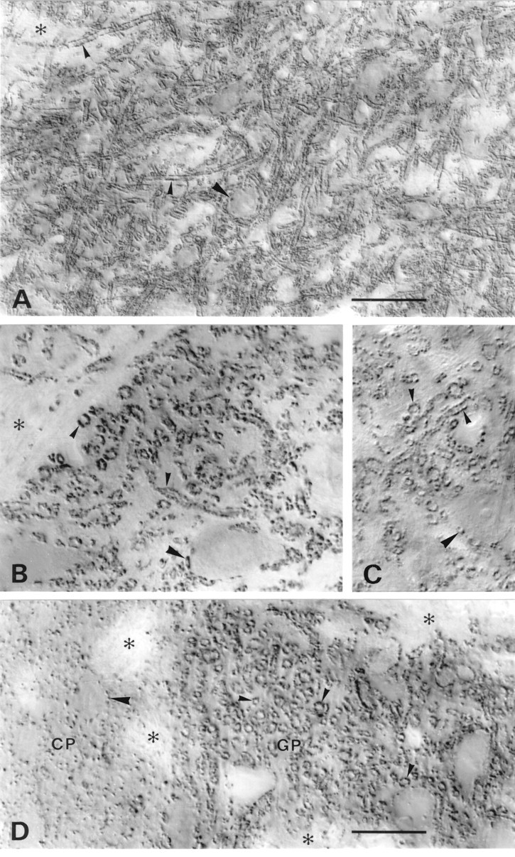
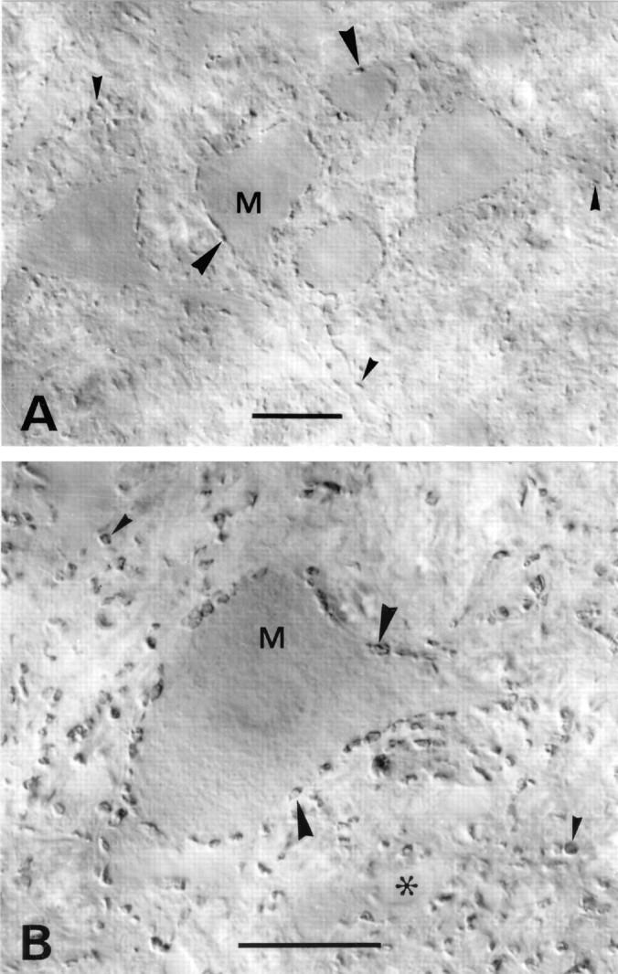
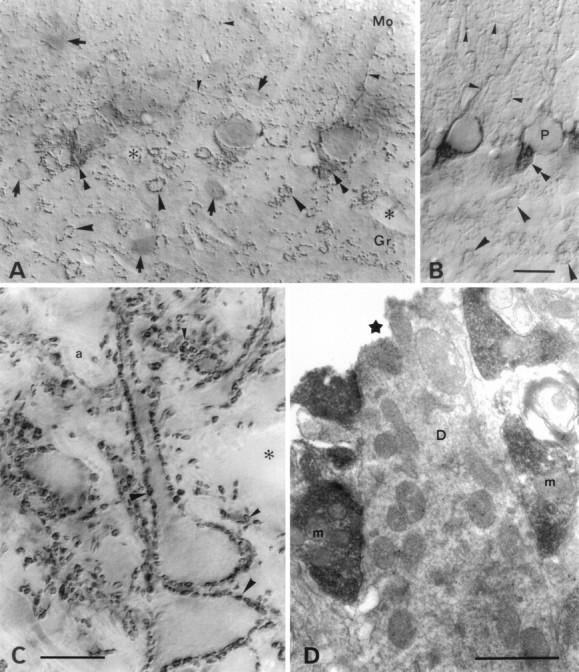
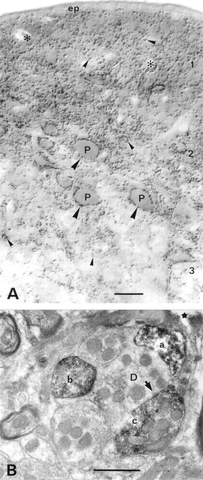
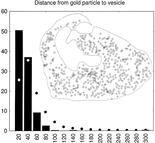
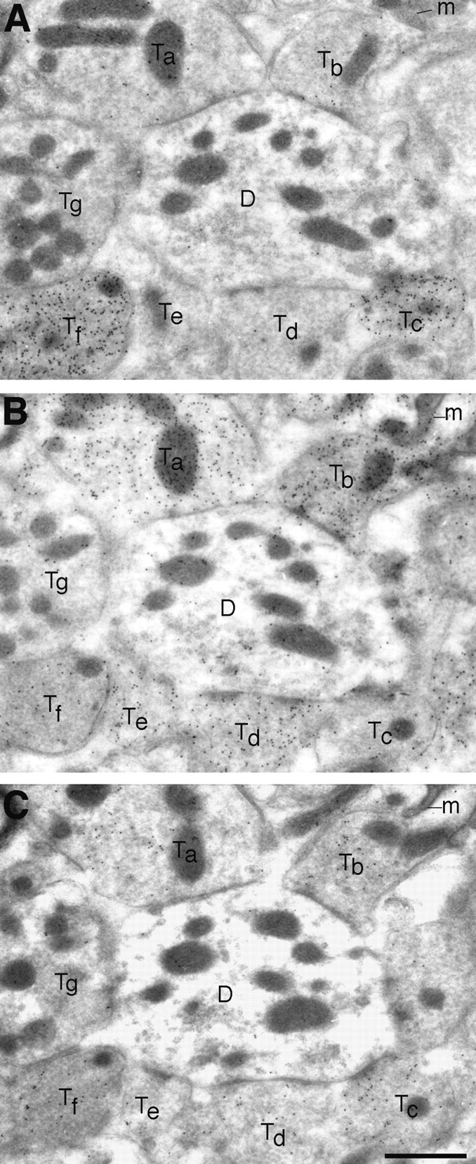
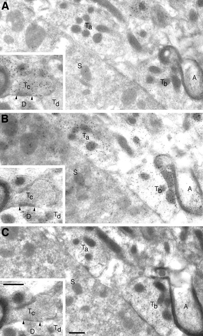
A transporter thought to mediate accumulation of GABA into synaptic vesicles has recently been cloned (McIntire et al., 1997). This vesicular GABA transporter (VGAT), the first vesicular amino acid transporter to be molecularly identified, differs in structure from previously cloned vesicular neurotransmitter transporters and defines a novel gene family. Here we use antibodies specific for N- and C-terminal epitopes of VGAT to localize the protein in the rat CNS. VGAT is highly concentrated in the nerve endings of GABAergic neurons in the brain and spinal cord but also in glycinergic nerve endings. In contrast, hippocampal mossy fiber boutons, which although glutamatergic are known to contain GABA, lack VGAT immunoreactivity. Post-embedding immunogold quantification shows that the protein specifically associates with synaptic vesicles. Triple labeling for VGAT, GABA, and glycine in the lateral oliva superior revealed a higher expression of VGAT in nerve endings rich in GABA, with or without glycine, than in others rich in glycine only. Although the great majority of nerve terminals containing GABA or glycine are immunopositive for VGAT, subpopulations of nerve endings rich in GABA or glycine appear to lack the protein. Additional vesicular transporters or alternative modes of release may therefore contribute to the inhibitory neurotransmission mediated by these two amino acids.


Figures

Fig. 1.
Fig. 1.
Specificity of antibodies to VGAT as demonstrated by immunoblotting after electrophoretic (SDS-PAGE) separation of proteins. Left, N2 antibody. Right, C1 antibody. Extracts of brain and VGAT-expressing PC12 cells (VGAT) show bands with a similar molecular mass (∼57 kDa). The bands are absent in liver and wild-type PC12 cells (wt). Positions of molecular mass standards are indicated on the left (83, 62, 47.5, 32.5, and 25 kDa, consecutively from the top).
Fig. 2.
Fig. 2.
Regional localization of VGAT shown by immunoperoxidase staining (with Triton) of closely spaced parasagittal Vibratome sections of rat brain. Antibodies N2 to the N-terminal fusion protein (A) and C1 to the C-terminal peptide (B) show the same distribution of immunoreactivity. Strong staining is shown in recognized targets of GABAergic nerve terminals (see Results). A, Amygdaloid nuclei; AD area dentata; AHi, amygdalohippocampal area; CA1, CA3, hippocampal subfields; Cbx, cortex cerebellaris;CI, colliculus inferior; CS colliculus superior; Cx, cortex cerebralis; Ce, central cerebellar nuclei (nucleus interpositus); CP, caudatoputamen; DC, dorsal nucleus cochlearis;EP, nucleus entopeduncularis; GP, globus pallidus; LOT, nucleus tractus olfactorius lateralis;N7, nucleus facialis; SNR, substantia nigra pars reticulata; Rt, nucleus reticularis thalami;Pir, cortex piriformis; Th, thalamic nuclei; Sp5, nucleus tractus spinalis nervi trigemini;Tu, tuberculum olfactorium; Ve, vestibular nuclei; VP, ventral pallidum;ZI, zona incerta. Scale bar, 2 mm.
Fig. 3.
Fig. 3.
Cellular localization of VGAT in hippocampus CA1 and area dentata by immunoperoxidase staining. A, CA1. Nerve terminals containing VGAT are distributed throughout the layers but are concentrated around the perikarya of pyramidal cells (small arrowheads) and interneurons (large arrowheads). The cytoplasm of interneuron perikarya shows immunoreactivity but less intense than that of the nerve endings. (The diffuse staining in the pyramidal layer is attributable to the presence of many stained nerve endings below the focal plane.) N2 antibody with Triton. B, Section adjacent to that in A, processed, photographed, and printed in the same conditions but after absorption of the N2 antibody with N-terminal fusion protein.C, Area dentata. VGAT-containing nerve terminals are distributed similarly as in CA1 on the granule cell bodies and in the neuropil. N2 antibody without Triton. O, P, R, Strata oriens, pyramidale, and radiatum of hippocampus, respectively;M, G, H, strata moleculare, granulare, and hilus (CA4) of area dentata, respectively. Asterisks, Blood vessels (emptied by perfusion). DIC optics (in this and subsequent light micrographs). Scale bar: A, B 50 μm; C, 20 μm.
Fig. 4.
Fig. 4.
VGAT in hippocampus CA3 and neocortex.A, CA3. Nerve endings immunoperoxidase-stained for VGAT are seen to outline unstained pyramidal cell bodies (arrowheads) and the initial parts of their axons (arrows) or are spread in the neuropil. Mossy fiber terminals in the stratum lucidum (LU) are not visualized. P, O, Strata pyramidale and oriens, respectively. B, CA3, stratum lucidum. Post-embedding immunogold labeling shows VGAT in a terminal with pleomorphic vesicles (T) but not in the large mossy fiber terminals (MT) making asymmetric synapses on a spine (S). C, Limb area of parietal cortex, layer 5. Strongly immunoreactive (peroxidase with Triton) nerve terminals are concentrated along pyramidal cell perikarya (large arrowheads) and are spread in the neuropil (small arrowheads). On the apical parts of the three large pyramidal cells shown, VGAT-containing boutons on the back or front of the cells can be seen en face (in part slightly out of focus).Asterisks, Blood vessels. Scale bars: A, 20 μm; B, 0.5 μm; C, 25 μm.
Fig. 5.
Fig. 5.
VGAT in basal ganglia and substantia nigra. A–C, Substantia nigra pars reticulata.D, Border between caudatoputamen (CP) and globus pallidus (GP). A,D, N2 antibody with Triton. B, N2 antibody without Triton. C, C1 antibody with Triton. VGAT-containing small nerve endings coat dendrites (small arrowheads) very densely and perikarya (large arrowheads) less completely. The staining pattern in substantia nigra and globus pallidus is typical of the GABAergic afferents from caudatoputamen. Antibodies to the N terminus (A, B, D) and C terminus (C) of VGAT show the same localization with (A, C, D) and without (B) Triton. Asterisks, Bundles of unstained nerve fibers. Scale bars: A, 50 μm;B–D, 20 μm. .
Fig. 6.
Fig. 6.
VGAT in motor nuclei. A, Medulla spinalis, ventral horn in an upper cervical segment. B, Nucleus facialis. Immunoreactive nerve terminals outline motoneuron perikarya (M, arrowheads). A high proportion of the terminals contacting these perikarya are known to contain glycine, alone or in addition to GABA. VGAT-containing terminals in the neuropil (small arrowheads) are sometimes seen to contact dendrites. Asterisk, Empty blood vessel. Scale bars: A, 25 μm; B, 20 μm.
Fig. 7.
Fig. 7.
VGAT and GAT-1 in cerebellum. A, VGAT; B, GAT-1 in the cortex cerebelli. VGAT is concentrated in nerve terminals, whereas GAT-1, a plasma membrane protein, is found in terminals and axons. Both proteins are localized in the mixed GABAergic–glycinergic terminals (deriving from Golgi cells) arranged in rosettes outlining glomeruli in the granular layer (large arrowheads), as well as in the predominantly GABAergic axons and terminals of the molecular layer (small arrowheads) (deriving from stellate cells and stellate basket cells). The axons and terminals from stellate cells can be seen forming a plexus in the molecular layer. Around the initial axon segments of Purkinje cells the axons and terminals of basket cells form a dense plexus called a pinceau (double arrowheads). There is some staining for VGAT in the cytoplasm of the perikarya of stellate, basket, and Golgi cells (arrows), and Purkinje cells (P). Mo, Gr, molecular and granular layers, respectively. Asterisks, Empty blood vessels. Parasagittal sections of hemisphere, lobus anterior. C, D, VGAT in nucleus interpositus (a central cerebellar nucleus). Light microscopic (C) and electron microscopic (D) immunoperoxidase staining shows VGAT-containing nerve terminals in the neuropil (small arrowheads) densely outlining perikarya and stem dendrites of the large immunonegative neurons (large arrowheads). Boutons in this position are mainly terminals of Purkinje cell axons. The nuclei are pierced by immunonegative, refringent bundles of myelinated axons (a). Terminals are often lightly stained in their centers; electron microscopy shows this to be attributable to centrally placed mitochondria (m). A postsynaptic dendrite (D) is immunonegative despite being opened to penetration of reagents at the Vibratome cut (star).Asterisks, Blood vessels. Scale bars: A, B, 25 μm; C, 20 μm; D, 1 μm.
Fig. 8.
Fig. 8.
VGAT in nucleus cochlearis dorsalis.A, Light microscopic immunoperoxidase staining shows VGAT-containing nerve terminals in the neuropil (small arrowheads), where some of them can be seen to outline dendrites. Perikarya of pyramidal cells (P, fusiform cells) in layer 2 are densely surrounded with VGAT-immunoreactive terminals (large arrowheads). The highest concentration of VGAT-positive nerve endings is found in the two superficial layers. (The diffuse darkening between the immunoreactive terminals here is attributable to numerous similar terminals below the focus plane.) The distribution of immunoreactivity is compatible with the combined distributions of GABA-containing and glycine-containing nerve endings (see Results).B, Electron microscopy confirms that the immunoreactive structures are nerve endings (a–c), one of which can be seen to form a symmetric synaptic contact (arrow).D, Dendrite; 1–3, layers of the nucleus;ep, ependyma. Asterisks, Blood vessels;star, surface of Vibratome section. Parasagittal sectioning plane. Scale bars: A, 25 μm;B, 0.5 μm.
Fig. 9.
Fig. 9.
VGAT in mainly glycinergic nerve endings in the LSO. Light microscopic immunoperoxidase staining at intermediate (A) and high (B) magnification is shown. Stained nerve endings (large arrowheads), partly of large size, densely outline perikarya and stem dendrites of unstained LSO neurons. These boutons are known to be mainly glycinergic. Some of the stained boutons are shown en face on the surface of perikarya graced by the section. Others can be seen to be continuous with stained axons (small arrowheads). The nucleus is pierced by tiny bundles of unstained myelinated axons (a). Asterisks, Empty blood vessel. Parasagittal sectioning plane. Scale bars, 20 μm.
Fig. 10.
Fig. 10.
Electron microscopic post-embedding immunogold localization of VGAT illustrated in different brain regions.A–D, Cortex cerebelli. E, Hippocampus CA1. F, Nucleus interpositus (a central cerebellar nucleus). G, Caudatoputamen. H, Nucleus cochlearis dorsalis. A, C, E–H, Antiserum N2. B, D, Antiserum C1. The antiserum N2 to the N terminus of VGAT labels both basket cell boutons (Ta) contacting Purkinje cell dendrites (D) in the molecular layer and Golgi cell boutons (Tc) in the granule cell layer of cerebellum. Only few particles can be detected in mossy fiber terminals (M), granule cell dendritic digits (Di), or other structures in the neuropil. In parallel sections, the antiserum C1 to the C terminus labels the same boutons (Tb, basket) or the same type of boutons (Td, Golgi) as does the antiserum to the N terminus. Boutons with pleomorphic vesicles (Te) making symmetric contacts (E, inset) on the somata (So) of hippocampal pyramidal cells are heavily labeled for VGAT. In the cerebellar nuclei and striatum, VGAT labeling is likewise in terminals (Tf, Tg), making symmetric synapses. Terminals (T′ in E,G) making asymmetric synapses on spines (S) and other structures in the neuropil are not labeled. In the cochlear nuclei, putative glycinergic terminals (Th) are also labeled for VGAT. D, So, Dendrites (or dendritic digits) and somata contacted by VGAT-containing boutons; arrowheads, synaptic membrane specializations. Scale bars: 0.5 μm; inset, 0.25 μm.
Fig. 11.
Fig. 11.
VGAT is associated with synaptic vesicles.A, Pre-embedding electron microscopic peroxidase immunocytochemistry suggests VGAT to be confined to the vesicle-rich areas (arrows) of the labeled terminals (T) and absent from the areas devoid of vesicles (stars), even when the latter are closer to the surface (s) of the Vibratome section (cerebellar nucleus with VGAT-containing terminals). B, C, The vesicular localization is confirmed by post-embedding immunogold labeling. The gold particles are restricted to vesicle-containing areas (arrows), whereas particles are rare in the rest of the terminal, containing neurofilaments (stars), mitochondria, or intervesicular cytoplasmic matrix. Note that at higher power (D), the plasma membrane appears clear of labeling, and that most of the particles are associated with the vesicles (basket cell terminals in cerebellar cortex).T, VGAT-labeled terminals; T′, VGAT-negative terminal; D, dendrites; A, myelinated axons; Pu, Purkinje cell body. Scale bars:A–C, 0.5 μm; D, 0.25 μm.
Fig. 12.
Fig. 12.
Quantification of the association of VGAT with synaptic vesicles (GABAergic basket cell terminals in the cerebellar cortex). Inset, Illustration of the recording of data, exemplified by the terminal shown as an electron micrograph in Figure11D; the center of each gold particle (+) and of each identifiable synaptic vesicle (○) is digitized, as well as the outline of the terminal, excluding mitochondria and (in other terminals) areas occupied by filaments (compare Fig.11B,C). Columns, Intercenter distances between each gold particle (394 particles in 12 terminals) and the nearest synaptic vesicle sorted into bins of 20 nm, they-axis showing percent of total in each bin (columns). Filled circles, Distances to the vesicle centers from points (1000 per terminal) randomly distributed over the parts of the terminals accessible to synaptic vesicles (see inset). The two distributions were statistically significantly different (p = 0.0005, continuity corrected χ2 test).
Fig. 13.
Fig. 13.
Triple labeling for GABA (A), glycine (B), and VGAT (C) in adjacent ultrathin sections of LSO. VGAT is localized in terminals selectively immunoreactive for GABA (Tc), as well as in ones showing mixed immunoreactivity for GABA and glycine (Tf) or selective immunoreactivity for glycine (Ta,Tb, Td). One small putative glycinergic terminal (Te) has only a low (perhaps insignificant) level of VGAT. A putative glutamatergic terminal (Tg) illustrates the low “background” levels of particle densities. The postsynaptic dendrite (D) has a slight glycine signal.m, Mitochondria. Scale bar, 1 μm.
Fig. 14.
Fig. 14.
Zero or low levels of VGAT in subpopulations of nerve terminals that are enriched with GABA or glycine. Adjacent ultrathin sections of LSO were incubated with antibodies to GABA (A), glycine (B), and VGAT (C). Two putatively “pure” glycinergic terminals (Taand Tb), synapsing on a neuronal soma (S), show differential VGAT labeling. AlthoughTb is enriched with VGAT,Ta is not. In A, contrast the lack of GABA immunoreactivity in vesicle-containing areas ofTa and Tb with the intense labeling of a nearby small terminal (at the top border). A myelinated axon (A) is immunoreactive for glycine. Insets, A GABA-labeled terminal (Tc), forming a symmetric synapse on a dendrite (D), is not labeled for glycine or for VGAT. Another terminal (Td) apposed to the same dendrite is unlabeled with all three antibodies. The postsynaptic neuronal soma (S) has a moderate glycine signal and a slight VGAT signal. Scale bars, 0.5 μm.

Similar articles

Cited by

References

    1. Barber R, Saito K. Light microscopic visualization of GAD and GABA-T in immunocytochemical preparations of rodent CNS. In: Roberts E, Chase TN, Tower DB, editors. GABA in nervous system function. Raven; New York: 1976. pp. 113–132.
    1. Berod A, Hartman BK, Pujol JF. Importance of fixation in immunohistochemistry: use of formaldehyde solutions at variable pH for the localization of tyrosine hydroxylase. J Histochem Cytochem. 1981;29:844–850. - PubMed
    1. Blackstad TW, Karagülle T, Ottersen OP. MORFOREL, a computer program for two dimensional analysis of micrographs of biological specimens, with emphasis on immunogold preparations. Comput Biol Med. 1990;20:15–34. - PubMed
    1. Burger PM, Hell J, Mehl E, Krasel C, Lottspeich F, Jahn R. GABA and glycine in synaptic vesicles: storage and transport characteristics. Neuron. 1991;7:287–293. - PubMed
    1. Ceranik K, Bender R, Geiger JP, Monyer H, Jonas P, Frotscher M, Lubke J. A novel type of GABAergic interneuron connecting the input and the output regions of the hippocampus. J Neurosci. 1997;17:5380–5394. - PMC - PubMed

Publication types

MeSH terms

LinkOut - more resources