. 1998 Nov 15;161(10):5313-20.

Apoptotic death of CD8+ T lymphocytes after immunization: induction of a suppressive population of Mac-1+/Gr-1+ cells

Affiliations

Apoptotic death of CD8+ T lymphocytes after immunization: induction of a suppressive population of Mac-1+/Gr-1+ cells

V Bronte et al. J Immunol. .

Abstract

Following an infection or immunization, a primary CD8+ T cell response generally rises then falls rapidly before giving rise to a "memory" response. When we immunized mice with recombinant viral immunogens optimized to enhance the lytic capability of CD8+ T cells, we measured a profound depression in Ag-specific effector function after early restimulation. Indeed, a "mirror image" cytolytic capability was observed: the most powerful immunogens, as measured by cytolytic capacity 6 days after immunization, elicited the weakest secondary immune response when evaluated following an additional 6 days after restimulation. To understand the mechanism of this suppression, we examined the fate of splenocytes immunized with a vaccinia virus encoding Ag and IL-2 then restimulated ex vivo. We found that these splenocytes underwent an apoptotic cell death, upon early restimulation, that was not dependent on the engagement of the FasR (CD95). Unlike previously described mechanisms of "propriocidal cell death" and "clonal exhaustion," the cell death we observed was not an inherent property of the CD8+ T cells but rather was due to a population of splenocytes that stained positive for both the Mac-1 and Gr-1 surface markers. Deletion of these cells in vitro or in vivo completely abrogated the observed suppression of cytolytic reactivity of Ag-specific CD8+ T cells. These observations could account for the apparent absence of Ag-specific immune responses after some current vaccination regimens employing powerful immunogens. Finally, our results may shed new light on a mechanism for the suppression of CD8+ T cell responses and its effect on vaccine efficacy and on immune memory.


Figures

FIGURE 1
FIGURE 1
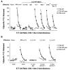
Immunization with powerful immunogens induces a profound loss of cytolytic capacity upon restimulation with peptide. IL-2-rVV immunization augments primary cytolytic responses. Two BALB/c mice were immunized i.v. with 5 × 10⁶ PFU/mouse of different rVV. After 6 days, the spleens were removed, pooled, and tested in a 6-h 51Cr release assay against CT26.WT tumor cells infected with VV-WT (A) or pulsed with the β-gal peptide (B). Secondary responses are lost in mice immunized with IL-2-rVV. The splenocytes used for the previous experiment were further incubated in vitro with 1 μg/ml of the synthetic β-gal peptide for 6 days and then assayed in a 6-h 51Cr release assay against the CT26.CL25 β-gal-positive clone (C) or the CT26.WT cells pulsed with the β-gal peptide (D). Purification of CD8+ cells restores specific reactivity in mice immunized with IL-2-rVV. CD8+ cells were enriched through affinity columns from splenocytes used in Fig. 1C and D. This enriched population was admixed with naive, CD8-depleted splenocytes from syngeneic mice to constitute about 10% of total cells in culture. After a 6-day in vitro restimulation with β-gal peptide, the mixture was tested in a 6-h 51Cr release assay against CT26.WT cells pulsed with the β-gal peptide (E). Cytotoxicity toward the CT26.WT cells or the irrelevant E22 target cells was always <5% even at the highest E:T ratio (not shown). The E:T ratio was 33:1, then diluted threefold (11:1, 4:1, 1:1). The experiment was repeated four times with similar results.
FIGURE 2
FIGURE 2
Impairment of immune response after boosting occurs in vivo and is long lasting. Five different BALB/c mice in each treatment group were injected i.v. with 5 × 10⁶ PFU of the rVV indicated at the bottom of the boxes. HBSS alone was used as vehicle control. Six days later, mice were boosted with two i.v. injections of PBS alone (not shown) or PBS containing 400 μg of either OVA or β-gal protein (top). Alternatively, mice were boosted with a single i.v. inoculation of 107 PFU/mouse of wild-type (FPV-WT) or β-gal-expressing FPV (bottom). On day 21 following the in vivo restimulation, mice were challenged i.v. with 5 × 10⁵ CT26.CL25 cells to establish pulmonary metastases. Twelve days later, mice were sacrificed, and pulmonary nodules were counted in a coded and blinded fashion. Two separate, independent experiments were performed under identical experimental conditions with overall similar results. An arbitrary value of 250 was assigned when metastases were too numerous to count. Data represent the mean ± SD of pulmonary metastases.
FIGURE 3
FIGURE 3
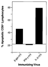
CD8+ T lymphocytes die an apoptotic death after immunization with IL-2-rVV in vivo and peptide restimulation in vitro. The cells used in this experiment are from the splenocytes used in the experiment shown in Fig. 1, C and D. Two spleens from BALB/c mice immunized 6 days earlier with different rVV were stimulated in vitro for 6 days with 1 μg/ml of β-gal peptide. One million cells were stained with FITC-conjugated mAb against the CD8 Ag. After staining, cells were fixed in ethanol and exposed to propidium iodide solution to allow detection of hypodiploid nuclei. Figures indicate the percentage of hypodiploid cells among CD8+ lymphocytes. The apoptosis percentage in cultures of spleens from mice immunized with NP-rVV, V69, was used as the background line. The experiment was repeated two additional times with similar results.
FIGURE 4
FIGURE 4
Suppression of CD8+ T cell function requires cell-cell contact. A suspension of three spleens from BALB/c mice infected 6 days earlier with 5 × 10⁶ PFU/mouse of IFN-γ-rVV was incubated with a β-gal peptide either alone (control) or together with splenocytes of mice similarly infected with IL-2-rVV (1:1). The latter were either admixed in the same well (cell contact) or separated by a semipermeable membrane (no cell contact). After 6 days, CTL activity in the cultures was assayed against the same panel of target cells used in previous experiments. For simplicity, only the cytolytic activity against CT26.CL25 at an E:T ratio of 10:1 is shown. Control wells containing 1:1 mixture of splenocytes from IFN-γ-rVV-infected mice and normal mice did not show any difference from control culture whether they were admixed or separated by the insert membrane (not shown).
FIGURE 5
FIGURE 5
Inhibition of CD8+ T cell killing is not Fas-dependent. Lymphoblasts obtained from ConA-stimulated splenocytes from C57BL/6J and MRL-lpr/lpr mice were evaluated for CD95 (Fas/Apo1) expression by cytofluorometric analysis (A, left and right, respectively). Mice were immunized i.v. with 5 × 10⁶ PFU/mouse of IL-2-rVV or IFN-γ-rVV (B). Six days later, the spleens from three different mice were removed, pooled, and tested in a 6-h 51Cr release assay against EL-4, EL-4 tumor cell line infected with VV-WT, or the β-gal-expressing line, E22 (Primary, top). The splenocytes used for the previous experiment where further incubated in vitro with 1 μg/ml of the H-2Kb-restricted, synthetic β-gal peptide for 6 days and then assayed in a 6-h 51Cr release assay against CT26.CL25, EL-4, or E22 (Secondary, bottom). The experiment was repeated with similar results.
FIGURE 6
FIGURE 6
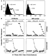
Immunization with IL-2-rVV results in an increase in the percentage of Mac-1+ and Gr-1+-double-positive splenocytes. Splenocyte pools used in the experiment shown in Fig. 7 were stained with FITC-anti-Gr-1 and phycoerythrin-anti-Mac-1 mAbs in the presence of an Ab blocking the Fcγ-RII/Fcγ-RIII (2.4G2). Upper panels show unseparated splenocytes from unimmunized mice (naïve) or mice immunized 6 days earlier with either IFN-γ-rVV or IL-2-rVV. Lower panels show the IL-2-rVV-immunized splenocytes after depletion with the indicated mAbs. Not shown are the results of the in vitro depletion with IgG2b mAb used as negative control. As for the in vivo data, depletion with this mAb did not change the percentages of double-positive cells seen in untreated controls. A secondary anti-rat IgG Ab revealed the number of positive cells to be <1% after depletion, confirming that the negativity was not due to Ab competition. Data are representative of four separate, independently performed experiments.
FIGURE 7
FIGURE 7
In vitro and in vivo depletion of either Mac-1+ or Gr-1+ cells restores CTL response in splenocyte cultures from mice immunized with IL-2-rVV. A, In vitro depletion of Mac-1+/Gr-1+ cells restores the immune response. Two to five BALB/c mice/group were inoculated with HBSS (naïve), 5 × 10⁶ PFU of IL-2 rVV, or 5 × 10⁶ PFU of IFN-γ-rVV. After 6 days, spleens were pooled together and depleted in vitro of Mac-1+ or Gr-1+ cells. IgG2b isotype-matched mAb was used as negative control of the depletion protocol. Splenocytes collected after depletion were cultured with β-gal peptide as described previously. The same panel of target cells was used to evaluate the cytolytic activity (A). As positive controls, the following experimental groups were also included: CD8+ splenocytes from mice inoculated with IL-2-rVV and cultured together with CD8-depleted naïve spleens (*); mice inoculated 6 days earlier with IFN-γ-rVV; and mice inoculated with IL-2-rVV 14 days before the in vitro restimulation. Data with E22 tumor cells were negative and are not shown for simplicity. The E:T ratio is 100:1 followed by threefold dilutions (33:1, 11:1, 4:1, 1:1, 0.3:1). The experiment was repeated four more times with similar results. B, In vivo depletion of Mac-1+/Gr-1+ cells restores the immune response. The experiment was performed as in A, with the exception that 200 μg Gr-1 mAb and the IgG2b controls were injected i.p. into mice on day 2 and 4 following immunization with the rVV (B).

Similar articles

Cited by

References

    1. Scott DW, Grdina T, Shi Y. T cells commit suicide, but B cells are murdered! J. Immunol. 1996;156:2352. - PubMed
    1. Lenardo MJ. Interleukin-2 programs mouse αβ T lymphocytes for apoptosis. Nature. 1991;353:858. - PubMed
    1. Critchfield JM, Racke MK, Zuniga-Pflucker JC, Cannella B, Raine CS, Goverman J, Lenardo MJ. T cell deletion in high antigen dose therapy of autoimmune encephalomyelitis. Science. 1994;263:1139. - PubMed
    1. Welsh RM, Selin LK, Razvi ES. Role of apoptosis in the regulation of virus-induced T cell responses, immune suppression, and memory. J. Cell. Biochem. 1995;59:135. - PubMed
    1. Abbas AK. Die and let live: eliminating dangerous lymphocytes. Cell. 1996;84:655. - PubMed

Publication types

MeSH terms

LinkOut - more resources