. 1998 Dec;18(12):7038-51.
doi: 10.1128/MCB.18.12.7038.

ASAP1, a phospholipid-dependent arf GTPase-activating protein that associates with and is phosphorylated by Src

Affiliations

ASAP1, a phospholipid-dependent arf GTPase-activating protein that associates with and is phosphorylated by Src

M T Brown et al. Mol Cell Biol. 1998 Dec.

Abstract

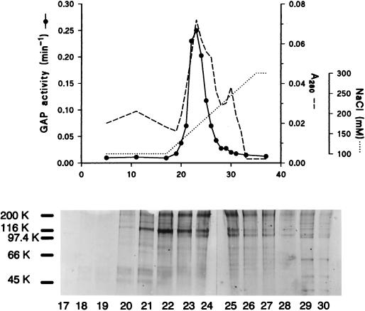
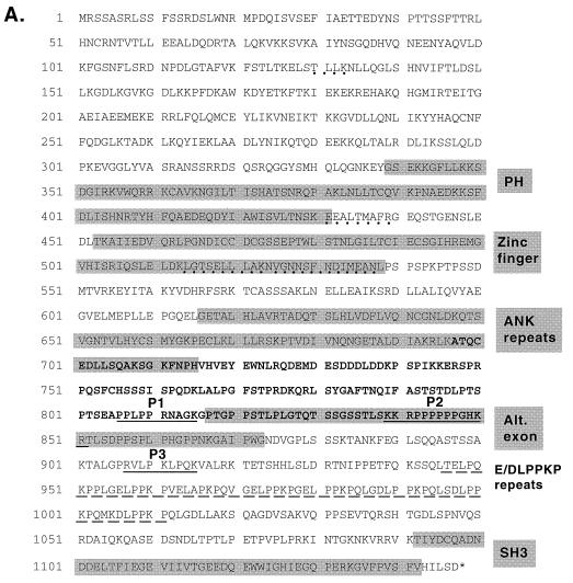
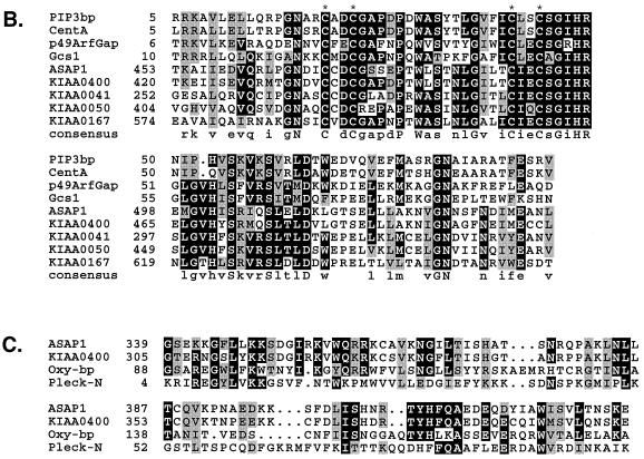
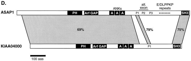
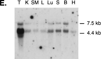
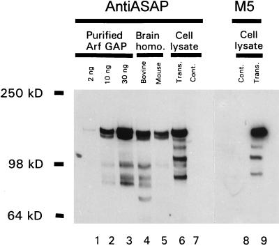
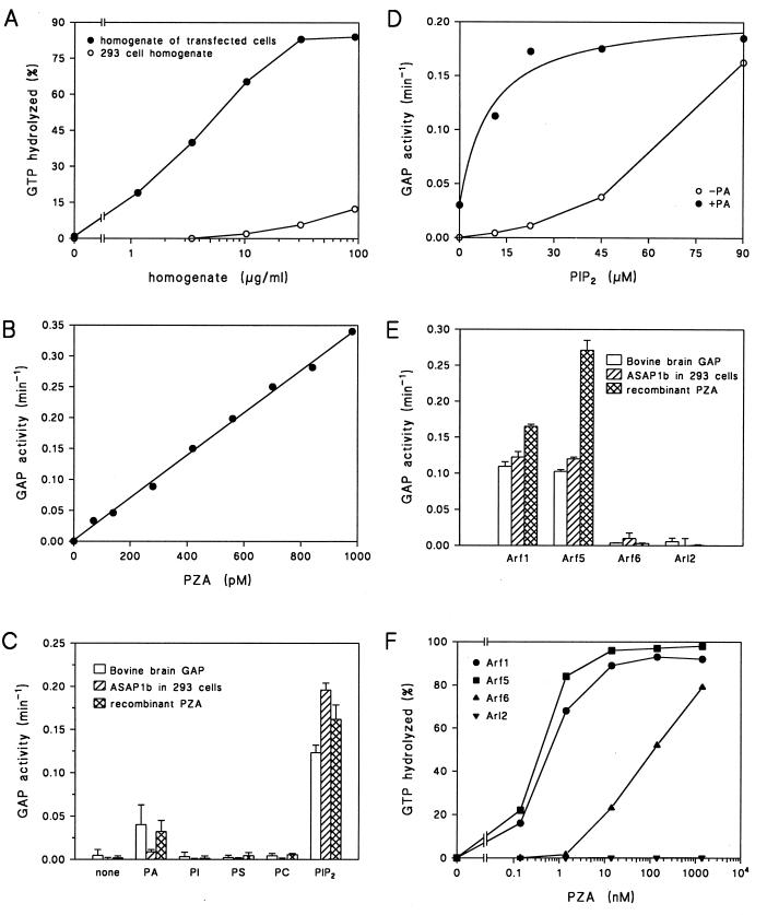
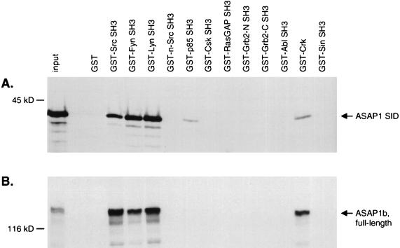
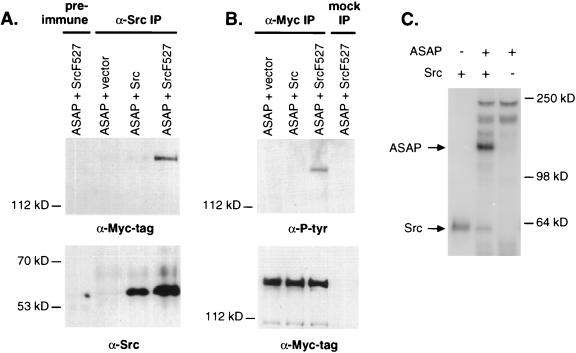
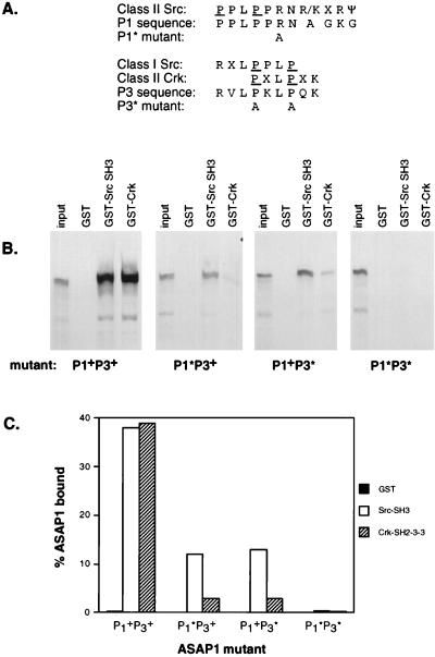
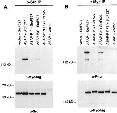
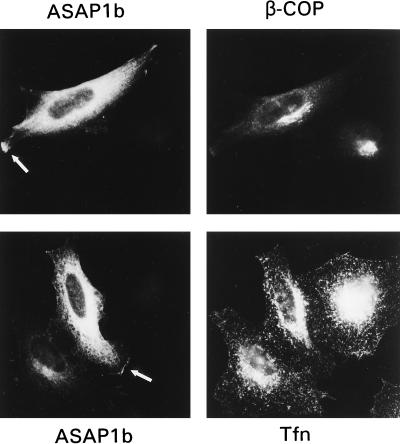
Membrane trafficking is regulated in part by small GTP-binding proteins of the ADP-ribosylation factor (Arf) family. Arf function depends on the controlled exchange and hydrolysis of GTP. We have purified and cloned two variants of a 130-kDa phosphatidylinositol 4, 5-biphosphate (PIP2)-dependent Arf1 GTPase-activating protein (GAP), which we call ASAP1a and ASAP1b. Both contain a pleckstrin homology (PH) domain, a zinc finger similar to that found in another Arf GAP, three ankyrin (ANK) repeats, a proline-rich region with alternative splicing and SH3 binding motifs, eight repeats of the sequence E/DLPPKP, and an SH3 domain. Together, the PH, zinc finger, and ANK repeat regions possess PIP2-dependent GAP activity on Arf1 and Arf5, less activity on Arf6, and no detectable activity on Arl2 in vitro. The cDNA for ASAP1 was independently identified in a screen for proteins that interact with the SH3 domain of the tyrosine kinase Src. ASAP1 associates in vitro with the SH3 domains of Src family members and with the Crk adapter protein. ASAP1 coprecipitates with Src from cell lysates and is phosphorylated on tyrosine residues in cells expressing activated Src. Both coimmunoprecipitation and tyrosine phosphorylation depend on the same proline-rich class II Src SH3 binding site required for in vitro association. By directly interacting with both Arfs and tyrosine kinases involved in regulating cell growth and cytoskeletal organization, ASAP1 could coordinate membrane remodeling events with these processes.


Figures

FIG. 1
FIG. 1
Polypeptides coeluting with GAP activity on anion exchange. Activity eluting from the Sephacryl S-300 column was adsorbed to a Resource Q column and eluted as described in Materials and Methods. Samples from the indicated fractions were electrophoresed in SDS on a 7.5% polyacrylamide gel, and proteins were visualized with a colloidal blue stain.
FIG. 2
FIG. 2
ASAP1 sequence, alignment with related proteins, and expression. (A) Predicted amino acid sequence of ASAP1a and ASAP1b. ASAP1 was conceptually translated from the cDNA sequence (GenBank accession no. AF075461 and AF075462). Shaded boxes indicate the PH, GCS-type zinc finger, ANK repeat, and SH3 domains, and the alternate exon found only in ASAP1a. Potential SH3 ligand sites P1, P2, and P3 are underlined with solid lines, E/DLPPKP repeats are underlined with a broken line, and peptide sequences of tryptic fragments from the purified bovine protein are underlined with a dotted line. The SID is indicated in boldface type. (B) Alignment of zinc-finger-containing domains. Conserved cysteine residues of the CXXCX16CXXC zinc finger motif are indicated by asterisks. Sequences were aligned by using PILEUP (University of Wisconsin Genetics Computer Group package). (C) Alignment of PH domains. PH domains were aligned by using PILEUP and secondary structure predictions. Databases and accession numbers: ArfGAP1 (GenBank U35776), GCS1 (Swiss Protein P35197), PIP3bp (DDBJ D89940), CentA (Centaurin-α; GenBank U51013), KIAA0400 (DDBJ AB007860), KIAA0041 (DDBJ D26069), KIAA0050 (DDBJ D30758), KIAA0167 (DDBJ D79989), Oxy-bp (oxysterol binding protein; Swiss Protein P22059), Pleck-N (N-terminal PH domain of pleckstrin; Swiss Protein P08567). (D) Schematic structure of ASAP1 and similarity to human KIAA0400. Numbers in boldface indicate the percentages of identity between the two proteins for the specified regions. Drawn to scale; bar = 100 amino acids. (E) Hybridization of ASAP1 cDNA to mouse poly(A) RNA. A radiolabeled probe corresponding to the ASAP1a SID was hybridized to poly (A) RNA on nylon membranes (obtained from Clontech) as recommended by Clontech. Each lane contains approximately 2 μg of RNA. H, heart; B, brain; S, spleen; Lu, lung; L, liver; SM, skeletal muscle; K, kidney; T, testis.
FIG. 2
FIG. 2
ASAP1 sequence, alignment with related proteins, and expression. (A) Predicted amino acid sequence of ASAP1a and ASAP1b. ASAP1 was conceptually translated from the cDNA sequence (GenBank accession no. AF075461 and AF075462). Shaded boxes indicate the PH, GCS-type zinc finger, ANK repeat, and SH3 domains, and the alternate exon found only in ASAP1a. Potential SH3 ligand sites P1, P2, and P3 are underlined with solid lines, E/DLPPKP repeats are underlined with a broken line, and peptide sequences of tryptic fragments from the purified bovine protein are underlined with a dotted line. The SID is indicated in boldface type. (B) Alignment of zinc-finger-containing domains. Conserved cysteine residues of the CXXCX16CXXC zinc finger motif are indicated by asterisks. Sequences were aligned by using PILEUP (University of Wisconsin Genetics Computer Group package). (C) Alignment of PH domains. PH domains were aligned by using PILEUP and secondary structure predictions. Databases and accession numbers: ArfGAP1 (GenBank U35776), GCS1 (Swiss Protein P35197), PIP3bp (DDBJ D89940), CentA (Centaurin-α; GenBank U51013), KIAA0400 (DDBJ AB007860), KIAA0041 (DDBJ D26069), KIAA0050 (DDBJ D30758), KIAA0167 (DDBJ D79989), Oxy-bp (oxysterol binding protein; Swiss Protein P22059), Pleck-N (N-terminal PH domain of pleckstrin; Swiss Protein P08567). (D) Schematic structure of ASAP1 and similarity to human KIAA0400. Numbers in boldface indicate the percentages of identity between the two proteins for the specified regions. Drawn to scale; bar = 100 amino acids. (E) Hybridization of ASAP1 cDNA to mouse poly(A) RNA. A radiolabeled probe corresponding to the ASAP1a SID was hybridized to poly (A) RNA on nylon membranes (obtained from Clontech) as recommended by Clontech. Each lane contains approximately 2 μg of RNA. H, heart; B, brain; S, spleen; Lu, lung; L, liver; SM, skeletal muscle; K, kidney; T, testis.
FIG. 3
FIG. 3
An antibody raised to mouse ASAP1 recognizes Arf GAP purified from bovine brain. The indicated quantity of purified bovine brain Arf GAP (lanes 1 to 3), a bovine brain homogenate (30 μg of protein), a mouse brain homogenate (25 μg of protein), and lysates (0.75 μg of protein) from COS7 cells transfected with a FLAG-tagged ASAP1b expression vector (trans) and COS7 cells that had not been transfected (cont) were fractionated by SDS-PAGE and transferred to nitrocellulose. The blots were probed with 3820J anti-ASAP1 antiserum (lanes 1 to 7) or a monoclonal antibody (M5) to the FLAG epitope (lanes 8 and 9).
FIG. 4
FIG. 4
Recombinant ASAP1 has Arf GAP activity. (A) Increased Arf GAP activity in lysates of cells overexpressing ASAP1b. The Arf GAP activity in lysates of 293 cells that were transfected with an expression vector for ASAP1b (closed circles) or cells that were not transfected (open circles) was determined. (B) Arf GAP activity of PZA. The recombinant protein PZA was incubated at the indicated concentrations with approximately 10 nM Arf1-GTP in the presence of crude phosphoinositides for 3 min at 30°C. (C) Phospholipid dependence of purified Arf GAP, recombinant protein ASAP1, and PZA. Arf-GTP was incubated with either 0.9 nM Arf GAP purified from bovine brain, 0.03 μg of protein from the lysate of 293 cells transiently transfected with a ASAP1b expression vector per ml, or 0.7 nM bacterially expressed recombinant protein PZA. PIP2, 180 μM PIP2; PA, 750 μM phosphatidic acid; PI, 720 μM phosphatidylinositol; PS, 720 μM phosphatidylserine; PC, 720 μM phosphatidylcholine. (D) PIP2-dependence of recombinant PZA. Arf-GTP was incubated with 0.7 nM PZA and the indicated concentrations of PIP2 in the presence or absence of 750 μM PA. (E) Substrate specificity of purified Arf GAP, recombinant protein ASAP1, and PZA. MyrArf1-GTP, myrArf5-GTP, myrArf6-GTP and Arl2-GTP were used as substrates in reactions containing 1 mg of crude phosphoinositide/ml as a source of PIP2 and 2 nM purified bovine brain Arf GAP, lysates of 293 cells transiently transfected with an expression vector for ASAP1b (0.03 μg of protein/ml), or 1.4 nM bacterially expressed PZA. The means ± standard deviations of quadruplicate determinations are shown for bovine Arf GAP and PZA. The means ± the ranges for duplicates are shown for the cell lysates. (F) Substrate specificity of PZA. Conditions were the same as for panel E, but the indicated concentrations of PZA were used.
FIG. 5
FIG. 5
In vitro association of ASAP1 with SH3 domains. Myc-tagged ASAP1a SID (A) or full-length ASAP1b (B) was labeled with [35S]methionine and mixed with ∼2 μg of immobilized GST-SH3 fusion proteins (see Materials and Methods). Bound proteins were eluted and separated by SDS-PAGE. The first lane of each gel contains 1/30 the amount of the radioactive protein that was added to each binding reaction. Grb2-N, N-terminal SH3 domain of mouse Grb2; Grb2-C, C-terminal Grb2 SH3 domain; p85, p85 subunit of PI3-kinase; Ras-GAP, p120 RasGAP.
FIG. 6
FIG. 6
ASAP1 associates with Src in 293 cells and becomes tyrosine phosphorylated both in Src-expressing cells and by Src in vitro. Lysates were prepared from 293 cells cotransfected with CS3+MT−ASAP1b and either LXSH, LXSH-c-Src, or LXSH-c-SrcF527. (A) Coimmunoprecipitation of ASAP1 with Src. Lysates were immunoprecipitated with 3060 anti-Src rabbit antiserum or preimmune rabbit serum. Twin blots were probed with either 9E10 anti-Myc monoclonal antibody (top panel) or 327 anti-Src monoclonal antibody (bottom panel). (B) Tyrosine phosphorylation of ASAP1 in vivo. 293 lysates were immunoprecipitated with either 9E10 or a mock antibody mix lacking 9E10. Identical blots were probed with anti-phosphotyrosine 4G10 (top panel) or 9E10 (bottom panel) monoclonal antibodies. (C) In vitro phosphorylation of native ASAP1 by Src. Recombinant c-Src (1 U) was incubated with 40 ng of purified ASAP1 from bovine brain and 5 μM [γ-32P]ATP.
FIG. 7
FIG. 7
ASAP1 proline-rich sequences are required for binding to Src and Crk in vitro. (A) P1 and P3 amino acid sequences, alignment with SH3 consensus binding motifs, and residues changed by site-directed mutagenesis. Underlined residues indicate conserved prolines in the canonical SH3 binding motif PXXP. (B) In vitro binding of ASAP1b to GST-Src SH3 and GST-Crk. Binding reactions were as described in Fig. 5 and Materials and Methods. The first lane of each of the four gels contains 1/30 the amount of [35S]methionine-labeled ASAP1b added to each binding reaction. (C) Quantitation of ASAP1 mutant binding efficiencies. The fraction of ASAP1 bound by immobilized GST fusion protein was quantitated by PhosphorImager analysis.
FIG. 8
FIG. 8
ASAP1 mutants show impaired Src association and tyrosine phosphorylation in mammalian cells. 293 cells were cotransfected with LXSH or LXSH-c-Src F527 and either CS3+MT, CS3+MT−ASAP1b, CS3+MT−ASAP1b-P1*, CS3+MT−ASAP1b−P3*, or CS3+MT−ASAP1b−P1*P3*. (A) Cell lysates were immunoprecipitated (IP) with 3060 anti-Src antiserum. Identical Western blots were probed with either 9E10 anti-Myc tag (top panel) or 327 anti-Src (bottom panel). (B) Lysates were immunoprecipitated with 9E10. 4G10 anti-phosphotyrosine (top panel) or 9E10 (bottom panel) was used to probe twin Western blots.
FIG. 9
FIG. 9
Localization of epitope-tagged ASAP1b in tissue culture cells. ASAP1b was detected in HeLa cells transfected with a FLAG-tagged ASAP1b expression vector by using either a rabbit polyclonal antibody 551 raised to ASAP1 (upper panels) or a monoclonal antibody against the FLAG tag (lower left panel). Arrows indicate ASAP1 that was detected at the cell edge, likely associated with the plasma membrane.

Similar articles

Cited by

References

    1. Alexandropoulos K, Baltimore D. Coordinate activation of c-Src by SH3- and SH2-binding sites on a novel p130Cas-related protein, Sin. Genes Dev. 1996;10:1341–1355. - PubMed
    1. Allen C M, Ely C M, Juaneza M A, Parsons S J. Activation of Fyn tyrosine kinase upon secretogogue stimulation of bovine chromaffin cells. J Neurosci Res. 1996;44:421–429. - PubMed
    1. Antonny B, Huber I, Paris S, Chabre M, Cassel D. Activation of ADP-ribosylation factor 1 GTPase-activating protein by phosphatidylcholine-derived diacylglycerols. J Biol Chem. 1997;272:30848–30851. - PubMed
    1. Aoe T, Cukierman E, Lee A, Cassel D, Peters P J, Hsu V W. The KDEL receptor, ERD2, regulates intracellular traffic by recruiting a GTPase-activating protein for ARF1. EMBO J. 1997;16:7305–7316. - PMC - PubMed
    1. Barnekow A, Jahn R, Schartl M. Synaptophysin: a substrate for the protein tyrosine kinase pp60c-src in intact synaptic vesicles. Oncogene. 1990;5:1019–1024. - PubMed

Publication types

MeSH terms

Associated data