. 1998 Nov 1;18(21):8637-47.
doi: 10.1523/JNEUROSCI.18-21-08637.1998.

Localization of myosin-Ibeta near both ends of tip links in frog saccular hair cells

Affiliations

Localization of myosin-Ibeta near both ends of tip links in frog saccular hair cells

J A García et al. J Neurosci. .

Abstract

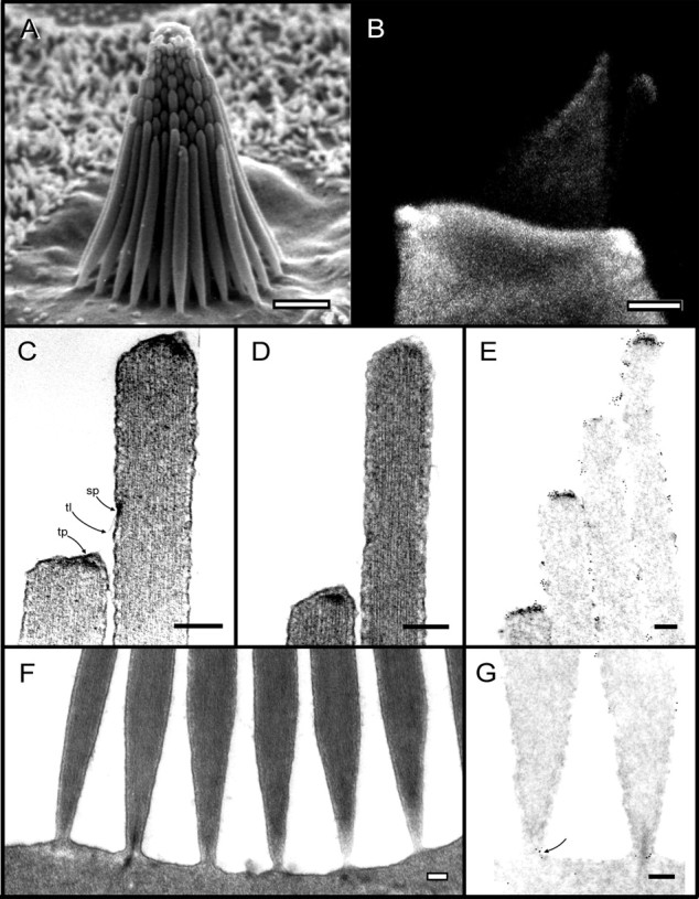
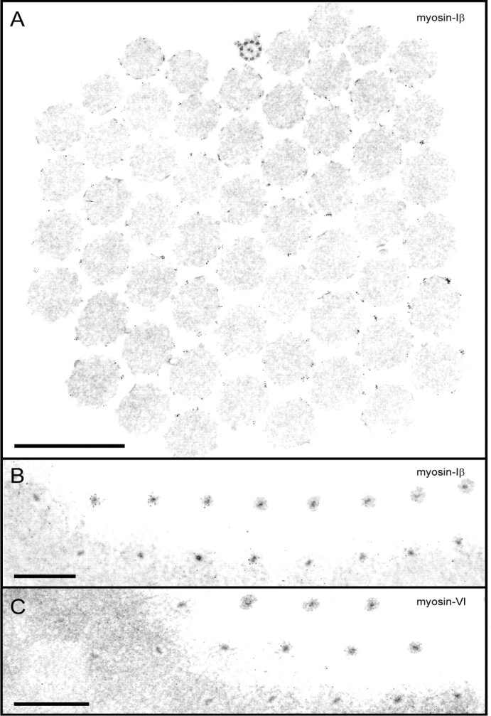
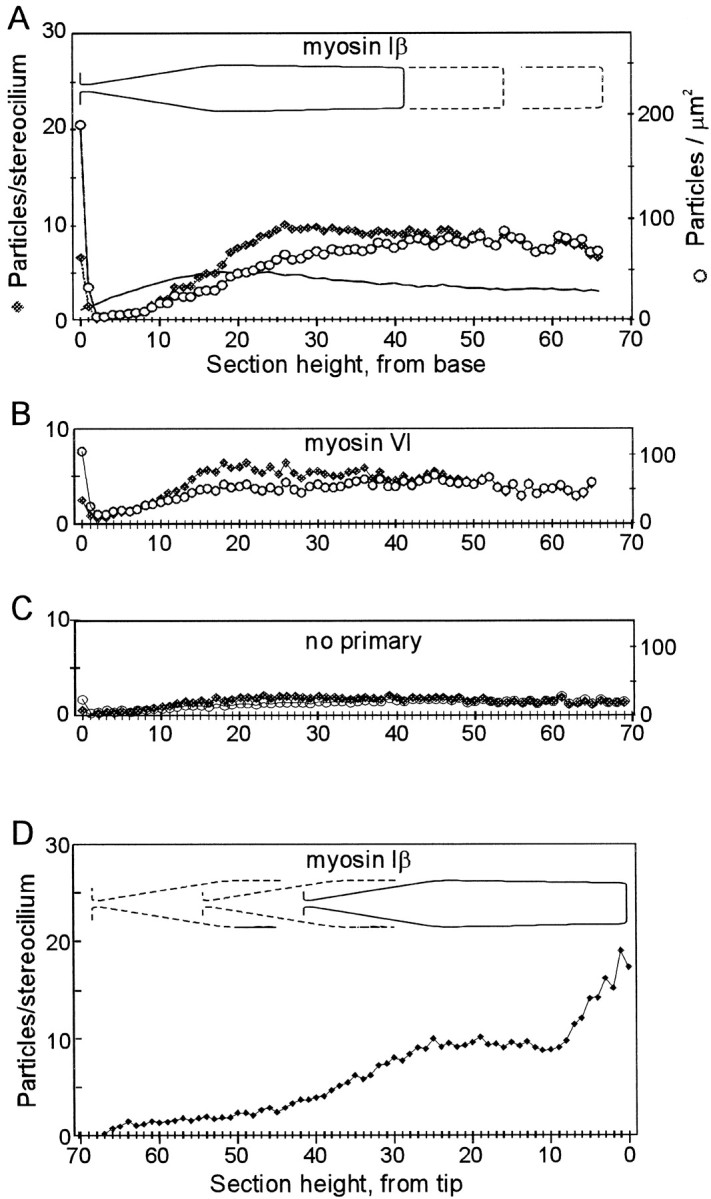
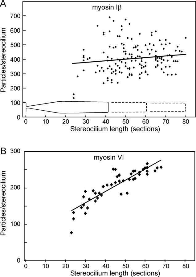
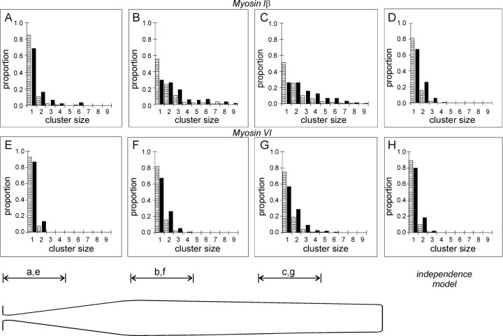
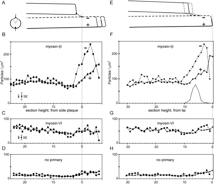
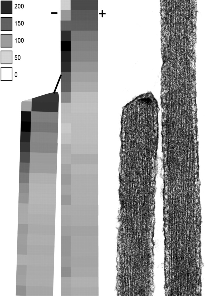
Current evidence suggests that the adaptation motor of mechanoelectrical transduction in vertebrate hair cells is myosin-Ibeta. Previously, confocal and electron microscopy of bullfrog saccular hair cells using an anti-myosin-Ibeta antibody labeled the tips of stereocilia. We have now done quantitative immunoelectron microscopy to test whether myosin-Ibeta is enriched at or near the side plaques of tip links, the proposed sites of adaptation, using hair bundles that were serially sectioned parallel to the macular surface. The highest particle density occurred at stereocilia bases, close to the cuticular plate. Also, stereocilia of differing lengths had approximately the same number of total particles, suggesting equal targeting of myosin-Ibeta to all stereocilia. Finally, particles tended to clump in clusters of two to five particles in the distal two-thirds of stereocilia, suggesting a tendency for self-assembly of myosin-Ibeta. As expected from fluorescence microscopy, particle density was high in the distal 1 micrometer of stereocilia. If myosin-Ibeta is the adaptation motor, a difference should exist in particle density between regions containing the side plaque and those excluding it. Averaging of particle distributions revealed two regions with approximately twice the average density: at the upper ends of tip links in a 700-nm-long region centered approximately 100 nm above the side plaque, and at the lower ends of tip links within the tip plaques. Controls demonstrated no such increase. The shortest stereocilia, which lack side plaques, showed no concentration rise on their sides. Thus, the specific localization of myosin-Ibeta at both ends of tip links supports its role as the adaptation motor.


Figures

Fig. 1.
Fig. 1.
Morphology of bullfrog saccular hair bundles and labeling with antibody to myosin-Iβ. A, A saccular bundle typical of those in this study contains ∼60 stereocilia in nine columns. Lengths vary from ∼4 μm at the shorter (negative) edge to ∼8 μm at the taller (positive) edge. B, Labeling of a dissociated hair cell with an antibody to myosin-Iβ. The secondary antibody was labeled with Cy3; the image is a summation of a stack of confocal images through the middle 2 μm of the cell.C, D, Two examples of the tip link (tl) and the osmiophilic densities marking the tip-insertion plaque (tp) and side-insertion plaque (sp). E, Immunogold labeling of the tips of stereocilia with the myosin-Iβ antibody. Gold particles are present along the sides of stereocilia but appear more concentrated at the tip plaques and in a region near the side plaques.F, Example of the basal tapers of stereocilia and their insertions into the cuticular plate. Stereocilia are ∼150 nm in diameter where their rootlets insert into the cuticular plate, thicken to a maximum diameter of ∼550 nm ∼2 μm above the cuticular plate, and then narrow to a diameter of 300–400 nm at their distal tips.G, Immunogold labeling of the bases of stereocilia. Particles appear around the rootlet at the narrowest part of the taper where the rootlet enters the cuticular plate (arrow) but are otherwise scarce in the taper region. Scale bars: A,B, 2 μm; C–G, 200 nm. (PanelA was copyrighted by J. A. Assad and D. P. Corey and was reproduced with permission.)
Fig. 2.
Fig. 2.
Immunogold labeling of stereocilia, sectioned horizontally. A, Myosin-Iβ antibody; section ∼2 μm above the cuticular plate. The orientation is provided by the conspicuous kinocilium at the top of the image so thattop is positive, bottom is negative, and tip links would run among stereocilia in a vertical column. Membranes were removed by detergent before labeling with primary antibody. Gold particles are visible around the actin cores of stereocilia but not within the cores. In sections of this height and higher, they often occur in clusters of two to six particles. B, Myosin-Iβ antibody; grazing section at the level of the cuticular plate. Rootlets are apparent as dense cores within stereocilia and are ringed with gold particles. Particles are concentrated but not clustered. Particles are not present a bit higher (top right) or a bit lower (bottom left) than the level of the apical surface. C, Myosin-VI antibody; grazing section at the level of the cuticular plate. Particles also appear concentrated around the stereocilia at the apical surface but do not ring the rootlet as tightly as for myosin-Iβ. Scale bars, 1 μm.
Fig. 3.
Fig. 3.
Distribution of gold particles along the lengths of stereocilia with stereocilia aligned by their bases (A–C) or by their tips (D). A, Myosin-Iβ antibody.Filled diamonds indicate the average number of gold particles per stereocilium in each section, averaged over all the stereocilia of six bundles. Open symbols indicate the density of particles per square micrometer of membrane, after correction for the taper of the stereocilia. The solid line shows the average diameter of stereocilia as a function of height, used for the area correction, with five on theleft axis representing 0.5 μm diameter.B, Myosin-VI antibody. Filled andopen symbols represent particles per section or per square micrometer for all stereocilia of one bundle. C, No primary antibody; two bundles. D, Myosin-Iβ antibody; average of six bundles with all stereocilia aligned by distal tips. Particle counts on the left half (sections 30–70 from the tip) are not particularly meaningful, because different regions of tapers were averaged together.
Fig. 4.
Fig. 4.
Number of particles in each stereocilium as a function of stereocilium length. A, Myosin-Iβ antibody. The line is a least-squares fit to the data and indicates no significant correlation of particle number with length; long stereocilia do not seem to have more myosin-Iβ than short stereocilia. B, Myosin-VI antibody. A least-squares fit indicates a significant correlation with length at the p < 10−15 level.
Fig. 5.
Fig. 5.
Clustering of gold particles at different heights. In each panel, the striped bar indicates the proportion of clusters containing the indicated numbers of particles, and the solid bar indicates the proportion of particles in a cluster of a certain size. A, Myosin-Iβ antibody; sections 1–10, counting from stereocilia bases.B, Sections 21–30. C, Sections 41–50.D, Apparent clustering assuming random juxtaposition of particles expected from the average density in sections 41–50 (from Fig. 3A). The clustering observed in Band C is apparently not random. E–H, Myosin-VI antibody; clusters scored at the same height. Clustering observed in F and G is not substantially different from that occurring at random.
Fig. 6.
Fig. 6.
Distribution of gold particles along the lengths of stereocilia, with stereocilia aligned by the inferred position of the side-insertion plaques (A–D) or by the tips (E–H). Data for the negative side section ( 16) are connected by dashed lines; those for the positive side section ( 56) are connected by solid lines. Sections are illustrated in cross-section inA (left). Particles per stereocilium were calculated for each sector of each section, and sections were normalized to full circumference; thus, 10 particles per stereocilia per section corresponds to a surface density of ∼90 particles/μm2. A, Illustration of alignment by side plaques. Stereocilia ended about six sections above the side plaque, on average. B, Antibody to myosin-Iβ. At each height, the density of particles in the negative side section was 60–90% higher than in the remaining circumference, over a length of ∼700 nm around the side plaques. C, Antibody to myosin-VI. D, No primary antibody. Distribution of protein A gold apparently reflects surface area. E, Illustration of alignment by tips. Inferred positions of side plaques are plotted in F and indicated by the filled circles. F, Antibody to myosin-Iβ. The density on the positive side rose abruptly in the most distal two sections and fell on the negative side. G, Antibody to myosin-VI.H, No primary antibody. No such dichotomy was observed for myosin-VI. Error bars represent the maximum mean ± SE for each sector.
Fig. 7.
Fig. 7.
Summary of the data from Figure 6 for myosin-Iβ immunoreactivity. The intensity scale indicates particles per square micrometer.
Fig. 8.
Fig. 8.
Tests of the specificity of myosin-Iβ immunoreactivity. A, Density of particles at morphologically identified side-insertion plaques. Filled bars represent the negative 16 sector; open bars represent the positive 56 sector. Particles were counted in one section each of 19 stereocilia of three bundles. The particle density at identified side plaques is higher than for all stereocilia averaged. B, Particle density for the shortest stereocilia, aligned by tips. The shortest stereocilium of each column (n = 53 from about nine columns in each of six bundles) was selected for averaging and analyzed as for Figure6. Whereas the positive sector showed a rise in density at the very tips that matched that of all stereocilia, the negative sector (which would not contain a side plaque) was not significantly different from the positive sector over most of the length. Both sectors showed a decline in the taper region, as expected from Figure 3. Error bars represent the maximum mean ± SE for each sector.

Similar articles

Cited by

References

    1. Assad JA, Corey DP. An active motor model for adaptation by vertebrate hair cells. J Neurosci. 1992;12:3291–3309. - PMC - PubMed
    1. Assad JA, Shepherd GMG, Corey DP. Tip-link integrity and mechanical transduction in vertebrate hair cells. Neuron. 1991;7:985–994. - PubMed
    1. Barylko B, Wagner MC, Reizes O, Albanesi JP. Purification and characterization of a mammalian myosin I. Proc Natl Acad Sci USA. 1992;89:490–494. - PMC - PubMed
    1. Burlacu S, Tap WD, Lumpkin EA, Hudspeth AJ. ATPase activity of myosin in hair bundles of the bullfrog’s sacculus. Biophys J. 1997;72:263–271. - PMC - PubMed
    1. Denk W, Holt JR, Shepherd GMG, Corey DP. Calcium imaging of single stereocilia in hair cells: localization of transduction channels at both ends of tip links. Neuron. 1995;15:1311–1321. - PubMed

Publication types

MeSH terms