GIPC, a PDZ domain containing protein, interacts specifically with the C terminus of RGS-GAIP
- PMID: 9770488
- PMCID: PMC22833
- DOI: 10.1073/pnas.95.21.12340
GIPC, a PDZ domain containing protein, interacts specifically with the C terminus of RGS-GAIP
Abstract
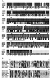
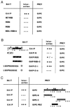
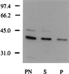
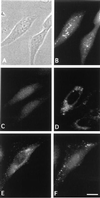
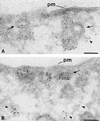
We have identified a mammalian protein called GIPC (for GAIP interacting protein, C terminus), which has a central PDZ domain and a C-terminal acyl carrier protein (ACP) domain. The PDZ domain of GIPC specifically interacts with RGS-GAIP, a GTPase-activating protein (GAP) for Galphai subunits recently localized on clathrin-coated vesicles. Analysis of deletion mutants indicated that the PDZ domain of GIPC specifically interacts with the C terminus of GAIP (11 amino acids) in the yeast two-hybrid system and glutathione S-transferase (GST)-GIPC pull-down assays, but GIPC does not interact with other members of the RGS (regulators of G protein signaling) family tested. This finding is in keeping with the fact that the C terminus of GAIP is unique and possesses a modified C-terminal PDZ-binding motif (SEA). By immunoblotting of membrane fractions prepared from HeLa cells, we found that there are two pools of GIPC-a soluble or cytosolic pool (70%) and a membrane-associated pool (30%). By immunofluorescence, endogenous and GFP-tagged GIPC show both a diffuse and punctate cytoplasmic distribution in HeLa cells reflecting, respectively, the existence of soluble and membrane-associated pools. By immunoelectron microscopy the membrane pool of GIPC is associated with clusters of vesicles located near the plasma membrane. These data provide direct evidence that the C terminus of a RGS protein is involved in interactions specific for a given RGS protein and implicates GAIP in regulation of additional functions besides its GAP activity. The location of GIPC together with its binding to GAIP suggest that GAIP and GIPC may be components of a G protein-coupled signaling complex involved in the regulation of vesicular trafficking. The presence of an ACP domain suggests a putative function for GIPC in the acylation of vesicle-bound proteins.
Figures


Similar articles
-
GIPC and GAIP form a complex with TrkA: a putative link between G protein and receptor tyrosine kinase pathways.Mol Biol Cell. 2001 Mar;12(3):615-27. doi: 10.1091/mbc.12.3.615. Mol Biol Cell. 2001. PMID: 11251075 Free PMC article.
-
GIPC recruits GAIP (RGS19) to attenuate dopamine D2 receptor signaling.Mol Biol Cell. 2004 Nov;15(11):4926-37. doi: 10.1091/mbc.e04-04-0285. Epub 2004 Sep 8. Mol Biol Cell. 2004. PMID: 15356268 Free PMC article.
-
A PDZ protein regulates the distribution of the transmembrane semaphorin, M-SemF.J Biol Chem. 1999 May 14;274(20):14137-46. doi: 10.1074/jbc.274.20.14137. J Biol Chem. 1999. PMID: 10318831
-
GIPC gene family (Review).Int J Mol Med. 2002 Jun;9(6):585-9. Int J Mol Med. 2002. PMID: 12011974 Review.
-
RGS17/RGSZ2 and the RZ/A family of regulators of G-protein signaling.Semin Cell Dev Biol. 2006 Jun;17(3):390-9. doi: 10.1016/j.semcdb.2006.04.001. Epub 2006 Apr 7. Semin Cell Dev Biol. 2006. PMID: 16765607 Review.
Cited by
-
Myosin VI is required for targeted membrane transport during cytokinesis.Mol Biol Cell. 2007 Dec;18(12):4750-61. doi: 10.1091/mbc.e07-02-0127. Epub 2007 Sep 19. Mol Biol Cell. 2007. PMID: 17881731 Free PMC article.
-
RGS3 interacts with 14-3-3 via the N-terminal region distinct from the RGS (regulator of G-protein signalling) domain.Biochem J. 2002 Aug 1;365(Pt 3):677-84. doi: 10.1042/BJ20020390. Biochem J. 2002. PMID: 11985497 Free PMC article.
-
GAIP interacting protein C-terminus regulates autophagy and exosome biogenesis of pancreatic cancer through metabolic pathways.PLoS One. 2014 Dec 3;9(12):e114409. doi: 10.1371/journal.pone.0114409. eCollection 2014. PLoS One. 2014. PMID: 25469510 Free PMC article.
-
Cloning and characterization of neuropilin-1-interacting protein: a PSD-95/Dlg/ZO-1 domain-containing protein that interacts with the cytoplasmic domain of neuropilin-1.J Neurosci. 1999 Aug 1;19(15):6519-27. doi: 10.1523/JNEUROSCI.19-15-06519.1999. J Neurosci. 1999. PMID: 10414980 Free PMC article.
-
A GIPC1-Palmitate Switch Modulates Dopamine Drd3 Receptor Trafficking and Signaling.Mol Cell Biol. 2016 Jan 19;36(6):1019-31. doi: 10.1128/MCB.00916-15. Mol Cell Biol. 2016. PMID: 26787837 Free PMC article.
References
Publication types
MeSH terms
Substances
Associated data
- Actions
- Actions
- Actions
Grants and funding
LinkOut - more resources
Full Text Sources
Other Literature Sources
Molecular Biology Databases
Research Materials
Miscellaneous

