

Bacterial stress responses to 1-megahertz pulsed ultrasound in the presence of microbubbles
- PMID: 9758821
- PMCID: PMC106580
- DOI: 10.1128/AEM.64.10.3927-3931.1998
Bacterial stress responses to 1-megahertz pulsed ultrasound in the presence of microbubbles
Abstract
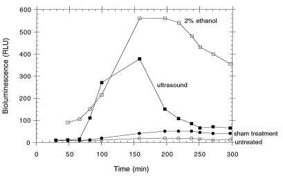
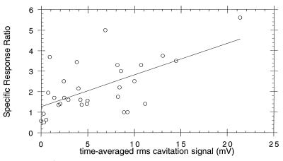
Members of a panel of stress-responsive biosensors have been used to study the effect of megahertz frequency ultrasound on Escherichia coli. Insonification causes acoustic cavitation, the collapse of oscillating microbubbles in solution, which can damage bacterial cells. A focused 1-MHz ultrasound transducer, capable of generating a spatial peak pulse average intensity of 500 W/cm2, was used to treat liquid bacterial cultures. Stress-responsive promoters fused to luxCDABE allowed the continuous measurement of light produced as a result of protein damage, DNA damage, oxidative stress, and membrane perturbation. A promoter responsive to ammonia limitation was not transcriptionally activated under test conditions. In contrast to bacteria in exponentially growing cultures, those in stationary-phase cultures were more resistant to the effects of ultrasound treatment. Quantification of the degree of acoustic cavitation due to symmetric bubble collapse was measured by a 20-MHz passive transducer, the output of which appears to be only partially correlated with cellular damage and survival. The methods and results summarized here provide the basis for further investigation into applications, including the purification of water samples.
Figures
Similar articles
-
Identification and quantification of toxic chemicals by use of Escherichia coli carrying lux genes fused to stress promoters.Appl Environ Microbiol. 1998 Nov;64(11):4346-52. doi: 10.1128/AEM.64.11.4346-4352.1998. Appl Environ Microbiol. 1998. PMID: 9797288 Free PMC article.
-
An Escherichia coli biosensor capable of detecting both genotoxic and oxidative damage.Appl Microbiol Biotechnol. 2004 Mar;64(1):46-52. doi: 10.1007/s00253-003-1418-0. Epub 2003 Aug 23. Appl Microbiol Biotechnol. 2004. PMID: 12937953
-
Detection of DNA damage by use of Escherichia coli carrying recA'::lux, uvrA'::lux, or alkA'::lux reporter plasmids.Appl Environ Microbiol. 1997 Jul;63(7):2566-71. doi: 10.1128/aem.63.7.2566-2571.1997. Appl Environ Microbiol. 1997. PMID: 9212407 Free PMC article.
-
Construction of a sodA::luxCDABE fusion Escherichia coli: comparison with a katG fusion strain through their responses to oxidative stresses.Appl Microbiol Biotechnol. 2003 Jan;60(5):577-80. doi: 10.1007/s00253-002-1168-4. Epub 2002 Dec 13. Appl Microbiol Biotechnol. 2003. PMID: 12536259
-
Mutagenesis and more: umuDC and the Escherichia coli SOS response.Genetics. 1998 Apr;148(4):1599-610. doi: 10.1093/genetics/148.4.1599. Genetics. 1998. PMID: 9560379 Free PMC article. Review.
Cited by
-
Combined Effect of Ultrasound and Low-Heat Treatments on E. coli in Liquid Egg Products and Analysis of the Inducted Structural Alterations by NIR Spectroscopy.Sensors (Basel). 2022 Dec 16;22(24):9941. doi: 10.3390/s22249941. Sensors (Basel). 2022. PMID: 36560311 Free PMC article.
-
Effect of ultrasound on survival and growth of Escherichia coli in cactus pear juice during storage.Braz J Microbiol. 2016 Apr-Jun;47(2):431-7. doi: 10.1016/j.bjm.2016.01.014. Epub 2016 Mar 2. Braz J Microbiol. 2016. PMID: 26991288 Free PMC article.
-
Inactivation of Planktonic Escherichia coli by Focused 2-MHz Ultrasound.Ultrasound Med Biol. 2017 Jul;43(7):1476-1485. doi: 10.1016/j.ultrasmedbio.2017.03.009. Epub 2017 Apr 26. Ultrasound Med Biol. 2017. PMID: 28454842 Free PMC article.
-
The evolution of the bacterial luciferase gene cassette (lux) as a real-time bioreporter.Sensors (Basel). 2012;12(1):732-52. doi: 10.3390/s120100732. Epub 2012 Jan 11. Sensors (Basel). 2012. PMID: 22368493 Free PMC article. Review.
-
Dispersing and Sonoporating Biofilm-Associated Bacteria with Sonobactericide.Pharmaceutics. 2022 May 30;14(6):1164. doi: 10.3390/pharmaceutics14061164. Pharmaceutics. 2022. PMID: 35745739 Free PMC article.
References
-
- Anderson D M, Nosenchuck D M, Reynolds G T, Walton A J. Mechanical stimulation of bioluminescence in the dinoflagellate Gonyaulux ployhedra Stein. J Exp Mar Biol Ecol. 1988;122:277–288.
-
- Apfel R E. Sonic effervescence: a tutorial on acoustic cavitation. J Acoust Soc Am. 1997;101:1227–1237.
-
- Armour E P, Corry P M. Cytotoxic effects of ultrasound in vitro dependence on gas content, frequency, radical scavengers, and attachment. Radiat Res. 1982;89:369–380. - PubMed
-
- Belkin, S. Unpublished results.
-
- Belkin S, Vollmer A C, Van Dyk T K, Smulski D R, Reed T R, LaRossa R A. Oxidative and DNA damaging agents induce luminescence in E. coli harboring lux fusions to stress promoters. In: Campbell A K, Kricka L J, Stanley P E, editors. Bioluminescence and chemiluminescence: fundamentals and applied aspects. Chichester, England: John Wiley and Sons; 1995. pp. 509–512.
Publication types
MeSH terms
Substances
LinkOut - more resources
Full Text Sources
Other Literature Sources
