

Prostate stem cell compartments: expression of the cell cycle inhibitor p27Kip1 in normal, hyperplastic, and neoplastic cells
- PMID: 9736039
- PMCID: PMC1853003
- DOI: 10.1016/S0002-9440(10)65632-5
Prostate stem cell compartments: expression of the cell cycle inhibitor p27Kip1 in normal, hyperplastic, and neoplastic cells
Abstract
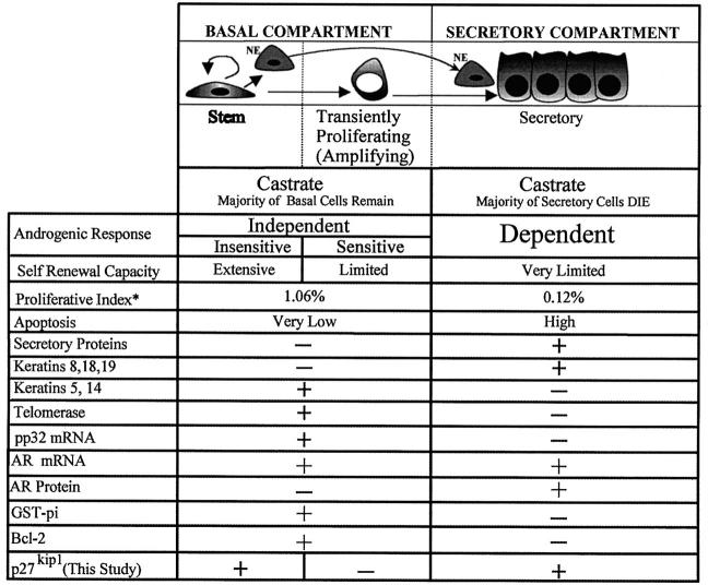
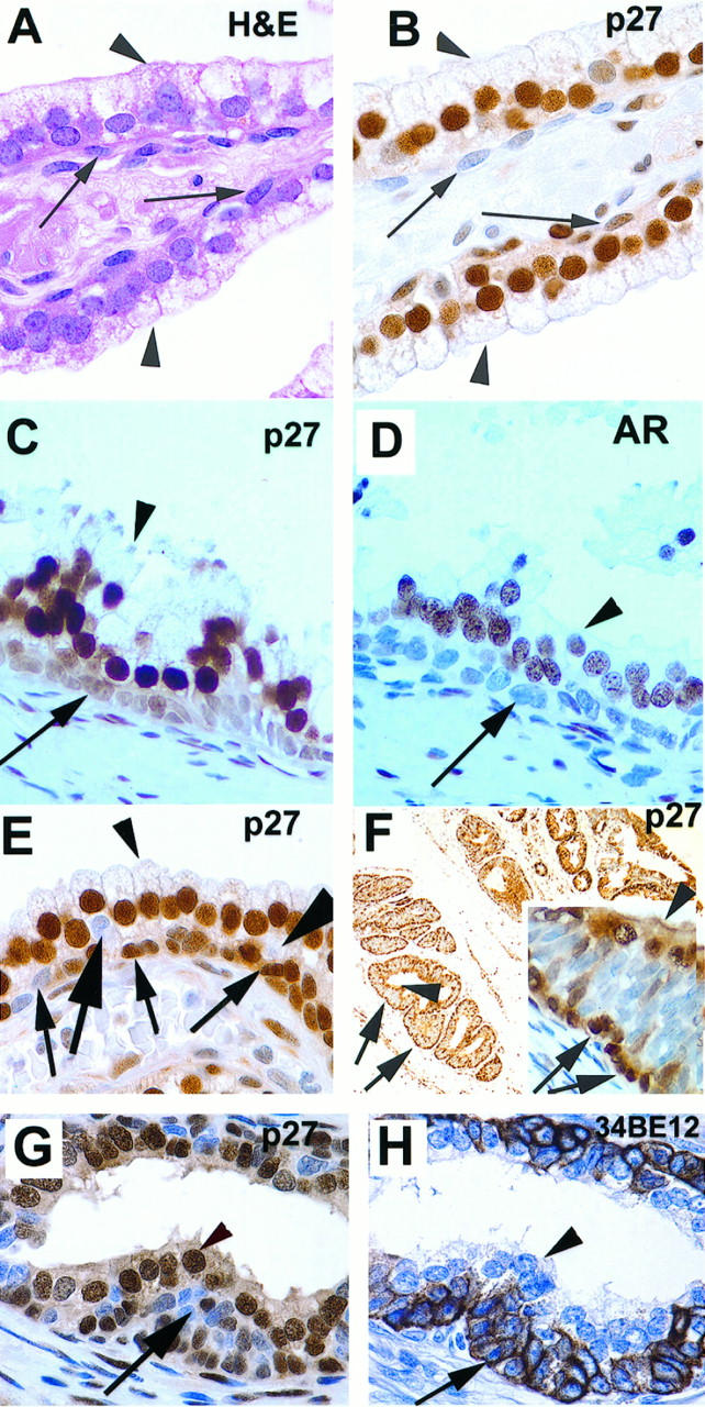
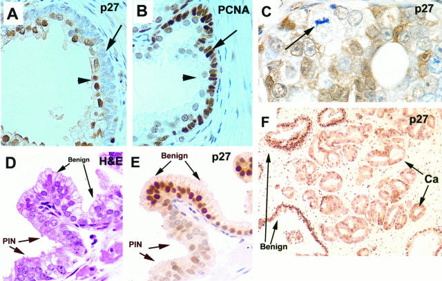
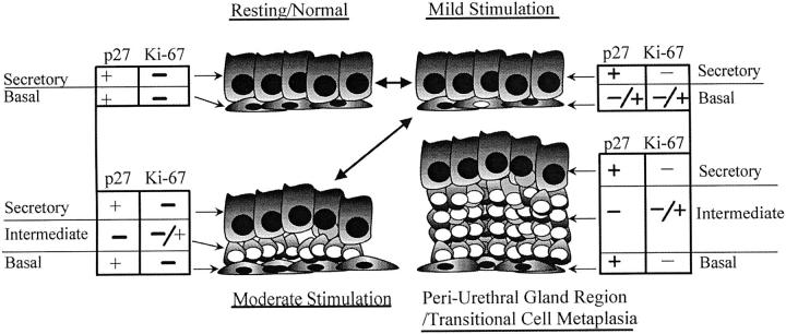
The stem cells of rapidly renewing tissues give rise to transiently proliferating cells, which in turn give rise to postmitotic terminally differentiated cells. Although the existence of a transiently proliferating compartment has been proposed for the prostate, little molecular anatomical evidence for its presence has been obtained to date. We used down-regulation of the cyclin-dependent kinase inhibitor p27Kip1 to identify cells capable of entering the proliferative phase of the cell cycle and, therefore, competent to fulfill the role of the transiently proliferating compartment. We examined the expression of p27Kip1 in relation to its role in the development of prostatic carcinoma. Formalin-fixed paraffin-embedded specimens from matched samples of normal-appearing prostate tissue, benign prostatic hyperplasia, high-grade prostatic intraepithelial neoplasia, primary adenocarcinomas, and pelvic lymph node metastases were evaluated by comparative immunohistochemistry against p27Kip1. In normal-appearing prostate epithelium, moderate to strong nuclear staining of p27Kip1 was present in greater than 85% of the terminally differentiated secretory cells. The normal basal cell compartment, believed to contain prostatic stem cells, showed distinctive p27Kip1 expression; acini in epithelial benign prostatic hyperplasia tissue contained more p27Kip1-negative basal cells than acini from non-benign prostatic hyperplasia tissue. A third layer of cells was identified that was sandwiched between the basal cells and the luminal cells, and this layer was consistently p27Kip1 negative. This intermediate layer was accentuated in the periurethral region, as well as in prostate tissue that had been subjected to prior combined androgen blockade. We hypothesize that, on appropriate additional mitogenic stimulation, cells in this layer, and other p27Kip1-negative basal cells, are competent for rapid entry into the cell cycle. Consistent with the fact that cancer cells are capable of cell division, all cases of high-grade prostatic intraepithelial neoplasia and invasive carcinoma also showed down-regulation of p27Kip1 as compared with the surrounding normal-appearing secretory cells. In pelvic lymph node metastases, p27Kip1 expression was also reduced. In summary, our results suggest that lack of nuclear p27Kip1 protein may delineate a potential transiently proliferating subcompartment within the basal cell compartment of the human prostate. In addition, these studies support the hypothesis that reduced expression of p27Kip1 removes a block to the cell cycle in human prostate epithelial cells and that dysregulation of p27Kip1 protein levels may be a critical early event in the development of prostatic neoplasia.
Figures
Similar articles
-
Levels of expression of p27KIP1 protein in human prostate and prostate cancer: an immunohistochemical analysis.Mod Pathol. 1999 Aug;12(8):751-5. Mod Pathol. 1999. PMID: 10463475
-
Expression of p27/Kip1 is down-regulated in human prostate carcinoma progression.J Pathol. 1999 Apr;187(5):563-6. doi: 10.1002/(SICI)1096-9896(199904)187:5<563::AID-PATH292>3.0.CO;2-3. J Pathol. 1999. PMID: 10398122
-
Apoptosis incidence and protein expression of p53, TGF-beta receptor II, p27Kip1, and Smad4 in benign, premalignant, and malignant human prostate.Hum Pathol. 2004 Mar;35(3):290-7. doi: 10.1016/j.humpath.2003.11.001. Hum Pathol. 2004. PMID: 15017584
-
Stem cell features of benign and malignant prostate epithelial cells.J Urol. 1998 Dec;160(6 Pt 2):2381-92. doi: 10.1097/00005392-199812020-00004. J Urol. 1998. PMID: 9817389 Review.
-
Differentiation pathways and histogenetic aspects of normal and abnormal prostatic growth: a stem cell model.Prostate. 1996 Feb;28(2):98-106. doi: 10.1002/(SICI)1097-0045(199602)28:2<98::AID-PROS4>3.0.CO;2-J. Prostate. 1996. PMID: 8604398 Review.
Cited by
-
Molecular pathways in prostate cancer.Nephrourol Mon. 2013 Jul 1;5(3):792-800. doi: 10.5812/numonthly.9430. Epub 2013 Jun 8. Nephrourol Mon. 2013. PMID: 24282788 Free PMC article. Review.
-
Disruption of prostate epithelial differentiation pathways and prostate cancer development.Front Oncol. 2013 Oct 31;3:273. doi: 10.3389/fonc.2013.00273. Front Oncol. 2013. PMID: 24199173 Free PMC article. Review.
-
Functional and Mechanistic Interrogation of BET Bromodomain Degraders for the Treatment of Metastatic Castration-resistant Prostate Cancer.Clin Cancer Res. 2019 Jul 1;25(13):4038-4048. doi: 10.1158/1078-0432.CCR-18-3776. Epub 2019 Mar 27. Clin Cancer Res. 2019. PMID: 30918020 Free PMC article.
-
The evolving landscape of prostate cancer stem cell: Therapeutic implications and future challenges.Asian J Urol. 2016 Oct;3(4):203-210. doi: 10.1016/j.ajur.2016.09.006. Epub 2016 Sep 20. Asian J Urol. 2016. PMID: 29264188 Free PMC article. Review.
-
Novel In Vivo model for combinatorial fluorescence labeling in mouse prostate.Prostate. 2015 Jun 15;75(9):988-1000. doi: 10.1002/pros.22984. Epub 2015 Mar 8. Prostate. 2015. PMID: 25753731 Free PMC article.
References
-
- Potten CS, Morris RJ: Epithelial stem cells in vivo. J Cell Science 1988, 10(Suppl):45-62 - PubMed
-
- Potten CS, Loeffler M: Stem cells: attributes, cycles, spirals, pitfalls and uncertainties: lessons for and from the crypt. Development 1990, 110:1001-1020 - PubMed
-
- Potten CS: Stem Cells. ed 1. London, Academic Press, 1997
-
- Isaacs JT, Coffey DS: Etiology and disease process of benign prostatic hyperplasia. Prostate 1989, 2(Suppl):33-50 - PubMed
-
- Bonkhoff H, Stein U, Remberger K: Multidirectional differentiation in the normal, hyperplastic, and neoplastic human prostate: simultaneous demonstration of cell-specific epithelial markers. Hum Pathol 1994, 25:42-46 - PubMed
Publication types
MeSH terms
Substances
Grants and funding
LinkOut - more resources
Full Text Sources
Other Literature Sources
Medical
