. 1998 Oct;72(10):7960-71.
doi: 10.1128/JVI.72.10.7960-7971.1998.

E1B 55-kilodalton-associated protein: a cellular protein with RNA-binding activity implicated in nucleocytoplasmic transport of adenovirus and cellular mRNAs

Affiliations

E1B 55-kilodalton-associated protein: a cellular protein with RNA-binding activity implicated in nucleocytoplasmic transport of adenovirus and cellular mRNAs

S Gabler et al. J Virol. 1998 Oct.

Abstract

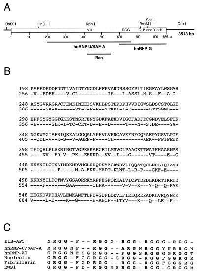
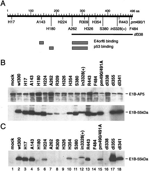
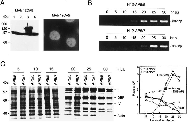
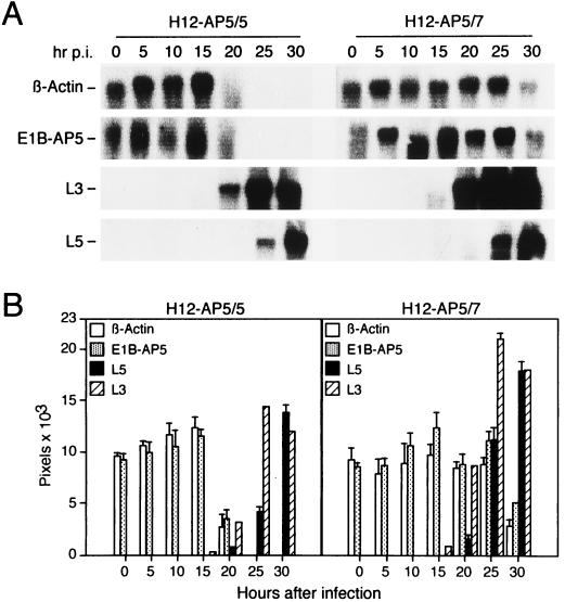
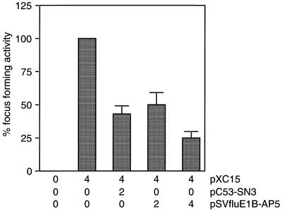
The adenovirus type 5 (Ad5) early 1B 55-kDa protein (E1B-55kDa) is a multifunctional phosphoprotein that regulates viral DNA replication and nucleocytoplasmic RNA transport in lytically infected cells. In addition, E1B-55kDa provides functions required for complete oncogenic transformation of rodent cells in cooperation with the E1A proteins. Using the far-Western technique, we have isolated human genes encoding E1B-55kDa-associated proteins (E1B-APs). The E1B-AP5 gene encodes a novel nuclear RNA-binding protein of the heterogeneous nuclear ribonucleoprotein (hnRNP) family that is highly related to hnRNP-U/SAF-A. Immunoprecipitation experiments indicate that two distinct segments in the 55-kDa polypeptide which partly overlap regions responsible for p53 binding are required for complex formation with E1B-AP5 in Ad-infected cells and that this protein interaction is modulated by the adenovirus E4orf6 protein. Expression of E1B-AP5 efficiently interferes with Ad5 E1A/E1B-mediated transformation of primary rat cells. Furthermore, stable expression of E1B-AP5 in Ad-infected cells overcomes the E1B-dependent inhibition of cytoplasmic host mRNA accumulation. These data suggest that E1B-AP5 might play a role in RNA transport and that this function is modulated by E1B-55kDa in Ad-infected cells.


Figures

FIG. 1
FIG. 1
E1B-AP5 cDNA maps, nucleotide sequence, and predicted amino acid sequence. (A) cDNA clones E1B-AP5/1 and E1B-AP5/2 were isolated with the [γ-32P]ATP-labeled GSTE1B-55kDa protein probe from a λgt11 HeLa cDNA expression library. cDNA clones E1B-AP5/3 to E1B-AP5/7 were isolated from the same library by rescreening with E1B-AP5/2 and later with a fragment from the 3′ end of E1B-AP5/3. The thick black bar on top represents the 2,568-bp open reading frame of E1B-AP5. Thin bars denote the 5′ and 3′ untranslated sequences, respectively. The locations of some unique restriction enzymes are indicated above the bars. (B) The complete E1B-AP5 cDNA sequence was generated by assembling restriction fragments from E1B-AP5/6 and E1B-AP5/7 in pBKRSV. Sequence was determined from each cDNA clone twice on both strands by sequence-derived oligonucleotide primers. The predicted amino acid sequence is shown in the single-letter code.
FIG. 2
FIG. 2
Sequence homologies to hnRNP-U/SAF-A, hnRNP-G, and Ran. (A) The regions that have been identified within the predicted amino acid sequence of E1B-AP5 are shown below in their relative positions along the E1B-AP5 polypeptide (open box). The locations of the NTP-binding consensus sequence (NTP), RGG boxes (RGG), and glutamine-, proline-, and tyrosine-rich region (Q, P and Y-rich) at the carboxy terminus are indicated. (B) Direct sequence comparison of the related sequences of E1B-AP5 and hnRNP-U/SAF-A. The dashes in the hnRNP-U/SAF-A sequence are identical amino acids in E1B-AP5, and dots indicate gaps in the hnRNP-U/SAF-A sequence alignment. (C) Alignment of E1B-AP5 with RGG box domains from several proteins. The consensus sequences of hnRNP-U/SAF-A, hnRNP-A1, nucleolin, fibrillarin, and EWS1 were derived from the sequences listed in reference .
FIG. 3
FIG. 3
Analysis of E1B-AP5 mRNA and protein expression. (A) Northern blot analysis. A 20-μg portion of total RNA isolated from the indicated human tissues and cell lines were subjected to electrophoresis through a 1.2% agarose gel containing formaldehyde, transferred to a nitrocellulose filter, and hybridized to a [α-32P]dCTP-labeled E1B-AP5 coding sequence probe from E1B-AP5/5. (Monoc, monocytes; Macrop, macrophages; Lmypho, lymphocytes; Fibrob, fibroblasts; HepG2, hepatocytes; HaCAT, keratinocytes; MelIm, melanoma). The locations of the 18S and 28S rRNAs are indicated. RNA loading was determined by staining the RNA with ethidium bromide prior to transfer to nitrocellulose filters. (B) Immunoprecipitation of E1B-AP5 and hnRNP-U/SAF-A proteins. Radiolabeled E1B-AP5 (lanes 1 to 3) and hnRNP-U/SAF-A (lanes 4 to 6) proteins were generated by in vitro translation and subjected to immunoprecipitation with the anti-E1B-AP5 antiserum (α-E1B-AP5; lanes 2 and 5) and a matched preimmune serum (pre; lanes 3 and 6). The precipitates were analyzed by SDS-PAGE and autoradiography. Lanes designated “input” received the same amount of in vitro-translated proteins added to each immunoprecipitation reaction mixture. The positions of markers are indicated. (C) Western blot analysis of E1B-AP5 protein expression. Total-cell extracts were prepared and subjected to SDS-PAGE, transferred to nitrocellulose filters, and probed with the rabbit anti-E1B-AP5 antiserum. The positions of markers are indicated on the right. (D) Indirect immunofluorescence analysis. A549 cells were probed with the rabbit anti-E1B-AP5 antiserum (α-E1B-AP5) followed by fluorescein-conjugated sheep anti-rabbit antibodies. Magnification, ×100.
FIG. 4
FIG. 4
In vitro and in vivo association of E1B-AP5 with E1B-55kDa. (A) E1B-AP5 binds to E1B-55kDa in vitro. In vitro-translated [35S]methionine-labeled RCC1 (lane 1), E1B-AP5 (lane 2), and hnRNP-U/SAF-A (lane 3) proteins were incubated with GSTE1B-55kDa, GST-p53, or GST alone, and proteins bound to washed beads were separated by SDS-PAGE and visualized by autoradiography. Molecular mass markers are indicated on the left in kilodaltons. (B) E1B-55kDa binds to E1B-AP5 in vivo. Subconfluent 293 cells grown on 90-mm-diameter culture dishes were transfected with plasmid pSVfluE1B-AP5 expressing epitope-tagged E1B-AP5 by calcium phosphate coprecipitation. At 36 h after transfection, total-cell extracts were prepared. ATP or GTP was added to a final concentration of 100 μM as indicated, and the extracts were subjected to immunoprecipitation with monoclonal antibody (MAb) 12CA5 followed by immunoblotting. E1B-55kDa was detected with anti-55-kDa rat monoclonal antibody 9C10. Lanes 5 and 6, designated “input,” received 1/20 of the amount of total-cell extract added to each immunoprecipitation reaction mixture.
FIG. 5
FIG. 5
E1B-AP5 binds to ribonucleotide homopolymers and ssDNA. (A) Quantitation of E1B-AP5 binding to ribonucleotide homopolymers. The percentage of input E1B-AP5 bound to RNA at the indicated NaCl concentrations was determined as described in the text. The mean from three independent experiments is presented. (B) Quantitation of E1B-AP5 binding to ssDNA. The percentage of input E1B-AP5 bound to ssDNA at the indicated NaCl concentrations was determined. The mean from three independent experiments is presented.
FIG. 6
FIG. 6
Analysis of the E1B-55kDa/E1B-AP5 protein interaction in mutant and wild-type virus-infected MCF-7 cells. (A) E1B-55kDa mutation sites. The thick black bar at the top represents the 496 residues of the 55-kDa polypeptide. Number 496 denotes the last amino acid. The insertion mutations and the point mutation pm490A/491A (pm490/1) are shown below in their relative positions along the E1B-55kDa polypeptide. The deletion in dl338 is denoted by a thin bar. The p53 and E4orf6 interaction domains (gray boxes) are shown according to their positions along the 55-kDa polypeptide below the E1B protein and were defined by Yew et al. (75) and Rubenwolf et al. (62), respectively. (B) Expression of E1B-55kDa and E1B-AP5 virus-infected cells. Whole-cell extracts used for the coimmunoprecipitation experiment containing 40 μg of protein were subjected to PAGE followed by Western blotting with anti-E1B-55kDa (2A6) hybridoma supernatant and anti-E1B-AP5 antiserum. (C) Coimmunoprecipitation of E1B-55kDa. E1B-55kDa bound to E1B-AP5 protein was coprecipitated with anti-E1B-AP5 rabbit antiserum from the same whole-cell extracts, resolved on SDS–10% polyacrylamide gels, and visualized by Western immunoblot analysis with the 2A6 anti-55-kDa monoclonal antibody.
FIG. 7
FIG. 7
Effects of stable expressed fluE1B-AP5 on viral DNA synthesis and host cell shutoff. (A) Expression of fluE1B-AP5 in H12-AP5/7 cells. Total-cell extracts were prepared from H1299 cells (lane 1) and puromycin-resistant cell clones H12-AP/5 (lane 2), H12-AP5/7 (lane 3), and H12-AP5/8 (lane 4), subjected to SDS-PAGE, and analyzed by immunoblotting with monoclonal antibody (MAb) 12CA5. The additional faster-migrating band present in all lanes is due to a cross-reactivity of a cellular protein with monoclonal antibody 12CA5. Indirect immunofluorescence of H12-AP5/7 cells probed with 12CA5 followed by fluorescein-conjugated sheep anti-rabbit antibody. Magnification, ×100. (B) Analysis of viral DNA synthesis. H12-AP5/5 or H12-AP5/7 cells were infected with wt300 virus at an infectivity of 200 PFU per cell. Cells were harvested at the indicated time points postinfection (hr p.i.), and viral DNA synthesis was determined by PCR. The DNA products were subjected to agarose gel electrophoresis and ethidium bromide staining. (C) Analysis of fiber (IV) and actin protein steady-state levels. Cells were infected with wt300 virus and labeled with [35S]methionine for 1 h at 5, 10, 15, 20, 25, and 30 h after infection. A total of 2 × 10⁵ trichloroacetic acid-precipitable counts from each time point was fractionated by SDS-PAGE, and proteins were visualized by autoradiography. Molecular mass markers are indicated on the left. Bands corresponding to cellular actin and several viral polypeptides are listed on the right. The levels of fiber (IV) and actin proteins were quantitated as described and plotted as a function of time. The steady-state level of the E1B-AP5 protein was quantitated after Western blot analysis of the same total-cell extracts (data not shown).
FIG. 8
FIG. 8
Effects of stable expressed fluE1B-AP5 on cytoplasmic mRNA accumulation in wt300-infected cells. (A) Northern blot analysis of β-actin, E1B-AP5, L3, and L5 mRNA species in infected H12-AP5/5 and H12-AP5/7 cells. Cytoplasmic RNA was prepared at the indicated time points after infection (hr p.i.). Equal quantities of these RNAs were subjected to electrophoresis, transferred to nitrocellulose membranes, and hybridized with [α-32P]dCTP-labeled β-actin, E1B-AP5, L3, and L5 DNA probes. The bands corresponding to the viral and cellular mRNAs are indicated at the left. (B) The levels of β-actin, E1B-AP5, L3, and L5 mRNAs were quantitated as described and plotted as a function of time. The mean and standard deviation is presented for three independent experiments.
FIG. 9
FIG. 9
E1B-AP5 inhibits Ad5 E1A/E1B-mediated focus formation. Primary BRK cells were transfected with the indicated amounts of plasmids (micrograms of DNA per 3 × 10⁶ cells). Focus-forming activity is presented as a percentage of E1A plus E1B activity. The average number of dense foci for pXC15 was 128 in four independent experiments.

Similar articles

Cited by

References

    1. Babich A, Feldman L T, Nevins J R, Darnell J E, Weinberger C. Effect of adenovirus on metabolism of specific host mRNAs: transport control and specific translation discrimination. Mol Cell Biol. 1983;3:1212–1221. - PMC - PubMed
    1. Babiss L E, Ginsberg H S, Darnell J J. Adenovirus E1B proteins are required for accumulation of late viral mRNA and for effects on cellular mRNA translation and transport. Mol Cell Biol. 1985;5:2552–2558. - PMC - PubMed
    1. Bar-Sagi D, Rotin D, Batzer A, Mandiyan V, Schlessinger J. SH3 domains direct cellular localization of signaling molecules. Cell. 1993;74:83–91. - PubMed
    1. Beltz G A, Flint S J. Inhibition of HeLa cell protein synthesis during adenovirus infection. J Mol Biol. 1979;131:353–373. - PubMed
    1. Binger M H, Flint S J. Accumulation of early and intermediate mRNA species during subgroup C adenovirus productive infections. Virology. 1984;136:387–403. - PubMed

Publication types

MeSH terms

Associated data