. 1998 Sep 7;142(5):1245-56.
doi: 10.1083/jcb.142.5.1245.

The cytoplasmic tail of rhodopsin acts as a novel apical sorting signal in polarized MDCK cells

Affiliations

The cytoplasmic tail of rhodopsin acts as a novel apical sorting signal in polarized MDCK cells

J Z Chuang et al. J Cell Biol. .

Abstract

All basolateral sorting signals described to date reside in the cytoplasmic domain of proteins, whereas apical targeting motifs have been found to be lumenal. In this report, we demonstrate that wild-type rhodopsin is targeted to the apical plasma membrane via the TGN upon expression in polarized epithelial MDCK cells. Truncated rhodopsin with a deletion of 32 COOH-terminal residues shows a nonpolar steady-state distribution. Addition of the COOH-terminal 39 residues of rhodopsin redirects the basolateral membrane protein CD7 to the apical membrane. Fusion of rhodopsin's cytoplasmic tail to a cytosolic protein glutathione S-transferase (GST) also targets this fusion protein (GST-Rho39Tr) to the apical membrane. The targeting of GST-Rho39Tr requires both the terminal 39 amino acids and the palmitoylation membrane anchor signal provided by the rhodopsin sequence. The apical transport of GST-Rho39Tr can be reversibly blocked at the Golgi complex by low temperature and can be altered by brefeldin A treatment. This indicates that the membrane-associated GST-Rho39Tr protein may be sorted along a yet unidentified pathway that is similar to the secretory pathway in polarized MDCK cells. We conclude that the COOH-terminal tail of rhodopsin contains a novel cytoplasmic apical sorting determinant. This finding further indicates that cytoplasmic sorting machinery may exist in MDCK cells for some apically targeted proteins, analogous to that described for basolaterally targeted proteins.


Figures

Figure 1
Figure 1
Rhodopsin topology showing COOH-terminal mutant constructs used to characterize the apical sorting domains. Rhodopsin is a seven-transmembrane protein with its COOH terminus facing the cytoplasmic side and its NH2 terminus facing the extracellular side of the lipid membrane. The zigzags indicate the palmitoyl membrane anchor sites. All mutants used in this study were generated using site-directed in vitro mutagenesis. Δ5, Δ22, and Δ32 are COOH-terminal deletion mutants in which the last 5, 22, and 32 amino acid residues are missing, respectively. In the Cys322Cys323 → Ser322Ser323 (pal) mutant, the cysteines at positions 322 and 323 were replaced with serines. The residues between amino acids 310 and 348 are labeled with single-letter amino acid designations.
Figure 2
Figure 2
Immunolocalization of wild-type (Wt) and mutant rhodopsins in stably transfected MDCK cells. Rhodopsin was detected by surface immunofluorescent labeling using mAb B6-30 as described in Materials and Methods. The immunofluorescent staining was analyzed by laser scanning confocal microscopy. Optical sections horizontal (X-Y, top) and perpendicular (Z, bottom) to the monolayer are shown. Note that the clear Transwell filter itself generates a weak background. Bar, 5 μm.
Figure 3
Figure 3
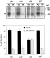
The steady-state surface distribution of rhodopsins expressed in stably transfected MDCK cells. The immunoprecipitated, biotinylated rhodopsin proteins were electrophoresed on 12% SDS-PAGE, transferred to a nitrocellulose filter, and probed with 125I-streptavidin as described in Materials and Methods. Note that rhodopsin is prone to forming higher-order aggregates (Sung et al., 1991). The arrow indicates the monomeric form. Blots were analyzed with a Molecular Dynamics PhosphorImager. The total rhodopsin expressed on the cell surface was taken to be 100%. Bars represent the mean ± SD of multiple determinations from more than three independent experiments (n = 5, 5, 6, and 4 for Wt, Δ22, Δ32, and pal, respectively). Signals derived from multimers were also taken into consideration in quantitation. pal = Cys322Cys323 → Ser322Ser323 mutant.
Figure 4
Figure 4
Apical targeting of CD7 by rhodopsin's COOH terminus. (A) Schematic diagrams of CD7, CD7–Rho7, and CD7–Rho39. CD7–Rho7 is a fusion protein in which the proximal seven residues of rhodopsin's cytoplasmic tail (N310-C316) is fused to the COOH terminus of CD7 (open box). CD7–Rho39 is a CD7 fusion protein containing rhodopsin's COOH-terminal 39 residues (N310-A348). TM, transmembrane domain; zigzag, palmitoyl membrane anchor; black box, rhodopsin sequence. (B) Polarized MDCK monolayers stably expressing CD7, CD7–Rho7, and CD7–Rho39 after surface immunofluorescent labeling with mAb T3-3A1, which recognizes the extracellular domain of CD7. Vertical optical sections obtained from confocal microscopic analysis are shown. (C) The levels of steady-state surface distribution of CD7 (∼40 kD), CD7–Rho7 (∼41 kD), and CD7–Rho39 (∼44 kD) were determined by the domain-selective biotinylation/immunoprecipitation assay. As was shown previously (Haller and Alper, 1993), an endogenous MDCK protein that migrates slightly slower than CD7–Rho7 and CD7–Rho39 on SDS-PAGE is also precipitated by mAb T3-3A1. Results shown are from a representative experiment chosen between four trials using more than two independent stable lines. Bar, 5 μm.
Figure 5
Figure 5
Targeting of wild-type rhodopsin from TGN to the apical surface. Vectorial delivery of wild-type rhodopsin to the cell surface was analyzed by a membrane targeting assay (see Materials and Methods). At various time points during the chase as indicated, 35S metabolically labeled, rhodopsin-expressing MDCK cells were biotinylated from either the apical (Ap) or basolateral (Bl) surface and immunoprecipitated with antirhodopsin mAb B6-30 as described. (A) A fraction of each immunoprecipitated sample was analyzed by 4–20% SDS-PAGE and is shown as total immunoprecipitate. (B) Biotinylated, radiolabeled immunoprecipitates were obtained by a subsequent reprecipitation with streptavidin agarose. (C) For quantitation, biotinylated, radiolabeled immunoprecipitates were treated with PNGase F, separated by SDS-PAGE, and visualized by autoradiography. No labeled material is precipitated from control MDCK cell lysates.
Figure 6
Figure 6
Targeting of CD7–Rho39 directly from the TGN to the apical surface. Targeting of CD7–Rho39 and CD7 was analyzed by the membrane targeting assay described in Fig. 5. In this experiment, the CD7–Rho39 fusion protein was precipitated by antirhodopsin COOH terminus mAb 1D4, and CD7 was precipitated by anti-CD7 mAb T3-3A1. (A and C) Autoradiograms of biotinylated, radiolabeled immunoprecipitates, which were analyzed by 12% SDS-PAGE. (B and D) Before the second streptavidin-agarose precipitation, fractions of total radiolabeled 1D4 or T3-3A1 immunoprecipitates were analyzed on SDS-PAGE to demonstrate that equal amounts of radiolabeled proteins were immunoprecipitated from each sample. Ap, apically biotinylated; Bl, basolaterally biotinylated.
Figure 7
Figure 7
The apical targeting of GST by addition of rhodopsin's COOH terminus. (A) Schematic diagrams of GST, GST–Rho39Tr, and GST–Rho39palTr. GST– Rho39Tr is a fusion protein in which a triple-repeat of the terminal 39 amino acids of rhodopsin (black boxes) was fused to the COOH terminus of GST (open box). GST– Rho39palTr is a similar fusion protein except that the 39 rhodopsin residues were derived from the Cys322Cys323 → Ser322Ser323 mutant so that the palmitoylation signal (zigzag) was removed. (B) Immunolocalization of GST, GST–Rho39Tr, and GST– Rho39palTr in MDCK cells. Permeabilized MDCK monolayers were labeled with anti-GST antibody followed by biotinylated anti–rabbit antibody and FITC-streptavidin. Optical sections perpendicular to the monolayer are shown. Both GST and GST–Rho39palTr show diffuse, nucleus-excluded cytoplasmic staining. (C) Subcellular distributions of GST–Rho39Tr and GST–Rho39palTr in MDCK cells. MDCK cell homogenates were fractionated into cytosolic (S100) and total cellular membrane (P100) fractions by centrifugation at 100,000 g for 45 min. Equal fractions of S100 and P100 were electrophoresed on SDS-PAGE and immunoblotted with mAb 1D4 followed by alkaline phosphatase anti– mouse IgG. NBT/BCIP substrates were used for color development. Bar, 5 μm.
Figure 8
Figure 8
TGN accumulation of GST– Rho39Tr in MDCK cells during a low-temperature block. MDCK cultures grown on coverslips were maintained either at 37°C (a and b) or removed to 20°C for 6 h (c and d) before PFA fixation. Fixed cells were permeabilized and double-labeled with anti-GST antibody (a and c) and anti– γ-adaptin antibody (b and d). Corresponding fluorescent-conjugated secondary antibodies were used for visualization. Confocal images of the xy plane are shown. Arrows indicate the perinuclear, Golgi-like staining. Bar, 5 μm.
Figure 9
Figure 9
Return of the apical surface localization of GST–Rho39Tr after release from low-temperature block. Monolayers of MDCK cells were incubated at 20°C for 6 h to accumulate newly synthesized GST–Rho39Tr in the Golgi complex. Cells were either immediately fixed by PFA (a and d) or transferred to medium containing cycloheximide (20 μg/ml) (b and e) for an additional 30 min at 37°C before fixation. For BFA treatment experiments, 1 μg/ml BFA was added to the cells in the last 15 min of the 20°C incubation and remained during the 37°C release (c and f). Fixed cells were permeabilized and then immunolabeled with anti-GST antibody, biotinylated anti–rabbit IgG, and streptavidin-FITC. Propidium iodide was included in the staining to show the nuclei in the z section (shown in gray in d–f). After the 20°C block, GST–Rho39Tr is concentrated in the Golgi complex, which is localized above the nucleus in the subapical membrane cytoplasm (d). Prominent labeling of GST–Rho39Tr is readily detectable on the apical surface 30 min after 37°C release in control cells (b and e). However, GST–Rho39Tr accumulates on the basolateral membrane as well as the apical membrane in the cells treated with BFA. Some punctate, intracellular staining of GST–Rho39Tr is also observed (c and f). The focal plane of c was set near the apical surface. Confocal images of both xy (top row) and z (bottom row) planes are shown. Bar, 5 μm.

Similar articles

Cited by

References

    1. Adamus G, Arendt A, Zam ZS, McDowell JH, Hargrave PA. Use of peptides to select for anti-rhodopsin antibodies with desired amino acid sequence specificities. Pept Res. 1988;1:42–47. - PubMed
    1. Applebury ML, Hargrave PA. Molecular biology of the visual pigments. Vision Res. 1986;26:1881–1895. - PubMed
    1. Arreaza G, Brown AD. Sorting and intracellular trafficking of a glycosylphosphatidylinositol-anchored protein and two hybrid transmembrane proteins with the same ectodomain in Madin-Darby canine kidney epithelial cells. J Biol Chem. 1995;270:23641–23647. - PubMed
    1. Bacallao R, Antony C, Dotti C, Karsenti E, Stelzer EHK, Simons K. The subcellular organization of Madin-Darby canine kidney cells during the formation of a polarized epithelium. J Cell Biol. 1989;109:2817–2832. - PMC - PubMed
    1. Balch WE, Glick BS, Rothman JE. Sequential intermediates in the pathway of intercompartmental transport in a cell-free system. Cell. 1984;39:525–536. - PubMed

Publication types