. 1998 Sep 7;142(5):1159-66.
doi: 10.1083/jcb.142.5.1159.

Dynamics of the genome during early Xenopus laevis development: karyomeres as independent units of replication

Affiliations

Dynamics of the genome during early Xenopus laevis development: karyomeres as independent units of replication

J M Lemaitre et al. J Cell Biol. .

Abstract

During Xenopus laevis early development, the genome is replicated in less than 15 min every 30 min. We show that during this period, DNA replication proceeds in an atypical manner. Chromosomes become surrounded by a nuclear membrane lamina forming micronuclei or karyomeres. This genomic organization permits that prereplication centers gather on condensed chromosomes during anaphase and that DNA replication initiates autonomously in karyomeres at early telophase before nuclear reconstruction and mitosis completion. The formation of karyomeres is not dependent on DNA replication but requires mitotic spindle formation and the normal segregation of chromosomes. Thus, during early development, chromosomes behave as structurally and functionally independent units. The formation of a nuclear envelope around each chromosome provides an in vivo validation of its role in regulating initiation of DNA replication, enabling the rate of replication to accelerate and S phase to overlap M phase without illegitimate reinitiation. The abrupt disappearance of this atypical organization within one cell cycle after thirteen divisions defines a novel developmental transition at the blastula stage, which may affect both the replication and the transcription programs of development.


Figures
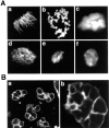
Figure 1
Figure 1
Nuclear reconstruction occurs from individualized chromosomes during early embryogenesis. (A) Changes in the reorganization of the chromatin is presented. DNA was analyzed by staining with Hoechst 33258 dye as described in Materials and Methods. Anaphase (a), telophase (b), interphase (c), and prophase (d) of nuclei before the 8,000-cell stage and telophase (e) and interphase (f) of nuclei after the 8,000-cell stage are shown. (B) Membranes associated with karyomeres (a) and newly formed nuclei (b) were detected by confocal microscopy using the lipid-staining dye DiOC6.
Figure 2
Figure 2
Karyomere formation is specific from the early embryogenesis. Xenopus embryos incubated at 21°C were taken from synchronized fertilized eggs at different times after fertilization. The kinetics and synchrony of cleavage were examined by light microscope during the first cell cycles; cycles were 30 min. The reorganization of the chromatin was analyzed by DNA staining with Hoescht 33258 dye as described in Materials and Methods. The plot shows the percentage of nuclei formed from karyomeres as a function of time after fertilization.
Figure 3
Figure 3
Lamins bind and surround each chromosome during the early embryonic cell cycles. Early embryos were prepared as described in Materials and Methods and submitted to indirect immunofluorescence visualization with a 687A7 monoclonal anti-lamin antibody (13). DNA was stained with propidium iodide. Green, lamins; red, DNA. A–F are successive stages of the organization of the nucleus during each cell cycle from anaphase (A) to prophase (F).
Figure 4
Figure 4
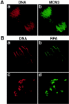
Prereplication centers are defined during anaphase. Early embryos were submitted to indirect immunofluorescence with XMCM3 (A) or RPA (B) antibodies. Red, DNA staining (propidium iodide); green, XMCM3 and RPA. Anaphase chromosomes at both poles (A, a and b) or one pole (B, a and b) and telophase karyomeres (B, c and d) are presented.
Figure 5
Figure 5
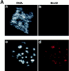
S phase occurs in karyomeres before nuclear formation and cell division in vivo. (A) Early embryos were incubated with BrdU for a 10-min pulse and prepared as described in Materials and Methods. BrdU incorporation was detected by indirect immunofluorescence after using an anti-BrdU antibody and DNA was visualized with Hoechst staining. BrdU incorporation is in red (b and d), and DNA in gray (a and c). Nuclei at anaphase (a and b) and early telophase (c and d) are shown. (B) Early embryos were examined by indirect immunofluorescence with PCNA and β tubulins antibodies. Green, PCNA (b) and β tubulins (d); red, DNA staining (propidium iodide) (a and c).
Figure 5
Figure 5
S phase occurs in karyomeres before nuclear formation and cell division in vivo. (A) Early embryos were incubated with BrdU for a 10-min pulse and prepared as described in Materials and Methods. BrdU incorporation was detected by indirect immunofluorescence after using an anti-BrdU antibody and DNA was visualized with Hoechst staining. BrdU incorporation is in red (b and d), and DNA in gray (a and c). Nuclei at anaphase (a and b) and early telophase (c and d) are shown. (B) Early embryos were examined by indirect immunofluorescence with PCNA and β tubulins antibodies. Green, PCNA (b) and β tubulins (d); red, DNA staining (propidium iodide) (a and c).
Figure 6
Figure 6
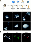
(A) An embryonic extract which mimics the early embryonic cell cycles. An extract was prepared from early embryos containing G2 synchronized nuclei, as described in Materials and Methods. The extracts were incubated for 30 min at 23°C after addition of a metaphase-arrested egg extract. Under these conditions, the nuclear membrane breaks down and nuclei arrest at the metaphase stage of mitosis. Completion of mitosis and S phase was induced by addition of 0.6 mM CaCl2. Samples were then placed onto slides and fixed as described as in Materials and Methods. (a) G2/prophase embryo nucleus; (b) metaphase; (c) anaphase, 15 min after Ca++ activation; (d) early telophase, 15–30 min after Ca++ activation; (e) mid-telophase 30 min after Ca++ activation; (f) prophase 45–60 min after Ca++ activation. DNA was stained by Hoechst 33258 and observed by video enhanced microscopy. (B) DNA replication occurs in karyomeres in vitro. An extract from embryos synchronized in G2 was induced to undergo mitosis by addition of MPF activity as described in Materials and Methods. Biotin-dUTP (20 μM) was then added and completion of mitosis and S phase was induced by further addition of 0.6 μM CaCl2. Samples were analyzed for biotin-dUTP incorporation 15–30 min after activation by Ca2+. Biotin in DNA was detected by indirect immunofluorescence with streptavidin Texas red (c) and RPA (b) immunostaining was detected with specific antibody. DNA was stained with Hoescht 33258 dye (a). Long and short arrows show condensed and decondensed chromatin respectively into independent karyomeres, respectively.
Figure 7
Figure 7
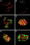
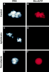
Karyomere formation occurs independently of DNA replication but requires spindle formation. An embryonic extract containing endogenous G2 nuclei was released into mitotic metaphase as in Materials and Methods (A and B). DNA replication was blocked by 50 μg/ml aphidicolin (C and D), and spindle formation was prevented by 5 μg/ml nocodazole (E and F). Biotin-dUTP (20 μM) was added and progression into S phase was induced by 0.6 mM CaCl2. DNA synthesis was followed by biotin-dUTP incorporation after 15–30 min of incubation in B, D, and F. DNA was stained by Hoescht 33258 (A, C, and E).
Figure 8
Figure 8
The cell cycle during early development and after MBT. At each cell cycle during the first twelve divisions, chromosomes, during their segregation to the poles, form karyomeres wherein DNA replication begins. A distinct transition occurs at the mid-blastula stage when karyomeres are no longer formed during segregation. The nucleus is then reconstructed from 2n chromosomes, and a classical cell cycle appears for the first time during development.

References

    1. Adchi Y, Laemmli UK. Study of the cell cycle-dependent assembly of the DNA pre-replication centres in Xenopusegg extracts. EMBO (Eur Mol Biol Organ) J. 1994;13:4153–4164. - PMC - PubMed
    1. Baker TA, Bell SP. Polymerases and the replisome: machines within machines. Cell. 1998;92:295–305. - PubMed
    1. Blow JJ, Laskey RA. Initiation of DNA replication in nuclei and purified DNA by a cell-free extract of Xenopuseggs. Cell. 1986;47:577–587. - PubMed
    1. Blow JJ, Laskey RA. A role for the nuclear envelope in controlling DNA replication within the cell cycle. Nature. 1988;332:546–548. - PubMed
    1. Bravo R, Frank R, Blundell PA, Macdonald-Bravo H. Cyclin/ PCNA is the auxiliary protein of DNA polymerase-delta. Nature. 1987;326:515–517. - PubMed

Publication types