. 1998 Sep 1;95(18):10579-83.
doi: 10.1073/pnas.95.18.10579.

Homologous pairing in stretched supercoiled DNA

Affiliations

Homologous pairing in stretched supercoiled DNA

T R Strick et al. Proc Natl Acad Sci U S A. .

Abstract

By using elastic measurements on single DNA molecules, we show that stretching a negatively supercoiled DNA activates homologous pairing in physiological conditions. These experiments indicate that a stretched unwound DNA locally denatures to alleviate the force-driven increase in torsional stress. This is detected by hybridization with 1 kb of homologous single-stranded DNA probes. The stretching force involved (approximately 2 pN) is small compared with those typically developed by molecular motors, suggesting that this process may be relevant to DNA processing in vivo. We used this technique to monitor the progressive denaturation of DNA as it is unwound and found that distinct, stable denaturation bubbles formed, beginning in A+T-rich regions.


Figures

Figure 1
Figure 1
An overview of the experiment, which takes place in four steps: (A) We apply a low stretching force (F < Fc) to the DNA and measure its extension as a function of supercoiling. In these conditions, a negatively supercoiled DNA forms writhed structures known as plectonemic supercoils (or plectonemes) and its extension decreases. (B) The negatively supercoiled DNA then is stretched with a force F > Fc. This disrupts plectonemes and locally denatures the molecule. (C) Single-stranded, 1-kb DNA fragments complementary to the sequence in the denaturation bubble thus can hybridize onto the exposed bases of the duplex. A double helix is formed on one strand of the denaturation bubble, and the other single strand is displaced. (D) We relax the stretching force. The hybridized exogenous strand now prevents the bubble from closing. The bubble absorbs a part of the negative supercoiling of the DNA, and as a result, fewer plectonemes are present in the molecule. This is detected when we measure the low force extension vs. supercoiling curve.
Figure 2
Figure 2
The fractional GC content of λ-DNA averaged over a few hundred base pairs (on shorter length scales, this value fluctuates more). This map was used to test for the localization of denaturation bubbles: 1-kb probe AT1 (consisting of a ssDNA fragment spanning base pairs 22,942–23,942) was selected for its high AT content (70%). AT2 (60% A+T) probed base pairs 34,000 to 35,000, and GC1 (50% A+T) covered base pairs 32,000 to 33,000. Probes AT2 and GC1 were selected because they have significantly different AT contents yet are physically very close to one another. All probes were prepared homologous to the same strand of λ-DNA.
Figure 3
Figure 3
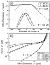
(A) Extension vs. supercoiling curves for a single DNA molecule in the standard solution. The DNA measured 16 μm as determined by a worm-like chain fit (21, 22) to its torsion-free force vs. extension curve (data not shown). The low force curve (F = 0.1 pN) obtained at 42°C is translated by −27 turns relative to the one obtained at 27°C. The high force curve (F = 1 pN and T = 27°C) is interesting in that the molecule contracts differently for n > 0 than for n < 0. In particular, the symmetry between overwinding and underwinding is broken at about nc ≈ −70 turns (where the experimental curve separates from the symmetrical continuous curve, obtained by reflecting the data for σ > 0 about σ = 0). (B) Force vs. extension curves obtained at 27°C. The σ = 0 data set was fit by the worm-like chain model with L = 16 μm and ξ = 45 nm. Note that the rigidity of the molecule is a sensitive function of the degree of supercoiling. In these conditions, the molecule undergoes an abrupt transition to an extended state at the critical force Fc ≈ 1 pN.
Figure 4
Figure 4
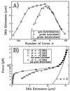
(A) Extension vs. supercoiling curves for a DNA molecule during the course of the hybridization experiment (T = 42°C). The low force curve obtained before hybridization (⋄) is symmetric and serves as the reference data set. After hybridization has occurred (at n = −500 and F = 2 pN), the curve obtained at low force (○) is no longer symmetric but is “broadened” by ≈100 turns relative to the prehybridization curve. +, After dehybridization is performed (see text), the molecule recovers its initial extension vs. supercoiling behavior. (B) Force vs. extension curves for a single DNA molecule (T = 42°C). Plain curve: worm-like chain model (22) for a torsionally relaxed (σ = 0) DNA with a persistence length ξ ≈ 50 nm and contour length L ≈ 16 μm. (○): DNA unwound by n = −300 turns (σ = −0.064). ⋄, DNA unwound by n = −200 turns (σ = −0.043). Note that at T = 42°C Fc = 0.7 pN. +, DNA unwound by n = −300 turns and containing a region hybridized to a 1-kb homologous single-stranded DNA. The elastic properties of the DNA are now similar to those for a DNA unwound by n = −200 turns. ×, After dehybridizing the probe, the duplex recovers its initial behavior.
Figure 5
Figure 5
Extension vs. supercoiling curves measured at low force (F ≈ 0.1 pN) for DNA incubated with (□) or without (○) a 1-kb hybridization probe. The labels indicate the conditions of incubation: the probe tested, the degree of unwinding of the DNA, and the stretching force applied. (A and B) Experiments performed with probe AT1. (C and D) Experiments peformed with probe AT2. (A) No hybridization takes place in these conditions, as F < Fc does not generate a denaturation bubble. (B) Because F > Fc can generate a denaturation bubble, hybridization has occurred as evidenced by the broadening between the two curves. (C) The stretching force is large enough to generate a denaturation bubble in the DNA, but this bubble does not encompass the region spanned by probe AT2. (D) With this degree of unwinding, a denaturation bubble has formed in the region spanned by AT2, allowing for hybridization.

Similar articles

Cited by

References

    1. Dunaway M, Ostrander E A. Nature (London) 1993;361:746–748. - PubMed
    1. Wu H-Y, Shyy S, Wang J C, Liu L F. Cell. 1988;53:433–440. - PubMed
    1. Patterton H-G, von Holt C. J Mol Biol. 1993;229:623–636. - PubMed
    1. Vinograd J, Lebowitz J, Watson R. Proc Natl Acad Sci USA. 1968;33:173–197.
    1. Vologodskii A V, Lukashin A V, Anshelevich V V, Frank-Kamenetskii M D. Nucleic Acids Res. 1979;6:967–982. - PMC - PubMed

Publication types

LinkOut - more resources