. 1998 Aug 24;142(4):1095-104.
doi: 10.1083/jcb.142.4.1095.

Functional gap junctions in the schwann cell myelin sheath

Affiliations

Functional gap junctions in the schwann cell myelin sheath

R J Balice-Gordon et al. J Cell Biol. .

Abstract

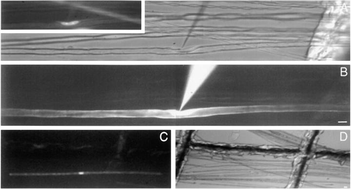
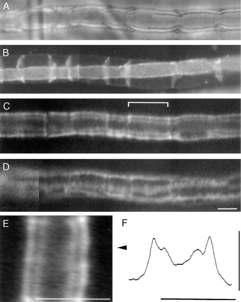
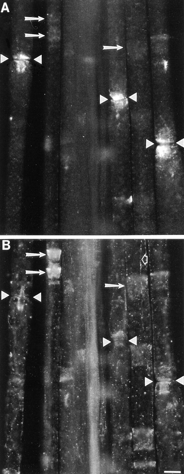
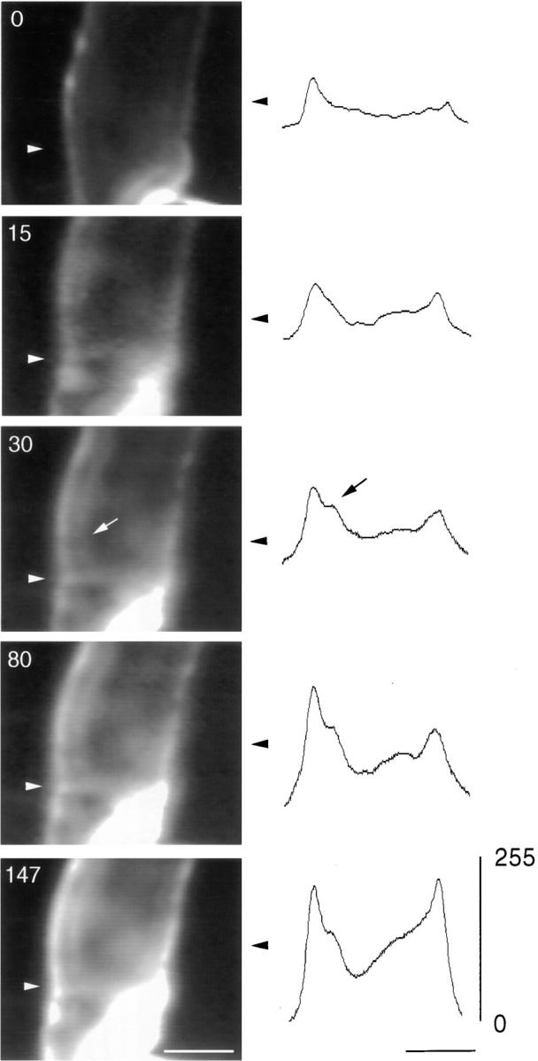
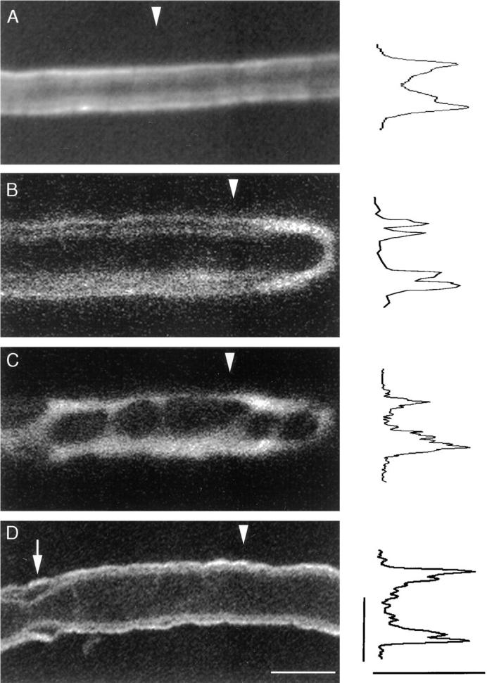
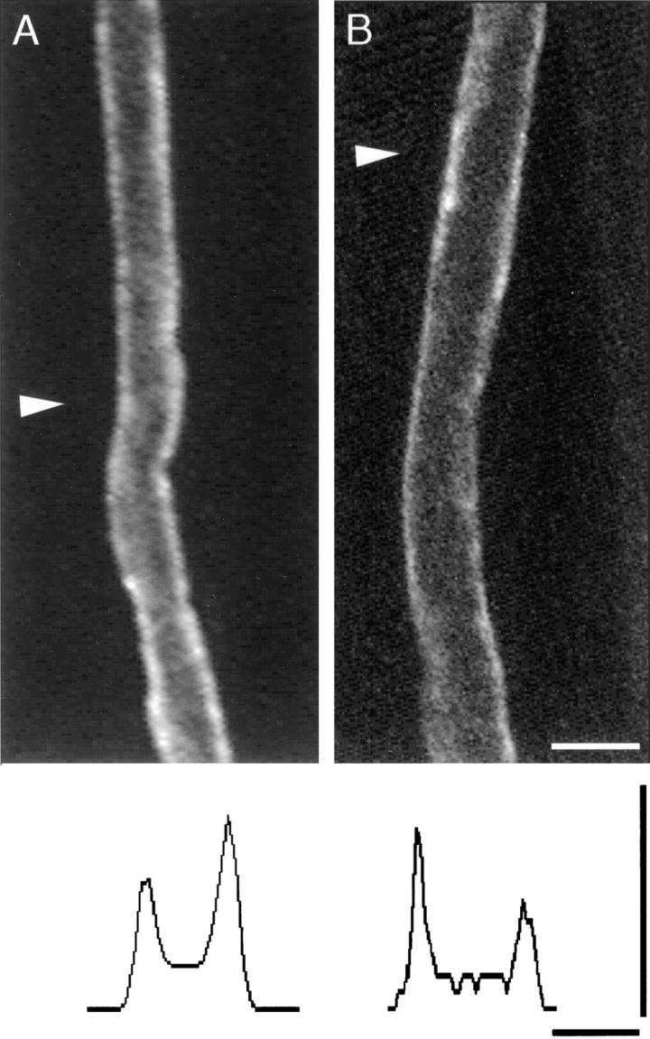
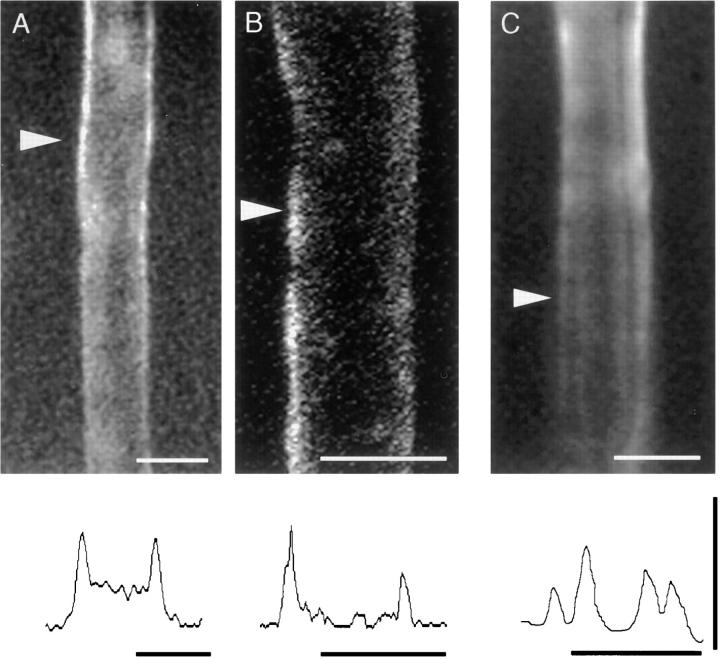
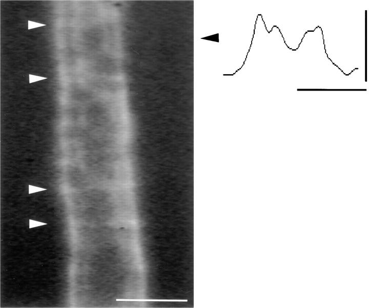
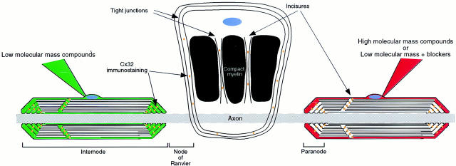
The Schwann cell myelin sheath is a multilamellar structure with distinct structural domains in which different proteins are localized. Intracellular dye injection and video microscopy were used to show that functional gap junctions are present within the myelin sheath that allow small molecules to diffuse between the adaxonal and perinuclear Schwann cell cytoplasm. Gap junctions are localized to periodic interruptions in the compact myelin called Schmidt-Lanterman incisures and to paranodes; these regions contain at least one gap junction protein, connexin32 (Cx32). The radial diffusion of low molecular weight dyes across the myelin sheath was not interrupted in myelinating Schwann cells from cx32-null mice, indicating that other connexins participate in forming gap junctions in these cells. Owing to the unique geometry of myelinating Schwann cells, a gap junction-mediated radial pathway may be essential for rapid diffusion between the adaxonal and perinuclear cytoplasm, since this radial pathway is approximately one million times faster than the circumferential pathway.


Figures

Figure 1
Figure 1
Dye injection into living, teased myelinated fibers from mouse sciatic nerve. Shown are images of a single fiber that was iontophoretically injected with 5,6-carboxyfluorescein. (A) Polarized light in combination with fluorescent bis-benzamide staining (inset) shows an electrode near a Schwann cell nucleus. (B) Diffusion of 5,6-carboxyfluorescein during iontophoretic injection. (C) Diffusion of dye from node to node was observed within 0.5–5 min (this image was captured at ∼1 min into injection). (D) After imaging of dye diffusion, injected fibers were mapped at lower magnification relative to grid lines (same field as C) to facilitate identification for confocal microscopic analysis. Bars, 10 μm.
Figure 2
Figure 2
Dye diffusion results in labeling of adaxonal and abaxonal cytoplasm. Shown are images taken after pressure injection (40 psi, 250-ms pulses, 2 Hz for 1–3 minutes) of 5,6-carboxyfluorescein into a myelinating Schwann cell. (A) Schwann cell perinuclear region and incisures visualized with polarized light. (B) After injection, the fiber was immunostained for MAG, which is localized to incisures and colocalizes with incisures identified with polarized light. (C) Image taken about midway through depth of the cell shows that dye occupies the outer and inner collar of Schwann cell cytoplasm, creating a double train track pattern indicative of the radial diffusion of dye through incisures. Bracket, region enlarged in E. (D) Image taken at ∼5 μm above the plane shown in G reveals fingers of cytoplasm on the surface of the cell; these are easily distinguished from the double train track pattern. (E) Enlargement of region indicated by brackets in C; arrowheads indicate position of the line across which intensity was mapped. (F) Histogram of intensity across line perpendicular to the long axis of the fiber at location indicated by arrowhead; scale is the same as in image shown in E. Doublet of peaks on either end of the histogram is the quantitative representation of the double train track pattern evident in the image shown in E. Vertical scale is 0–255 intensity levels. Bars, 10 μm.
Figure 3
Figure 3
Cx32 and E-cadherin immunoreactivity in paranodes and incisures. Teased fibers from an adult rat sciatic nerve fixed for 30 min in Zamboni's fixative were immunostained with a monoclonal antibody against Cx32 (A; fluorescein optics) and a rabbit antiserum against E-cadherin (B; rhodamine optics). Cx32 and E-cadherin colocalize at paranodes, which flank nodes of Ranvier (apposed arrowheads), as well as incisures, some of which are marked (arrows). E-cadherin also stains mesaxons, one of which is seen in this focal plane (open arrow). Note that although E-cadherin and Cx32 immunoreactivity colocalize, E-cadherin staining is more pronounced at incisures, whereas Cx32 staining is more pronounced at paranodes. In addition, the subcellular distributions of E-cadherin and Cx32 immunoreactivity within paranodes appear to differ. Bar, 10 μm.
Figure 4
Figure 4
Rapid diffusion of 5,6-carboxyfluorescein across incisures to the inner/adaxonal collar of Schwann cell cytoplasm. Right panels, from the same fiber at the onset (0 s), and 15, 30, 80, and 147 s after the onset of iontophoretic injection. The locations of an incisure are indicated with an arrowhead and were confirmed by viewing the fiber with polarized light. At the onset of injection, dye immediately fills the outer collar of Schwann cell cytoplasm. By 15 s, an incisure is labeled and a faint train track is apparent on one side of the myelin sheath. The train track pattern and incisure are more apparent at 30 s. At longer times (80 and 147 s), more incisures become filled and the train track pattern is seen further away from the injection site, but in this case is only clearly visualized on the left side of the fiber. Left panels, quantitative analysis of pattern of dye distribution. The intensity of pixels in a line perpendicular to the long axis of the fiber was mapped at the position indicated by the black arrowheads in the corresponding photomicrograph. The light microscopic images of dye diffusion were obtained using manual gain settings of the SIT camera so that changes in the line histogram intensity mapped across the same region of the fiber over time could be compared in terms of absolute intensity (right panels; 0–255 intensity levels). A doublet is apparent in the left side of the histogram by 30 s after the onset of injection (black arrow; corresponding location in the 30-s image is indicated by white arrow). Within the doublet, the intensity of the first peak in the doublet representing the outside collar of cytoplasm and the intensity of the second peak increased over the first minutes of injection in parallel. Thus, the train track pattern of labeling is consistent with the diffusion of 5,6-carboxyfluorescein from the outer/abaxonal to the inner/adaxonal cytoplasm across incisures. Bars, 5 μm.
Figure 5
Figure 5
Injected low molecular mass compounds fill the outer/ abaxonal and inner/adaxonal collars of Schwann cell cytoplasm. A–D are from the same fiber after injection with 5,6-carboxyfluorescein and neurobiotin. Intensity profiles are illustrated for a line perpendicular to the long axis of the fiber at the location indicated by the white arrowhead in each panel; the scale bar for the intensity histogram (bottom right) is 0–255 intensity levels. (A) Double line of 5,6-carboxyfluorescein observed immediately after a perinuclear injection (left edge). Note doublet in the peak of the intensity histogram representing each side of the fiber. (B) Subsequent confocal analysis of the same fiber showed a similar double train track pattern of 5,6-carboxyfluorescein within the Schwann cell along the length of the fiber. A single confocal plane, taken at near the resolution limit of the confocal microscope, midway through the depth of the cell adjacent to a node of Ranvier (right edge) is shown; note that this region is brighter than the double train track as there is more cytoplasm in this location. There is a doublet present in each intensity peak shown at right of photomicrograph. (C) A single confocal plane of ∼5 μm above the plane shown in B demonstrates the filling of fingers of Schwann cell cytoplasm. Note absence of doublet in intensity peaks. (D) After confocal imaging, the same fiber was processed to visualize neurobiotin and was then reanalyzed by confocal microscopy. A single confocal plane is shown, midway through the fiber, demonstrating that neurobiotin diffusion also results in a double train track pattern, although the pattern is somewhat distorted by tissue processing. A doublet is apparent in each peak of the intensity histogram on each side of the fiber. Arrow, location of an incisure; visualization with polarized light confirmed this. Bars, 10 μm.
Figure 6
Figure 6
High molecular mass compounds do not diffuse across incisures. (A) Absence of a double line of dye staining 2 h after pressure injection of 3,000-Da rhodamine-conjugated dextran. (B) Subsequent confocal analysis of the same fiber did not reveal a double line pattern anywhere in the z projection of the cell; shown is a single confocal plane midway through the cell. Intensity profiles (marked by white arrowhead) illustrated at the bottom of each panel (scale, 0–255 intensity levels) confirm the absence of a doublet in any of the intensity peaks. In many cases, dye appeared to pool in the outer collar of cytoplasm near incisures but did not fill them (for example, upper left-hand edge of fiber above white arrowhead). Bars, 10 μm.
Figure 7
Figure 7
Pharmacologic blockade of gap junctions with AGA prevents the diffusion of 5,6-carboxyfluorescein across incisures. Intensity profiles are illustrated for each panel across a line perpendicular to the long axis of the fiber at the location indicated by the white arrowhead (scale, 0–255 intensity levels). (A) Filling of the outer, but not the inner, collar of cytoplasm 1 h after iontophoretic injection of 5,6-carboxyfluorescein after preincubation in 75 μM AGA. (B) Subsequent confocal analysis of 5,6-carboxyfluorescein in the same fiber shown in A confirmed the absence of a double train track pattern of staining; shown is a single confocal plane midway through the z projection. (C) Preincubation of a myelinated fiber in 0.15% DMSO for 1 h did not abolish a train track pattern of staining After injection of 5,6-carboxyfluorescein. The train track pattern is visible on the right-hand side of the fiber and also as a doublet in the intensity peak for this region. Bars, 10 μm.
Figure 8
Figure 8
Evidence for functional gap junctions in the incisures of cx32-null mice. Shown is a portion of a myelinated fiber from the sciatic nerve of a cx32-null mouse after injection of 5,6-carboxyfluorescein; the location of incisures is marked with white arrowheads. The intensity profile is illustrated for this fiber (scale, 0–255 intensity levels) across a line perpendicular to its long axis at the location indicated by the black arrowhead. As in myelinating Schwann cells from wild-type mice, a train track pattern is apparent on at least one side of the axon. Bar, 10 μm.
Figure 9
Figure 9
Schematic view of dye diffusion in myelinating Schwann cells following perinuclear dye injection. The left Schwann cell has been injected with a low molecular mass compound (e.g., 5,6-carboxyfluorescein); the right Schwann cell has been injected with a high molecular mass compound (e.g., 3,000-Da rhodamine dextran) or a low molecular mass compound in the presence of gap junction blockers. The middle Schwann cell has been unrolled to visualize regions of compact myelin, incisures and paranodes. Not to scale.

Similar articles

Cited by

References

    1. Anzini P, Neuberg DH-H, Schachner M, Nelles E, Willecke K, Zielasek J, Toyka K, Suter U, Martini R. Structural abnormalities and deficient maintenance of peripheral nerve myelin in mice lacking the gap junction protein connexin32. J Neurosci. 1997;17:4545–4561. - PMC - PubMed
    1. Balice-Gordon, R.J. 1998. In vivo approaches to neuromuscular junction structure and function. In Methods in Cell Biology. C.P. Emerson and H.L. Sweeney, editors. Academic Press, San Diego, CA. 323–348. - PubMed
    1. Berg, H.C. 1983. Random Walks in Biology. Princeton University Press, Princeton, NJ. 142 pp.
    1. Bergoffen J, Scherer SS, Wang S, Oronzi-Scott M, Bone L, Paul DL, Chen K, Lensch MW, Chance P, Fischbeck K. Connexin mutations in X-linked Charcot-Marie-Tooth disease. Science. 1993;262:2039–2042. - PubMed
    1. Berthold, C.-H., and M. Rydmark. 1995. Morphology of normal peripheral axons. In The Axon. S.G. Waxman, J.D. Kocsis, and P.K. Stys, editors. Oxford University Press, New York. 13–48.

Publication types

MeSH terms