

doi: 10.1101/gad.12.16.2463.
Nucleolar localization of early tRNA processing
Affiliations
- PMID: 9716399
- PMCID: PMC317091
- DOI: 10.1101/gad.12.16.2463
Item in Clipboard
Nucleolar localization of early tRNA processing
Genes Dev.
.
Abstract
There is little information as to the location of early tRNA biosynthesis. Using fluorescent in situ hybridization in the budding yeast, Saccharomyces cerevisiae, examples of nuclear pre-tRNAs are shown to reside primarily in the nucleoli. We also probed the RNA subunit of RNase P. The majority of the signal from RNase P probes was nucleolar, with less intense signals in the nucleoplasm. These results demonstrate that a major portion of the tRNA processing pathway is compartmentalized in nucleoli with rRNA synthesis and ribosomal assembly. The spatial juxtaposition suggests the possibility of direct coordination between tRNA and ribosome biosynthesis.
Figures
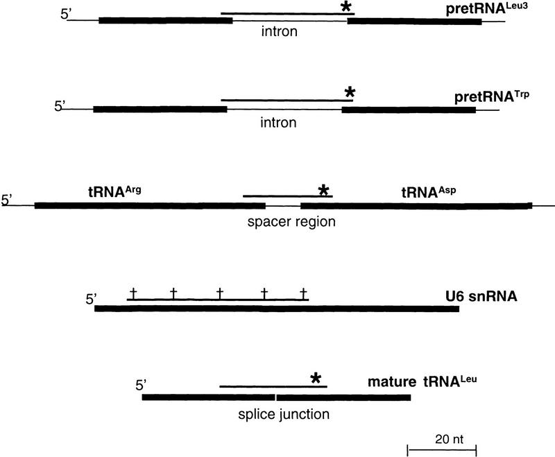
Schematic depiction of fluorescent probes. Oligodeoxynucleotide probes that anneal to several small RNAs are used in the experiments shown in Fig. 2. The positions at which the fluorescent probes bind to their target RNAs are shown relative to the full-length primary transcripts. 5′-Fluorescein labels are denoted by asterisks, with internal CY3 labels denoted by daggers. The fluorescent antisense RNA probes used to detect U14 snoRNA have been described previously (Samarsky et al. 1998).
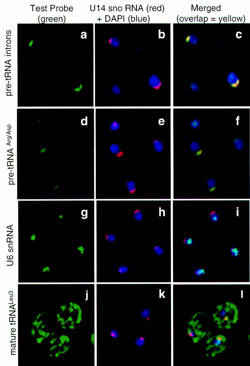
Nuclear tRNA precursors are found predominantly in the nucleolus of S. cerevisiae. In situ hybridization of fluorescently labeled oligonucleotide probes to endogenous RNAs was carried out as described in the text. The left panel of each triplet shows staining with the probe specific for the RNA named at the left of the panels. The center panels show staining of the same cells as at left but with a probe directed against U14 snoRNA (red) and DNA detection with DAPI (blue). Merging of the left and center panels is shown at right, with overlap between the green and red signals shown in yellow. Overlap between green and blue appears as blue–green. (a–c) Introns to pre-tRNALeu3 and pre-tRNATrp are probed simultaneously (a,c). (d–f) The intergenic spacer of pre-tRNAArg/tRNAAsp dimeric transcripts is probed (d,f). (g–i) U6 snRNA is probed (g,i). (j–l) Mature tRNALeu3 is probed (j,l).
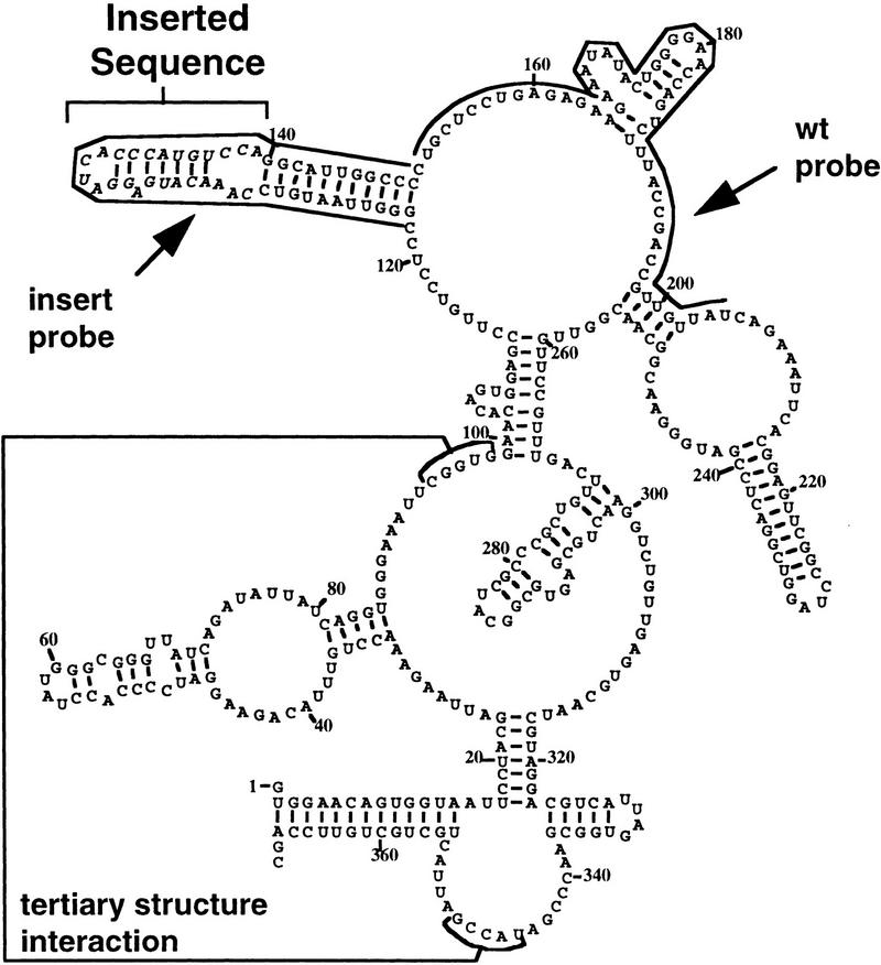
Fluorescent probes for wild-type RNase P RNA and an inserted sequence. The proposed secondary structure of the S. cerevisiae RNase P RNA (RPR1 RNA) subunit is shown, including the long-distance base-pairing that contributes to tertiary structure. The positions of the artificial 20-nucleotide insertion are italicized and indicated by a bracket. CY3-labeled complementary oligonucleotide probe positions to either the wild-type RPR1 sequence or the inserted sequence are indicated by lines along the sequence.
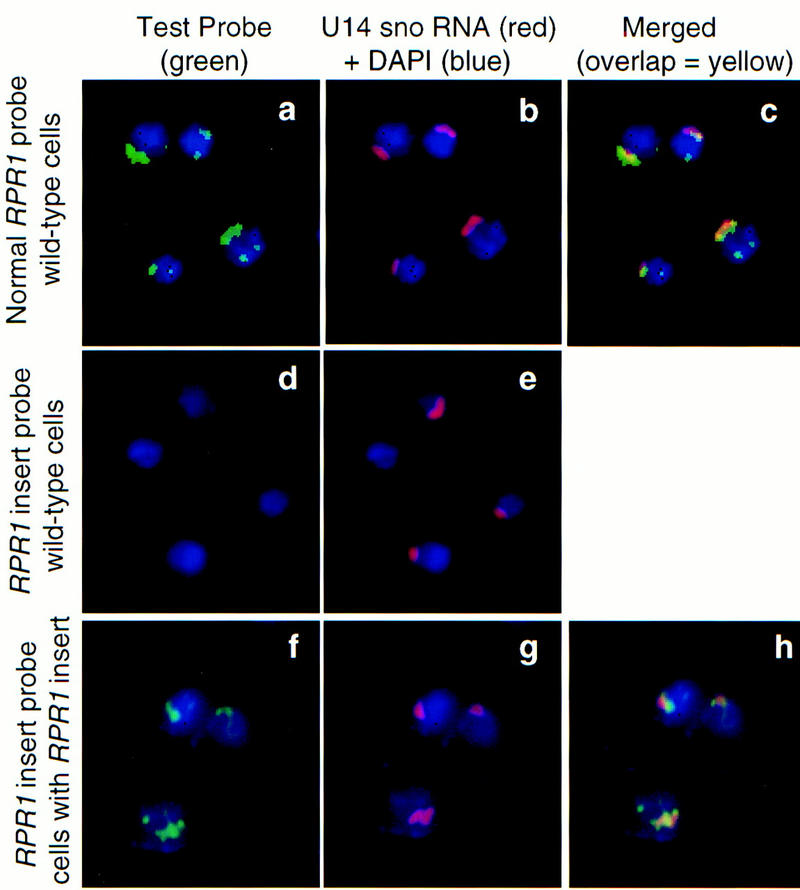
RNase P is located primarily in the nucleolus in S. cerevisiae. All panels are stained for nuclear DNA with DAPI (blue). Probes for RNase P RNA or an artificial insert in RNase P RNA (Fig. 3) are shown in green in the left panels. The center panels show probes of the same cells for U14 snoRNA (red). The right panels show the left and center panels merged, with overlap between the green and red signals in yellow. (No merged panel is given for the center set, as there is no RNA signal in the left panel.) (a–c) The mature domain of wild-type RNase P RNA is probed (RPR1 RNA, a,c). (d–e) The probe to an artificial insert in RPR1 RNA is applied to wild-type cells, in which, as expected, it gives no signal above background. (f–h) The artificial insert probe is used (f,h) with cells having the modified RPR1 allele containing inserted sequences complementary to the probe.
Similar articles
-
Rpp1, an essential protein subunit of nuclear RNase P required for processing of precursor tRNA and 35S precursor rRNA in Saccharomyces cerevisiae.Genes Dev. 1997 Nov 1;11(21):2926-37. doi: 10.1101/gad.11.21.2926. Genes Dev. 1997. PMID: 9353260 Free PMC article.
-
RNase MRP and rRNA processing.Mol Biol Rep. 1995-1996;22(2-3):69-73. doi: 10.1007/BF00988708. Mol Biol Rep. 1995. PMID: 8901490 Review.
-
An RNase P RNA subunit mutation affects ribosomal RNA processing.Nucleic Acids Res. 1996 Aug 15;24(16):3158-66. doi: 10.1093/nar/24.16.3158. Nucleic Acids Res. 1996. PMID: 8774895 Free PMC article.
-
An essential protein-binding domain of nuclear RNase P RNA.RNA. 2001 Apr;7(4):565-75. doi: 10.1017/s1355838201001996. RNA. 2001. PMID: 11345435 Free PMC article.
-
tRNA transfers to the limelight.Genes Dev. 2003 Jan 15;17(2):162-80. doi: 10.1101/gad.1049103. Genes Dev. 2003. PMID: 12533506 Review. No abstract available.
Cited by
-
TFIIIC localizes budding yeast ETC sites to the nuclear periphery.Mol Biol Cell. 2012 Jul;23(14):2741-54. doi: 10.1091/mbc.E11-04-0365. Epub 2012 Apr 11. Mol Biol Cell. 2012. PMID: 22496415 Free PMC article.
-
The Saccharomyces cerevisiae TRT2 tRNAThr gene upstream of STE6 is a barrier to repression in MATalpha cells and exerts a potential tRNA position effect in MATa cells.Nucleic Acids Res. 2004 Sep 30;32(17):5206-13. doi: 10.1093/nar/gkh858. Print 2004. Nucleic Acids Res. 2004. PMID: 15459290 Free PMC article.
-
Conventional and nonconventional roles of the nucleolus.Int Rev Cytol. 2002;219:199-266. doi: 10.1016/s0074-7696(02)19014-0. Int Rev Cytol. 2002. PMID: 12211630 Free PMC article. Review.
-
Stochastic transcription in the p53-mediated response to DNA damage is modulated by burst frequency.Mol Syst Biol. 2019 Dec;15(12):e9068. doi: 10.15252/msb.20199068. Mol Syst Biol. 2019. PMID: 31885199 Free PMC article.
-
Defects in tRNA processing and nuclear export induce GCN4 translation independently of phosphorylation of the alpha subunit of eukaryotic translation initiation factor 2.Mol Cell Biol. 2000 Apr;20(7):2505-16. doi: 10.1128/MCB.20.7.2505-2516.2000. Mol Cell Biol. 2000. PMID: 10713174 Free PMC article.
References
-
- Carter KC, Bowman D, Carrington W, Fogarty K, McNeil JA, Fay FS, Lawrence JB. A three-dimensional view of precursor messenger RNA metabolism within the mammalian nucleus. Science. 1993;259:1330–1335. - PubMed
Publication types
MeSH terms
Substances
Grants and funding
LinkOut - more resources
Full Text Sources
Other Literature Sources
Molecular Biology Databases