. 1998 Sep;18(9):5600-8.
doi: 10.1128/MCB.18.9.5600.

Sir proteins, Rif proteins, and Cdc13p bind Saccharomyces telomeres in vivo

Affiliations

Sir proteins, Rif proteins, and Cdc13p bind Saccharomyces telomeres in vivo

B D Bourns et al. Mol Cell Biol. 1998 Sep.

Abstract

Although a surprisingly large number of genes affect yeast telomeres, in most cases it is not known if their products act directly or indirectly. We describe a one-hybrid assay for telomere binding proteins and use it to establish that six proteins that affect telomere structure or function but which had not been shown previously to bind telomeres in vivo are indeed telomere binding proteins. A promoter-defective allele of HIS3 was placed adjacent to a chromosomal telomere. Candidate proteins fused to a transcriptional activation domain were tested for the ability to activate transcription of the telomere-linked HIS3 gene. Using this system, Rif1p, Rif2p, Sir2p, Sir3p, Sir4p, and Cdc13p were found to be in vivo telomere binding proteins. None of the proteins activated the same reporter gene when it was at an internal site on the chromosome. Moreover, Cdc13p did not activate the reporter gene when it was adjacent to an internal tract of telomeric sequence, indicating that Cdc13p binding was telomere limited in vivo. The amino-terminal 20% of Cdc13p was sufficient to target Cdc13p to a telomere, suggesting that its DNA binding domain was within this portion of the protein. Rap1p, Rif1p, Rif2p, Sir4p, and Cdc13p activated the telomeric reporter gene in a strain lacking Sir3p, which is essential for telomere position effect (TPE). Thus, the telomeric association of these proteins did not require any of the chromatin features necessary for TPE. The data support models in which the telomere acts as an initiation site for TPE by recruiting silencing proteins to the chromosome end.


Figures
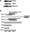
FIG. 1
FIG. 1
Schematic representation of strains and proteins. (A) Chromosomal context of reporter genes. Strains with HIS-Tel contained the promoter-defective allele of HIS3 integrated with its TATA element ∼50 bp from the start of the telomeric C1-3A/TG1-3 tract at the chromosome VII-L telomere. HIS3 was inserted within ADH4 in such a way that the ∼20 kb of DNA that is normally distal of ADH4 was deleted (23). HIS-Int contains the same HIS3 allele integrated at the same site within ADH4 but without deletion of distal DNA such that the reporter gene was ∼20 kb from the chromosome VII-L telomere. HIS-Int-CA was the same as HIS-Int except that it contained a ∼276-bp tract of C1-3A/TG1-3 DNA ∼50 bp distal to the HIS3 promoter. For all three strains, there is a copy of URA3 proximal to HIS3 that served as the selectable marker during transformation. (B) Structures of proteins that tested positive in the one-hybrid assay. Bars below the proteins indicate regions of the proteins tested in the studies described here. The 462 amino acids of the 827-amino-acid Rap1p that were tested contain the DNA binding domain but not the endogenous transcriptional activation domain. For Rif1p, 367 amino acids of the 1,915-amino-acid protein were expressed in the fusion protein, which included the region that interacts with Rap1p by two-hybrid analysis (28). For Sir3p, the carboxyl 532 amino acids of the 978-amino-acid protein were expressed. By two-hybrid analysis, amino acids 307 to 978 of Sir3p are sufficient for its interaction with Rap1p, Sir3p, and Sir4p (56). By in vitro analysis, the region of Sir3p sufficient for Sir4p interaction was delimited further (amino acids 622 to 978 [75]). Fusion proteins contained the entire ORF for Sir4p (1,358 amino acids), Sir2p (563 amino acids), and Rif2p (395 amino acids). The 924-amino-acid Cdc13p was tested both as a full-length fusion protein and as three fusion proteins containing 251 amino acids (Cdc13N-Actp), 257 amino acids (Cdc13M-Actp), or 416 amino acids (Cdc13C-Actp).
FIG. 2
FIG. 2
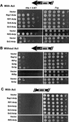
Rap1, Rif1, Sir4, Sir3, Rif2, and Sir2 fusion proteins activated HIS3 at a telomere (HIS-Tel) but not at an internal site on the chromosome (HIS-Int). HIS-Tel (A and B) or HIS-Int (C) cells expressing various proteins either fused to the activation domain (A and C) or without an activation domain (B) were spotted in 10-fold serial dilutions onto test plates that selected for the telomere interaction (left) or control plates (right). Here and in all other figures, test and control plates both contained galactose to induce expression of the fusion proteins. Here and in all subsequent figures, cells presented in the same photograph were assayed on the same plate (i.e., the vector control was always on the same plate as the protein being tested). Each fusion protein was tested in a range of 3-AT concentrations. In this and other figures, the 3-AT concentration showing the greatest growth difference between the control (vector alone) and the cells expressing the fusion protein is shown. In panels A and B, the concentration of 3-AT was 5 (Sir2), 20 (Rif2), or 10 (all other fusion proteins) mM. In panel C, plates had no 3-AT except for Rif2-Actp, which had 20 mM 3-AT. The HIS3 gene in HIS-Int was functional since cells containing vector alone often formed a few colonies at the highest dilution. Consistent with published reports (31), expression of some fusion proteins inhibited growth (see, especially, Sir2p with or without activation domain, control plates). Plates were incubated at 30°C for 4 (control plates), 8 (5 mM 3-AT), 7 (10 mM 3-AT), or 6 (20 mM 3-AT) days.
FIG. 3
FIG. 3
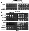
Rap1p, Rif1p, Sir4p, and Rif2p interactions with a telomere did not require Sir3p. HIS-Tel sir3Δ strains carrying vector alone (top rows) or expressing various fusion proteins were spotted in 10-fold serial dilutions onto test plates that select for the telomere interaction (left; 50 mM 3-AT) or control plates (right). Plates were incubated at 30°C for 2 (control plates, top), 4 (control plates, bottom), 6 (test plates, bottom), or 10 (test plates, top) days.
FIG. 4
FIG. 4
The amino-terminal portion of Cdc13p activated HIS-Tel. The amino-terminal 251 amino acids of Cdc13p (or vector alone) were expressed as a fusion protein in HIS-Tel with or without the transcriptional activation domain (A), in HIS-Int with the activation domain (B), or in sir3::LYS2 HIS-Tel with the activation domain (C). Cells were spotted in 10-fold serial dilutions on test plates that select for the telomere interaction (left) or control plates (right). Test plates in panels A and B had no 3-AT; test plates in panel C contained 50 mM 3-AT. Plates were incubated at 30°C for 5 (test plates, A and B), 3 (control plates), or 13 (test plates, C) days.
FIG. 5
FIG. 5
Rap1p, Rif1p, Rif2p, Sir3p, and Sir4p, but not Cdc13p or Sir2p, bind an internal tract of telomeric DNA in a wild-type strain. The indicated fusion proteins were expressed in HIS-Int-CA cells. Cells were spotted in 10-fold serial dilutions on control plates (right) or test plates (left) containing 10 (top), 0 (middle), or 20 (bottom) mM 3-AT. Control plates were incubated for 4 days, and test plates were incubated for 6 (top and bottom) or 16 (middle) days.
FIG. 6
FIG. 6
Sir4p requires Sir3p to bind an internal tract of C1-3A/TG1-3 DNA. The indicated fusion proteins were expressed in HIS-Int-CA sir3 strains. Cells were spotted in 10-fold serial dilutions onto test plates (left) or control plates (right). The 3-AT concentration was 35 (A) or 50 (B) mM. Control plates were incubated for 4 days, and test plates were incubated for 17 (A) or 13 (B) days.
FIG. 7
FIG. 7
Schematic representation of proteins associated with chromosomal telomeres and subtelomeric regions in vivo in a wild-type (A) or sir3 (B) strain. The physical presence of Rap1p at telomeres in vivo was first shown in reference and confirmed herein. The presence of Sir2p, Sir3p, and Sir4p on subtelomeric nucleosomes is from references and . The ovals labeled 2, 3, and 4 represent Sir2p, Sir3p, and Sir4p. Although one molecule of each telomere binding protein is indicated, nothing is known about the stoichiometry of telomere binding proteins except that there are 10 to 20 molecules of Rap1p per telomere (20, 87).

References

    1. Adams A K, Holm C. Specific DNA replication mutations affect telomere length in Saccharomyces cerevisiae. Mol Cell Biol. 1996;16:4614–4620. - PMC - PubMed
    1. Alexandre C, Grueneberg D A, Gilman M Z. Studying heterologous transcription factors in yeast. Methods Companion Methods Enzymol. 1993;5:147–155.
    1. Aparicio O M, Billington B L, Gottschling D E. Modifiers of position effect are shared between telomeric and silent mating-type loci in S. cerevisiae. Cell. 1991;66:1279–1287. - PubMed
    1. Boulton S J, Jackson S P. Components of the Ku-dependent non-homologous end-joining pathway are involved in telomeric length maintenance and telomeric silencing. EMBO J. 1998;17:1819–1828. - PMC - PubMed
    1. Boulton S J, Jackson S P. Identification of a Saccharomyces cerevisiae Ku80 homologue: roles in DNA double strand break rejoining and in telomeric maintenance. Nucleic Acids Res. 1996;24:4639–4648. - PMC - PubMed

Publication types

MeSH terms