

Dynamics of the TOM complex of mitochondria during binding and translocation of preproteins
- PMID: 9710610
- PMCID: PMC109111
- DOI: 10.1128/MCB.18.9.5256
Dynamics of the TOM complex of mitochondria during binding and translocation of preproteins
Abstract
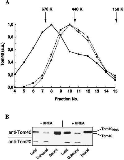
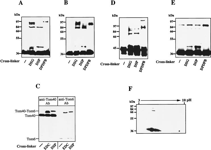
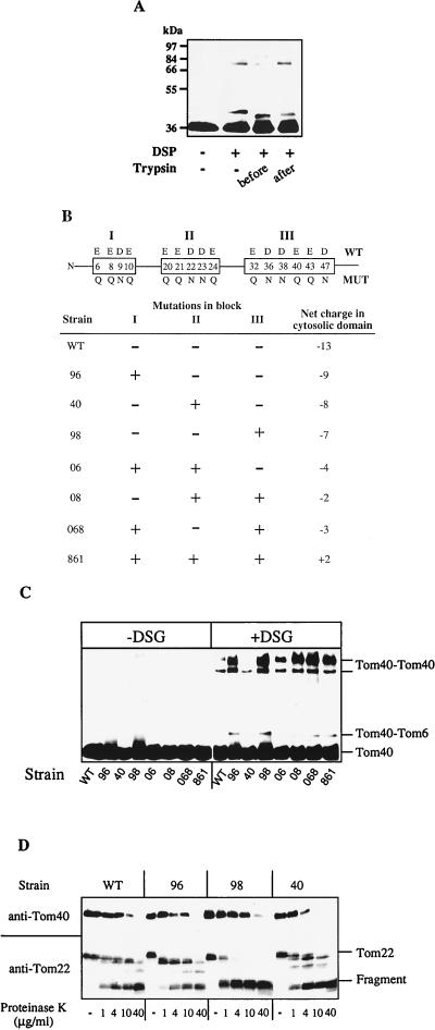
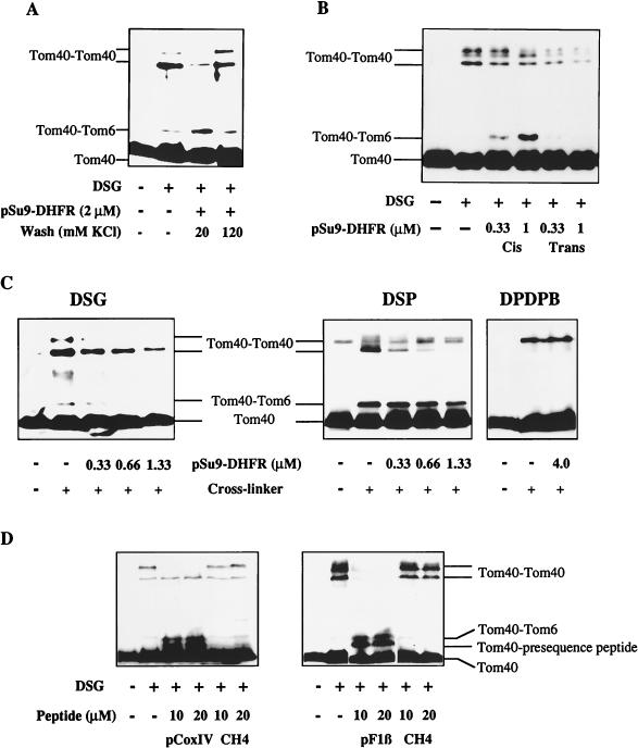
Translocation of preproteins across the mitochondrial outer membrane is mediated by the TOM complex. This complex consists of receptor components for the initial contact with preproteins at the mitochondrial surface and membrane-embedded proteins which promote transport and form the translocation pore. In order to understand the interplay between the translocating preprotein and the constituents of the TOM complex, we analyzed the dynamics of the TOM complex of Neurospora crassa and Saccharomyces cerevisiae mitochondria by following the structural alterations of the essential pore component Tom40 during the translocation of preproteins. Tom40 exists in a homo-oligomeric assembly and dynamically interacts with Tom6. The Tom40 assembly is influenced by a block of negatively charged amino acid residues in the cytosolic domain of Tom22, indicating a cross-talk between preprotein receptors and the translocation pore. Preprotein binding to specific sites on either side of the outer membrane (cis and trans sites) induces distinct structural alterations of Tom40. To a large extent, these changes are mediated by interaction with the mitochondrial targeting sequence. We propose that such targeting sequence-induced adaptations are a critical feature of translocases in order to facilitate the movement of preproteins across cellular membranes.
Figures
Similar articles
-
Mitochondrial protein import. Tom40 plays a major role in targeting and translocation of preproteins by forming a specific binding site for the presequence.J Biol Chem. 1997 Jul 25;272(30):18725-31. doi: 10.1074/jbc.272.30.18725. J Biol Chem. 1997. PMID: 9228044
-
Assembly of Tom6 and Tom7 into the TOM core complex of Neurospora crassa.J Biol Chem. 2001 May 25;276(21):17679-85. doi: 10.1074/jbc.M009653200. Epub 2001 Mar 6. J Biol Chem. 2001. PMID: 11278536
-
Recognition of preproteins by the isolated TOM complex of mitochondria.EMBO J. 2000 Sep 15;19(18):4895-902. doi: 10.1093/emboj/19.18.4895. EMBO J. 2000. PMID: 10990453 Free PMC article.
-
The mitochondrial import machinery for preproteins.Crit Rev Biochem Mol Biol. 2001;36(3):291-336. doi: 10.1080/20014091074200. Crit Rev Biochem Mol Biol. 2001. PMID: 11450972 Review.
-
The mitochondrial import machinery: preprotein-conducting channels with binding sites for presequences.Biochim Biophys Acta. 2002 Sep 2;1592(1):15-24. doi: 10.1016/s0167-4889(02)00260-4. Biochim Biophys Acta. 2002. PMID: 12191764 Review.
Cited by
-
Role of MMM1 in maintaining mitochondrial morphology in Neurospora crassa.Mol Biol Cell. 2000 Sep;11(9):2961-71. doi: 10.1091/mbc.11.9.2961. Mol Biol Cell. 2000. PMID: 10982393 Free PMC article.
-
Quaternary structure of the mitochondrial TIM23 complex reveals dynamic association between Tim23p and other subunits.Mol Biol Cell. 2008 Jan;19(1):159-70. doi: 10.1091/mbc.e07-07-0669. Epub 2007 Oct 24. Mol Biol Cell. 2008. PMID: 17959826 Free PMC article.
-
New insights into the structure and dynamics of the TOM complex in mitochondria.Biochem Soc Trans. 2024 Apr 24;52(2):911-922. doi: 10.1042/BST20231236. Biochem Soc Trans. 2024. PMID: 38629718 Free PMC article. Review.
-
Structural requirements of Tom40 for assembly into preexisting TOM complexes of mitochondria.Mol Biol Cell. 2001 May;12(5):1189-98. doi: 10.1091/mbc.12.5.1189. Mol Biol Cell. 2001. PMID: 11359915 Free PMC article.
-
A twin arginine signal peptide and the pH gradient trigger reversible assembly of the thylakoid [Delta]pH/Tat translocase.J Cell Biol. 2002 Apr 15;157(2):205-10. doi: 10.1083/jcb.200202048. Epub 2002 Apr 15. J Cell Biol. 2002. PMID: 11956224 Free PMC article.
References
-
- Bjellqvist B, Sanchez J C, Pasquali C, Ravier F, Paquet N, Frutiger S, Hughes G J, Hochstrasser D. Micropreparative two-dimensional electrophoresis allowing the separation of samples containing milligram amounts of proteins. Electrophoresis. 1993;14:1375–1378. - PubMed
-
- Brix J, Dietmeier K, Pfanner N. Differential recognition of preproteins by the purified cytosolic domains of the mitochondrial import receptors Tom20, Tom22, and Tom70. J Biol Chem. 1997;272:20730–20735. - PubMed
-
- Crowley K S, Liao S, Worrell V E, Reinhart G D, Johnson A E. Secretory proteins move through the endoplasmic reticulum membrane via an aqueous, gated pore. Cell. 1994;78:461–471. - PubMed
-
- Daum G, Böhni P C, Schatz G. Import of proteins into mitochondria: cytochrome b2 and cytochrome c peroxidase are located in the intermembrane space of yeast mitochondria. J Biol Chem. 1982;257:13028–13033. - PubMed
Publication types
MeSH terms
Substances
LinkOut - more resources
Full Text Sources
Molecular Biology Databases