. 1998 Sep;18(9):5021-31.
doi: 10.1128/MCB.18.9.5021.

Sendai virus Y proteins are initiated by a ribosomal shunt

Affiliations

Sendai virus Y proteins are initiated by a ribosomal shunt

P Latorre et al. Mol Cell Biol. 1998 Sep.

Abstract

The Sendai virus P/C mRNA expresses eight primary translation products by using a combination of ribosomal choice and cotranscriptional mRNA editing. The longest open reading frame (ORF) of the mRNA starts at AUG104 (the second initiation site) and encodes the 568-amino-acid P protein, an essential subunit of the viral polymerase. The first (ACG81), third (ATG114), fourth (ATG183), and fifth (ATG201) initiation sites are used to express a C-terminal nested set of polypeptides (collectively named the C proteins) in the +1 ORF relative to P, namely, C', C, Y1, and Y2, respectively. Leaky scanning accounts for translational initiation at the first three start sites (a non-ATG followed by ATGs in progressively stronger contexts). Consistent with this, changing ACG81/C' to ATG (GCCATG81G) abrogates expression from the downstream ATG104/P and ATG114/C initiation codons. However, expression of the Y1 and Y2 proteins remains normal in this background. We now have evidence that initiation from ATG183/Y1 and ATG201/Y2 takes place via a ribosomal shunt or discontinuous scanning. Scanning complexes appear to assemble at the 5' cap and then scan ca. 50 nucleotides (nt) of the 5' untranslated region before being translocated to an acceptor site at or close to the Y initiation codons. No specific donor site sequences are required, and translation of the Y proteins continues even when their start codons are changed to ACG. Curiously, ATG codons (in good contexts) in the P ORF, placed either 16 nt upstream of Y1, 29 nt downstream of Y2, or between the Y1 and Y2 codons, are not expressed even in the ACGY1/ACGY2 background. This indicates that ATG183/Y1 and ATG201/Y2 are privileged start sites within the acceptor site. Our observations suggest that the shunt delivers the scanning complex directly to the Y start codons.


Figures

FIG. 1
FIG. 1
Schematic representation of the SeV P/C mRNA. The 1,943-nt-long SeV P/C mRNA is shown as a horizontal line, with the various ORFs as boxes. The P ORF is shown as a shaded box, the C ORF (position +1 relative to P, encoding the C′, C, Y1, and Y2 proteins) is shown as an open box, and the internal V ORF (position −1 relative to P) is shown as a black box. The V and W proteins are generated by cotranscriptional editing of the P/C mRNA, during which a +1 G (for V) and +2 Gs (for W) are inserted at nucleotide position 1053. This fuses these ORFs to the N-terminal half of P. The 81-nt 5′ UTR is also indicated. Numbers refer to nucleotide positions relative to the 5′ end of the P/C mRNA.
FIG. 2
FIG. 2
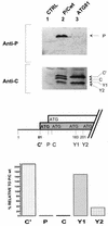
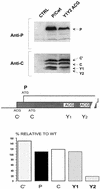
Mutation of ACG81 to ATG81 eliminates leaky scanning but not Y expression. The first initiation codon, ACG81/C′, was changed to ATG, thereby changing the C′ start site context from weak to strong (middle panel). The ATG81/C′ (ATG81) plasmid construct and the parental P/Cwt were transfected into HeLa cells which had been infected with a vaccinia virus recombinant (vTF7-3) expressing T7 RNA polymerase. As a control, cells were also mock transfected (CTRL). At 24 h posttransfection, cytoplasmic extracts were prepared by scraping the cells into lysis buffer. Extracts were resolved on both SDS–10% polyacrylamide gels (for the P protein) and SDS–15% polyacrylamide gels (for the C proteins). Protein expression was then detected by immunoblotting with an anti-P monoclonal antibody and a rabbit anti-C polyclonal antiserum (upper panel), using the CSPD light detection system (Boehringer). The blots were quantitated by densitometry, and expression of the C′, C, P, Y1, and Y2 proteins in the ATG81 construct was plotted as a percentage relative to expression of P/Cwt (lower panel).
FIG. 3
FIG. 3
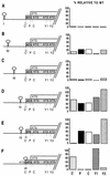
Effect of stable stem-loop structures introduced into the 5′ UTR. Palindromic sequences predicted to form thermodynamically stable stem-loop structures (ΔG > −50 kcal/mol) were inserted into the 5′ UTR of the P/C mRNA at the positions indicated on the left. In constructions A to D the stem-loop was introduced into the P/Cwt background. In construction E, the C′ initiation codon was changed to GCG, and in construction F, the stem-loop was inserted at the NcoI site generated by changing the C′ ACG to ATG (ATG81/C′). This in turn left ATG codons in the C ORF flanking the inserted stem-loop. These clones were expressed in HeLa cells, and C′, P, C, Y1, and Y2 protein expression was determined by immunoblotting (see the legend to Fig. 2). The blots were quantitated by densitometry, and protein expression was plotted as a percentage relative to the levels detected in cells transfected with P/Cwt.
FIG. 4
FIG. 4
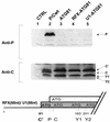
Replacement of the P/C mRNA 5′ UTR does not effect the shunt. Starting from the ATG81/C′ plasmid, the 80-nt 5′ UTR of the P/C mRNA was replaced with the 5′ UTR of the cellular RFX (68 nt) and U1 (86 nt) mRNAs (lower panel). These clones (referred to as RFX-ATG81 and U1-ATG81, respectively), P/Cwt, and ATG81/C′ (ATG81) were transfected into HeLa cells infected with a vaccinia virus recombinant expressing T7 RNA polymerase (vTF7-3). As a control, cells were also mock transfected (CTRL). At 24 h posttransfection, cytoplasmic extracts were prepared by scraping the cells into lysis buffer. Expression of the P and C proteins was determined by immunoblotting with an anti-P monoclonal antibody and a polyclonal anti-C antiserum as outlined in the legend to Fig. 2 (upper panel).
FIG. 5
FIG. 5
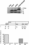
Displacement of the Y start sites downstream. The C, Y1, and Y2 initiation codons were displaced 63 nt downstream by the introduction of a spacer sequence between ATG104/P and ATG114/C within the background ATG81/C′ (middle panel) (see Materials and Methods). This spacer duplicated the sequences between the C and Y1 ATG codons (indicated by the small rectangles in the middle panel). The numbers in parentheses refer to the positions in the P/Cwt background. This clone (referred to as ATG81+SPACER), P/Cwt, and ATG81/C′ (ATG81) were transfected into HeLa cells infected with a vaccinia virus recombinant expressing T7 RNA polymerase (vTF7-3). Cytoplasmic extracts were resolved on an SDS–15% polyacrylamide gel, and C protein expression was determined by immunoblotting with a polyclonal anti-C antiserum by using the CSPD detection system (upper panel). The blot was quantitated by densitometry, and the level of each protein expressed in the ATG81+SPACER construct was plotted as a percentage relative to that expressed in ATG81/C′ (lower panel).
FIG. 6
FIG. 6
The Y proteins can initiate from a non-ATG codon. Both the Y1 and Y2 ATG start codons were changed to ACG (middle panel). This plasmid construct and the parental P/Cwt were transfected into HeLa cells infected with a vaccinia virus recombinant (vTF7-3) expressing T7 RNA polymerase. As a control, cells were also mock transfected. At 24 h posttransfection, cytoplasmic extracts were prepared by scraping the cells into lysis buffer. Expression of the P and C proteins was determined by immunoblotting with an anti-P monoclonal antibody and a polyclonal anti-C antiserum as outlined in the legend to Fig. 2 (upper panel). The blots were quantitated by densitometry, and the level of each protein expressed in the mutant construct was plotted as a percentage relative to that expressed in the parental P/Cwt (lower panel).
FIG. 7
FIG. 7
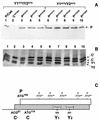
ATGs introduced into the P ORF around the Y start sites are silent. Single ATG start codons in good contexts were introduced at positions 131, 167, and 194 in the P/Cwt clone (Y1ATG/Y2ATG) and at positions 131, 167, 194, 230, and 287 in the construct P/C-Y1ACG/Y2ACG (C). These constructs along with P/Cwt (lanes 1) and ATG81/C′ (C′ATG81) (lanes 2) were expressed in HeLa cells as outlined in the legend to Fig. 2, and protein expression was monitored by immunoblotting with an anti-P monoclonal antibody (A) and a rabbit polyclonal anti-C serum (B).
FIG. 8
FIG. 8
The inserted ATGs are recognized by a linear-scanning complex. A BamHI site was introduced at position 116 in the P/C-Y1ACG/Y2ACG constructs containing single ATG start codons at positions 131, 167, 194, 230, and 287 (Fig. 7). By using this site, all of the sequences upstream of position 116 were deleted, leaving 30 nt of plasmid sequence between the end of the T7 promoter and the start of the P/C gene (lower panel). These clones were expressed in HeLa cells along with P/C-Y1ACG/Y2ACG (P/C ACG Y1/Y2) and a mock-transfected control (see Fig. 2). Expression of full-length P protein (upper panel, lane 2) and N-terminally truncated P protein initiated from the inserted ATGs (referred to as ΔP) was detected by immunoblotting with an anti-P monoclonal antibody recognizing a C-terminal epitope (upper panel).

Similar articles

Cited by

References

    1. Andino R, Rieckhof G E, Achacosa P L, Baltimore D. Poliovirus RNA synthesis utilises an RNP complex formed around the 5′-end RNA. EMBO J. 1993;12:3587–3598. - PMC - PubMed
    1. Böck R, Kolakofsky D. Positions +5 and +6 can be major determinants of the efficiency of non-AUG initiation codons for protein synthesis. EMBO J. 1994;13:3608–3617. - PMC - PubMed
    1. Bonneville J-M, Hohn T, Pfeiffer P. Reverse transcription in the plant virus, cauliflower mosaic virus. In: Domingo E, Holland J J, Ahlquist P, editors. RNA Genetics. Boca Raton, Fla: CRC Press; 1988. pp. 23–42.
    1. Chevrier D, Vézina C, Bastille J, Linhard C, Sonenberg N, Boileau G. Higher order structures of the 5′-proximal region decrease the efficiency of the porcine pro-opiomelanocortin mRNA. J Biol Chem. 1988;263:902–910. - PubMed
    1. Curran J, Kolakofsky D. Scanning independent ribosomal initiation of the Sendai virus X protein. EMBO J. 1988;7:2869–2874. - PMC - PubMed

Publication types