

Generation of polyclonal rabbit antisera to mouse melanoma associated antigens using gene gun immunization
- PMID: 9692858
- PMCID: PMC1951532
- DOI: 10.1016/s0022-1759(98)00036-2
Generation of polyclonal rabbit antisera to mouse melanoma associated antigens using gene gun immunization
Abstract
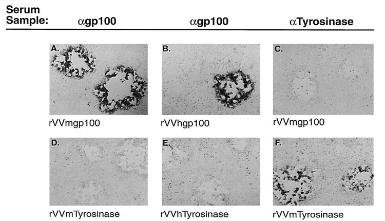
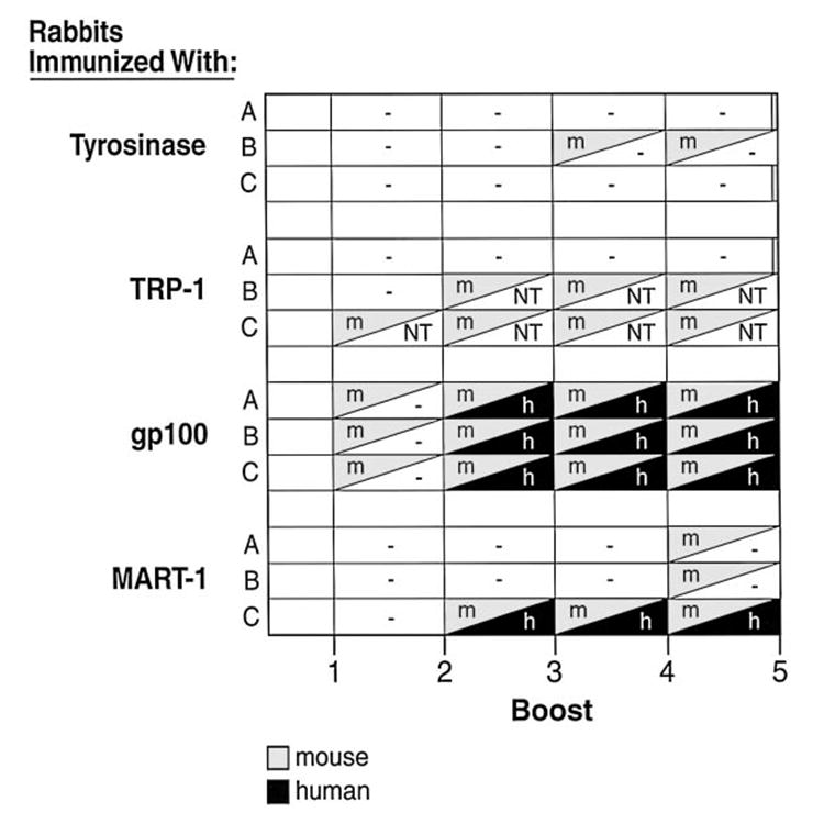
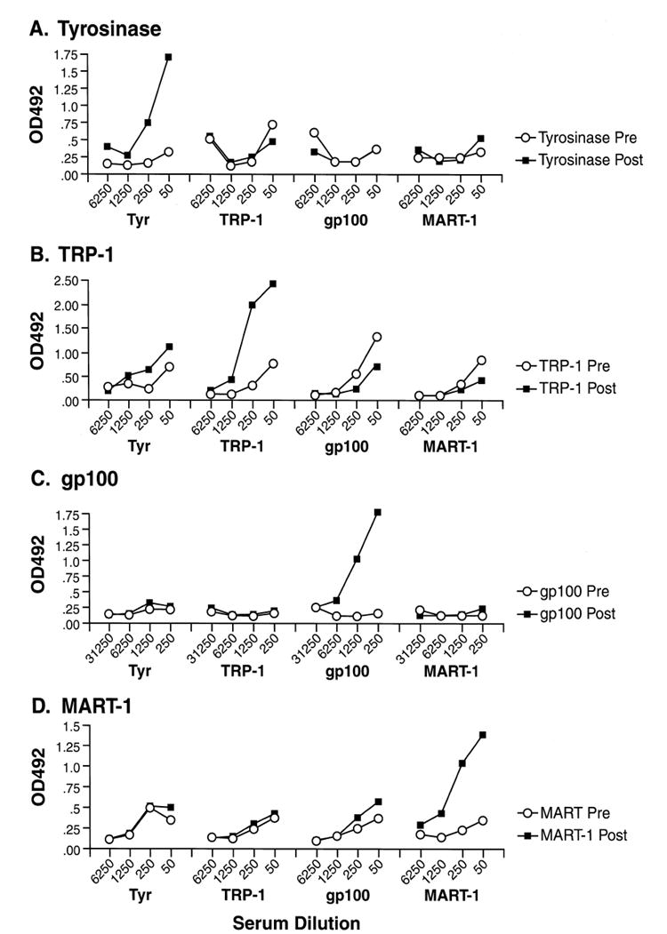
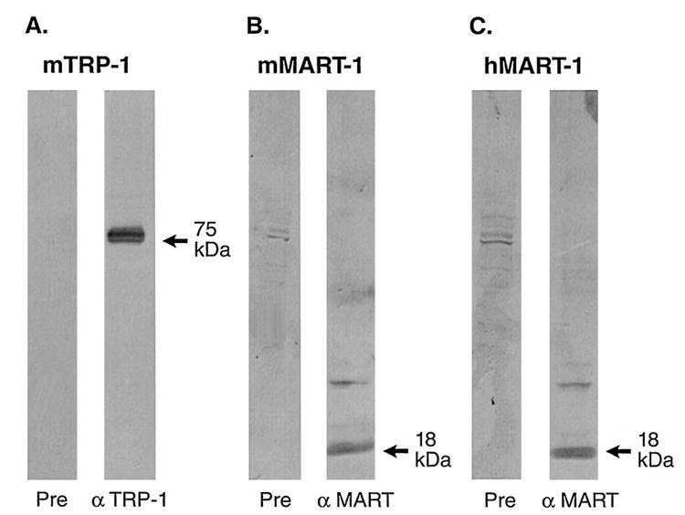
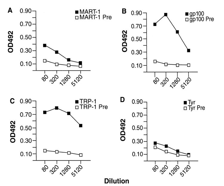
Lymphocytes from patients with melanoma have been used to clone melanoma associated antigens which are, for the most part, nonmutated melanocyte tissue differentiation antigens. To establish a mouse model for the use of these 'self' antigens as targets for anti-tumor immune responses, we have employed the mouse homologues of the human melanoma antigens Tyrosinase, Tyrosinase Related Protein-1 (TRP-1), gp100, and MART-1. We sought to generate antisera against these proteins for use in the construction of experimental recombinant and synthetic anti-cancer vaccines, and for use in biologic studies. Using genes cloned from the B16 mouse melanoma or from murine melanocytes, we immunized rabbits with plasmid DNAs coated onto microscopic gold beads that were then delivered using a hand-held, helium-driven 'gene gun'. This strategy enabled us to generate polyclonal rabbit sera containing antibodies that specifically recognized each antigen, as measured by immunostaining of vaccinia virus infected cells. The sera that we generated specifically for TRP-1, gp100, and MART-1 recognized extracts of the spontaneous murine melanoma, B16. The identities of the recognized proteins was confirmed by Western blot analysis. The titers and specificities of these antisera were determined using ELISA. Interestingly, serum samples generated against murine MART-1 and gp100 developed antibodies that were cross-reactive with the corresponding human homologues. Recognition of human gp100 and murine Tyrosinase appeared to be dependent upon conformational epitopes since specificity was lost upon denaturation of the antigens. These antisera may be useful in the detection, purification and characterization of the mouse homologues of recently cloned human tumor associated antigens and may enable the establishment of an animal model of the immune consequences of vaccination against 'self antigens.
Figures
Similar articles
-
Cloning and characterization of the genes encoding the murine homologues of the human melanoma antigens MART1 and gp100.J Immunother. 1997 Jan;20(1):15-25. doi: 10.1097/00002371-199701000-00002. J Immunother. 1997. PMID: 9101410 Free PMC article.
-
Cloning, expression and tissue distribution of the murine homologue of the melanocyte lineage-specific antigen gp100.Melanoma Res. 1997 Dec;7(6):463-70. doi: 10.1097/00008390-199712000-00004. Melanoma Res. 1997. PMID: 9464618
-
Vaccination with a recombinant vaccinia virus encoding a "self" antigen induces autoimmune vitiligo and tumor cell destruction in mice: requirement for CD4(+) T lymphocytes.Proc Natl Acad Sci U S A. 1999 Mar 16;96(6):2982-7. doi: 10.1073/pnas.96.6.2982. Proc Natl Acad Sci U S A. 1999. PMID: 10077623 Free PMC article.
-
The immunogenic properties of melanoma-associated antigens recognized by cytotoxic T lymphocytes.Exp Clin Immunogenet. 1998;15(1):19-32. doi: 10.1159/000019050. Exp Clin Immunogenet. 1998. PMID: 9619397 Review.
-
Melanoma-associated antigens recognized by cytotoxic T lymphocytes.APMIS. 1998 Jul;106(7):665-79. doi: 10.1111/j.1699-0463.1998.tb00210.x. APMIS. 1998. PMID: 9740504 Review.
Cited by
-
Type I Interferons are essential for the efficacy of replicase-based DNA vaccines.Vaccine. 2006 Jun 12;24(24):5110-8. doi: 10.1016/j.vaccine.2006.04.059. Epub 2006 May 6. Vaccine. 2006. PMID: 16725231 Free PMC article.
-
Developing recombinant and synthetic vaccines for the treatment of melanoma.Curr Opin Oncol. 1999 Jan;11(1):50-7. doi: 10.1097/00001622-199901000-00012. Curr Opin Oncol. 1999. PMID: 9914879 Free PMC article. Review.
-
gp100/pmel 17 is a murine tumor rejection antigen: induction of "self"-reactive, tumoricidal T cells using high-affinity, altered peptide ligand.J Exp Med. 1998 Jul 20;188(2):277-86. doi: 10.1084/jem.188.2.277. J Exp Med. 1998. PMID: 9670040 Free PMC article.
-
Building better vaccines: how apoptotic cell death can induce inflammation and activate innate and adaptive immunity.Curr Opin Immunol. 2000 Oct;12(5):597-603. doi: 10.1016/s0952-7915(00)00148-5. Curr Opin Immunol. 2000. PMID: 11007365 Free PMC article. Review.
-
Alphavirus-based DNA vaccine breaks immunological tolerance by activating innate antiviral pathways.Nat Med. 2003 Jan;9(1):33-9. doi: 10.1038/nm813. Epub 2002 Dec 23. Nat Med. 2003. PMID: 12496961 Free PMC article.
References
-
- Attanasio R, Pehler K, Scinicariello F. DNA-based immunization induces anti-CD4 antibodies directed primarily to native epitopes. FEMS Immunol Med Microbiol. 1997;17:207. - PubMed
-
- Barry MA, Barry ME, Johnston SA. Production of monoclonal antibodies by genetic immunization. Biotechniques. 1994;16:616. - PubMed
MeSH terms
Substances
Grants and funding
LinkOut - more resources
Full Text Sources
Other Literature Sources
Medical
Molecular Biology Databases
Research Materials
