Mouse adenovirus type 1 early region 1A is dispensable for growth in cultured fibroblasts
- PMID: 9658071
- PMCID: PMC109774
- DOI: 10.1128/JVI.72.8.6325-6331.1998
Mouse adenovirus type 1 early region 1A is dispensable for growth in cultured fibroblasts
Abstract
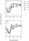
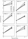
Mouse adenovirus type 1 (MAV-1) mutants with deletions of conserved regions of early region 1A (E1A) or with point mutations that eliminate translation of E1A were used to determine the role of E1A in MAV-1 replication. MAV-1 E1A mutants expressing no E1A protein grew to titers comparable to wild-type MAV-1 titers on mouse fibroblasts (3T6 fibroblasts and fibroblasts derived from Rb+/+, Rb+/-, and Rb-/- transgenic embryos). To test the hypothesis that E1A could induce a quiescent cell to reenter the cell cycle, fibroblasts were serum starved to stop DNA replication and cellular replication and then infected with the E1A mutant and wild-type viruses. All grew to equivalent titers. Steady-state levels of MAV-1 early mRNAs (E1A, E1B, E2, E3, and E4) from 3T6 cells infected with wild-type or E1A mutant virus were examined by Northern analysis. Steady-state levels of mRNAs from the mutant-infected cells were comparable to or greater than the levels found in wild-type virus infections for most of the early regions and for two late genes. The E2 mRNA levels were slightly reduced in all mutant infections relative to wild-type infections. E1A mRNA was not detected from infections with the MAV-1 E1A null mutant, pmE109, or from infections with similar MAV-1 E1A null mutants, pmE112 and pmE113. The implications for the lack of a requirement of E1A in cell culture are discussed.
Figures


Similar articles
-
E1A-CR3 interaction-dependent and -independent functions of mSur2 in viral replication of early region 1A mutants of mouse adenovirus type 1.J Virol. 2005 Mar;79(6):3267-76. doi: 10.1128/JVI.79.6.3267-3276.2005. J Virol. 2005. PMID: 15731221 Free PMC article.
-
Interaction of mouse adenovirus type 1 early region 1A protein with cellular proteins pRb and p107.Virology. 1996 Oct 1;224(1):184-97. doi: 10.1006/viro.1996.0520. Virology. 1996. PMID: 8862413
-
Bovine adenovirus type 3 E1B(small) protein is essential for growth in bovine fibroblast cells.Virology. 2001 Sep 30;288(2):264-74. doi: 10.1006/viro.2001.1104. Virology. 2001. PMID: 11601898
-
Genetically modified adenoviruses against gliomas: from bench to bedside.Neurology. 2004 Aug 10;63(3):418-26. doi: 10.1212/01.wnl.0000133302.15022.7f. Neurology. 2004. PMID: 15304571 Review.
-
How the Rb tumor suppressor structure and function was revealed by the study of Adenovirus and SV40.Virology. 2009 Feb 20;384(2):274-84. doi: 10.1016/j.virol.2008.12.010. Epub 2009 Jan 17. Virology. 2009. PMID: 19150725 Review.
Cited by
-
E1A-CR3 interaction-dependent and -independent functions of mSur2 in viral replication of early region 1A mutants of mouse adenovirus type 1.J Virol. 2005 Mar;79(6):3267-76. doi: 10.1128/JVI.79.6.3267-3276.2005. J Virol. 2005. PMID: 15731221 Free PMC article.
-
Establishment of a Simple and Efficient Reverse Genetics System for Canine Adenoviruses Using Bacterial Artificial Chromosomes.Viruses. 2020 Jul 16;12(7):767. doi: 10.3390/v12070767. Viruses. 2020. PMID: 32708703 Free PMC article.
-
Circumventing antivector immunity: potential use of nonhuman adenoviral vectors.Hum Gene Ther. 2014 Apr;25(4):285-300. doi: 10.1089/hum.2013.228. Epub 2014 Mar 25. Hum Gene Ther. 2014. PMID: 24499174 Free PMC article. Review.
-
Development of Novel Adenoviral Vectors to Overcome Challenges Observed With HAdV-5-based Constructs.Mol Ther. 2016 Feb;24(1):6-16. doi: 10.1038/mt.2015.194. Epub 2015 Oct 19. Mol Ther. 2016. PMID: 26478249 Free PMC article. Review.
-
Requirement of Sur2 for efficient replication of mouse adenovirus type 1.J Virol. 2004 Dec;78(23):12888-900. doi: 10.1128/JVI.78.23.12888-12900.2004. J Virol. 2004. PMID: 15542641 Free PMC article.
References
-
- Ball A O, Beard C W, Redick S D, Spindler K R. Genome organization of mouse adenovirus type 1 early region 1: a novel transcription map. Virology. 1989;170:523–536. - PubMed
-
- Beard C W, Ball A O, Wooley E H, Spindler K R. Transcription mapping of mouse adenovirus type 1 early region 3. Virology. 1990;175:81–90. - PubMed
Publication types
MeSH terms
Substances
Grants and funding
LinkOut - more resources
Full Text Sources
Other Literature Sources

