. 1998 Jul 7;95(14):8245-50.
doi: 10.1073/pnas.95.14.8245.

Propagation and recovery of intact, infectious Epstein-Barr virus from prokaryotic to human cells

Affiliations

Propagation and recovery of intact, infectious Epstein-Barr virus from prokaryotic to human cells

H J Delecluse et al. Proc Natl Acad Sci U S A. .

Abstract

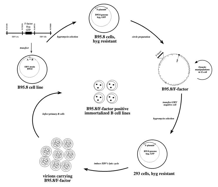
With current techniques, genetic alterations of herpesviruses are difficult to perform, mostly because of the large size of their genomes. To solve this problem, we have designed a system that allows the cloning of any gamma-herpesvirus in Escherichia coli onto an F factor-derived plasmid. Immortalized B cell lines were readily established with recombinant Epstein-Barr virus (EBV), demonstrating that the F factor-cloned EBV genome has all the characteristics of wild-type EBV. Because any genetic modification is possible in E. coli, this experimental approach opens the way to the genetic analysis of all EBV functions. Moreover, it is now feasible to generate attenuated EBV strains in vitro such that vaccine strains can be designed. Because we incorporated the genes for hygromycin resistance and green fluorescent protein onto the E. coli cloned EBV genome, the still open question of the EBV target cells other than B lymphocytes will be addressed.


Figures

Figure 1
Figure 1
Schematic overview of the EBV shuttle system. A linearized DNA fragment consisting of an F factor plasmid and two flanking regions. A and B of EBV are transfected into the B95.8 cell line, which is latently infected with EBV. Homologous recombinations occur via the regions A and B to generate a B95.8 cointegrate that encompasses the gene for hygromycin resistance (hyg) and green fluorescent protein (GFP) together with the F factor replicon. Cells that contain such a cointegrate B95.8/F factor survive under hygromycin selection. Preparation of circular DNA from these cells and its transfection into an appropriate E. coli strain establishes the B95.8/F factor molecule in E. coli for further genetic modifications. The B95.8/F factor DNA can be amplified and isolated from E. coli in microgram quantities to be used for transfection into EBV-negative cells, i.e., 293. Upon hygromycin selection, cell lines that carry the B95.8/F factor molecule as extrachromosomal copies can be established. Induction of the lytic phase of EBV’s life cycle yields viruses that carry the B95.8/F factor molecule as genetic information. Infection of primary B cells leads to their immortalization and B95.8/F factor-positive B cell lines.
Figure 2
Figure 2
Gardella gel analysis of individual, hygromycin-resistant B95.8 cell clones that were generated after transfection of the linearized F factor plasmid DNA shown in Fig. 1. After Southern blotting the blot was probed with an F factor-specific probe. Four of seven hygromycin-resistant cell lines showed clear signals, indicating a successful recombination between endogenous, wild-type B95.8 DNA molecules and the transfected linearized plasmid DNA carrying the F factor replicon. The two discrete bands correspond to the circular and linear forms of the B95.8/F factor DNA molecules. Linear EBV molecules are indicative of spontaneous lytic EBV replication, which is common in B95.8 cells.
Figure 3
Figure 3
Restriction fragment analysis of B95.8/F factor DNA in comparison with B95.8 virion DNA. Circular B95.8/F factor DNA molecules were extracted from the hygromycin-resistant B95.8 cell lines and rescued in E. coli strain DH10B. DNA was purified from the chloramphenicol-resistant E. coli clones and digested with NdeI and BamHI. The restriction pattern of the B95.8/F factor DNA was compared with the one obtained with linear wild-type B95.8 DNA extracted from viral capsids. Both restriction patterns were identical with the exception of distinct DNA fragments. New fragments were generated by the integration of the F factor into the B95.8/F factor molecule (11.2 and 4.8 kbp in size after digestion with BamHI; 14.3, 12.8, 2.8, and 2.3 kbp in size after digestion with NdeI). Fragments that represent the genomic terminal fragments in wild-type B95.8 virion DNA are fused in B95.8/F factor DNA to form a new terminal fusion fragment. One of the terminal fragments in B95.8 DNA generated by digestion with NdeI is too small to be visible on this gel.
Figure 4
Figure 4
(Top Left) GFP expression in 293 B95.8/F factor-positive cell lines. Living cells were examined with an inverse microscope under UV light. In both cases, the bright green color of all cells demonstrates a high level of expression of the GFP gene, which is part of the F factor backbone, as shown schematically in Fig. 1. (Top Right) Expression of the viral capsid antigen (VCA) in 293 carrying the B95.8/F factor DNA after induction of the lytic cycle. Fixed cells were incubated with an antibody against VCA and a second anti-mouse antibody coupled to the Cy5 fluorochrome. Stained cells were exposed to UV light. (Middle) GFP expression in cells incubated with supernatants from induced 293 cell lines carrying the B95.8/F factor DNA. Raji cells (1 × 10⁵) were incubated with 0.5 ml of supernatant from BZLF1-transfected 293 cells carrying the B95.8/F factor. Approximately 50% of the cells were GFP-positive, indicating a virus titer of at least 105 infectious viruses per ml. GFP fluorescence was investigated 48 hr after infection (Left). (Middle Right) Phase-contrast light microscopy. (Bottom) Primary human B cells were infected with supernatants from BZLF1 transfected 293 cells carrying the B95.8/F factor. As a consequence, immortalized B cell lines were generated that were investigated for GFP expression about 6 weeks after infection. (Bottom Left) UV light exposure. (Bottom Right) Phase-contrast light microscopy.
Figure 5
Figure 5
Gardella gel analysis of hygromycin-resistant 293 cell lines transfected with the B95.8/F factor plasmid DNA. After Southern blotting the blot was probed with an F factor-specific probe. Half of the tested clones proved to be positive after hybridization. As already observed in the B95.8/F factor clones, spontaneous replication occurs in these positive cell lines as demonstrated by the presence of linear genomic virion DNA.
Figure 6
Figure 6
Southern blot analysis of immortalized B cell lines that were established with virus stocks derived from 293 cells carrying the B95.8/F factor molecule. Total cell DNA from the immortalized B cell lines was digested with BamHI, separated by electrophoresis, blotted, and hybridized with a probe specific for the pMBO131 plasmid. DNAs extracted from the EBV-negative cell line HeLa served as a negative control. The positive control consisted of 100 pg of the pMBO plasmid lacking the genes for GFP and hygromycin resistance, which was digested together with 10 μg HeLa DNA. All cell lines tested were found to carry the F factor, although the number of B95.8/F factor copies varied in the immortalized B cell lines, as expected.

Similar articles

Cited by

References

    1. Hammerschmidt W, Sugden B. Nature (London) 1989;340:393–397. - PubMed
    1. Tomkinson B, Robertson E, Yalamanchili R, Longnecker R, Kieff E. J Virol. 1993;67:7298–7306. - PMC - PubMed
    1. Kempkes B, Spitkovssky D, Jansen-Dürr P, Ellwart J W, Delecluse H-J, Rottenberger C, Kremmer E, Bornkamm G W, Hammerschmidt W. EMBO J. 1995;14:88–96. - PMC - PubMed
    1. Cohen J I, Wang F, Mannick J, Kieff E. Proc Natl Acad Sci USA. 1989;86:9558–9562. - PMC - PubMed
    1. Smiley J. Nature (London) 1980;285:333–335. - PubMed

Publication types

MeSH terms

Substances

LinkOut - more resources