. 1998 Jul 7;95(14):7898-903.
doi: 10.1073/pnas.95.14.7898.

The mouse and human genes encoding the recognition component of the N-end rule pathway

Affiliations

The mouse and human genes encoding the recognition component of the N-end rule pathway

Y T Kwon et al. Proc Natl Acad Sci U S A. .

Abstract

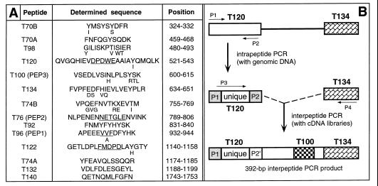
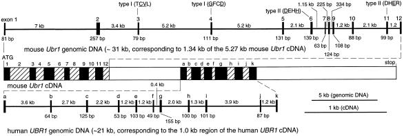
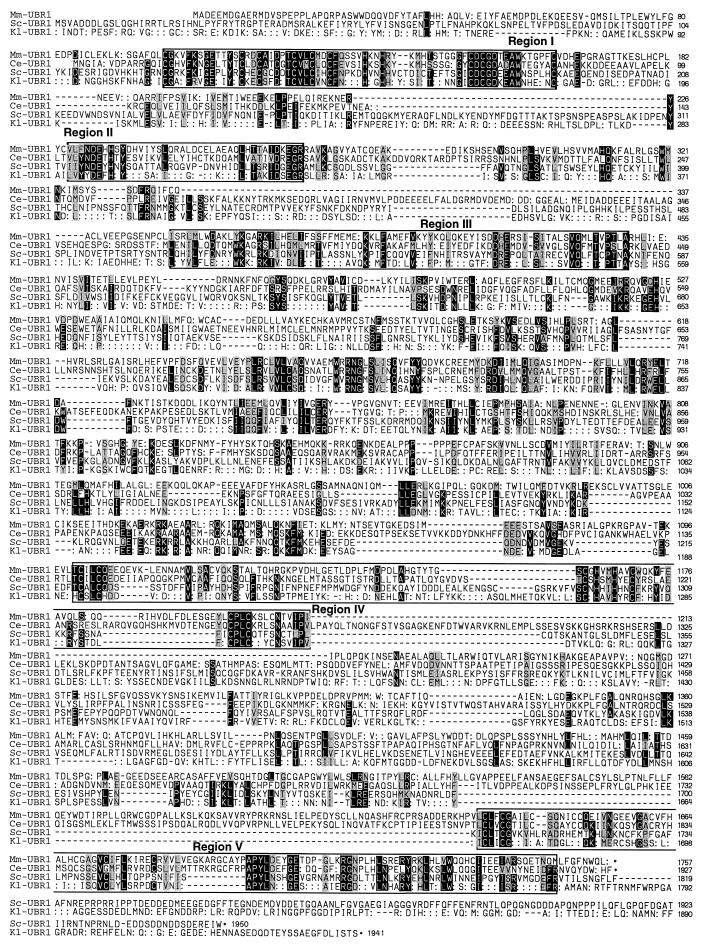
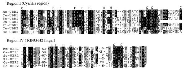
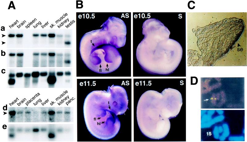
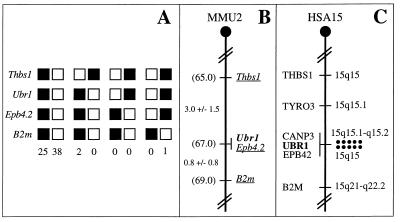
The N-end rule relates the in vivo half-life of a protein to the identity of its N-terminal residue. The N-end rule pathway is one proteolytic pathway of the ubiquitin system. The recognition component of this pathway, called N-recognin or E3, binds to a destabilizing N-terminal residue of a substrate protein and participates in the formation of a substrate-linked multiubiquitin chain. We report the cloning of the mouse and human Ubr1 cDNAs and genes that encode a mammalian N-recognin called E3alpha. Mouse UBR1p (E3alpha) is a 1,757-residue (200-kDa) protein that contains regions of sequence similarity to the 225-kDa Ubr1p of the yeast Saccharomyces cerevisiae. Mouse and human UBR1p have apparent homologs in other eukaryotes as well, thus defining a distinct family of proteins, the UBR family. The residues essential for substrate recognition by the yeast Ubr1p are conserved in the mouse UBR1p. The regions of similarity among the UBR family members include a putative zinc finger and RING-H2 finger, another zinc-binding domain. Ubr1 is located in the middle of mouse chromosome 2 and in the syntenic 15q15-q21.1 region of human chromosome 15. Mouse Ubr1 spans approximately 120 kilobases of genomic DNA and contains approximately 50 exons. Ubr1 is ubiquitously expressed in adults, with skeletal muscle and heart being the sites of highest expression. In mouse embryos, the Ubr1 expression is highest in the branchial arches and in the tail and limb buds. The cloning of Ubr1 makes possible the construction of Ubr1-lacking mouse strains, a prerequisite for the functional understanding of the mammalian N-end rule pathway.


Figures

Figure 1
Figure 1
Peptides of rabbit UBR1p (E3α) and isolation of the mouse Ubr1 cDNA. (A) Amino acid sequences of tryptic peptides of the purified rabbit UBR1p (see Materials and Methods). The alternative sets of peptide names, T-based and PEP1-PEP3 (in parentheses), refer to two different preparations of E3α. The sequences of T120, T76, T96, and T122 that were encoded by DNA sequences identified through intrapeptide PCR are underlined. Residues deduced from the mouse Ubr1 cDNA that differed from those inferred through peptide sequencing are indicated in a smaller font. The peptides’ positions in the deduced sequence of mouse UBR1p are indicated. (B) The intrapeptide/interpeptide-PCR cloning strategy. The products of the initial intrapeptide PCR, derived from rabbit genomic DNA, were used to carry out interpeptide PCRwith a rabbit liver cDNA library (CLONTECH). The resulting 392-bp fragment of the rabbit Ubr1 cDNA was used to isolate, using PCR and a λgt11 mouse liver cDNA library, the corresponding 392-bp mouse Ubr1 cDNA fragment. This fragment then was used to screen the same cDNA library, yielding a 2.4-kb fragment of the mouse Ubr1 cDNA that encoded several of the peptide-derived sequences of the rabbit UBR1p. The encoded sequence was also significantly similar to that of the N-terminal region of S. cerevisiae Ubr1p (13) and contained the putative start (ATG) codon of the mouse Ubr1 ORF. To isolate the rest of the 5′ region of the Ubr1 cDNA, 5′-rapid amplification of cDNA ends (RACE)–PCR (20) was performed with poly(A)+ RNA from mouse L cells and a primer from the 2.4-kb DNA fragment. 3′-RACE–PCR (20) was used to amplify a downstream region of Ubr1 cDNA. The resulting DNA fragment (nucleotides 2,470–3,467) then was used to screen a λgt10 mouse cDNA library from MEL-C19 cells. Five overlapping cDNA isolates (MR16, MR17, MR19, MR20, and MR23) that together spanned the entire Ubr1 cDNA were mapped and subcloned into Bluescript II SK+ (Stratagene), yielding the plasmid MR26, which contained the entire ORF of Ubr1. The ORF region of Ubr1 cDNA was sequenced on both strands at least twice, using independently derived cDNA clones.
Figure 2
Figure 2
The mouse and human Ubr1 cDNAs and genes. Thick horizontal lines represent genomic DNA. The upper one is a ≈31-kb fragment of the mouse Ubr1 gene that corresponds to a 1.34-kb region of mouse Ubr1 cDNA (nucleotides 115–1,454). Vertical rectangles represent exons. Their lengths, and the lengths of the introns, are indicated, respectively, below and above the horizontal line. In a composite diagram of the Ubr1 cDNA, the exons are depicted as alternatively shaded rectangles. For exon 1, only its translated region is indicated. Shown belowthe cDNA diagram is a ≈21-kb fragment of the human UBR1 gene, corresponding to 1.0 kb of the indicated region of the human UBR1 cDNA (nucleotides 2,218–3,227 of the mouse Ubr1 cDNA sequence). The mouse and human Ubr1 exons are denoted, respectively, by numbers and letters. Also indicated are the exon locations of some of the type 1 and type 2 substrate-binding sites of N-recognin (the essential amino acid residues are underlined) (A. Webster, M. Ghislain, and A.V., unpublished data; see the main text). Not shown are the 114-bp 5′-untranslated region (UTR) and the 1,010 bp 3′-UTR of the mouse Ubr1 cDNA. To isolate mouse Ubr1, a library of mouse genomic DNA fragments in a BAC vector (see Materials and Methods) was screened with a fragment of the mouse Ubr1 cDNA (nucleotides 105–1,333) as a probe, yielding seven BAC clones, of which BAC3 and BAC4 contained the entire Ubr1 gene. The exon/intron organization of the first 31 kb (≈1/4) of the mouse Ubr1 gene was determined by using exon-specific PCR primers to produce ≈40 genomic DNA fragments of the BAC3 insert that ranged in size from 1.3 to 18 kb. Regions encompassing the exon/intron junctions then were sequenced by using intron-specific primers. Fragments of the human genomic UBR1 DNA were isolated by using primers derived from the 1.0-kb fragment of the human UBR1 cDNA, the Expand High Fidelity PCR System (Roche Molecular Biochemicals, Indianapolis, IN), and genomic DNA from human 293 cells. The resulting four fragments were subcloned into pCR2.1 (Invitrogen), yielding the plasmids HR8, HR6–4, HR2–25, and HR7–2, whose partially overlapping inserts encompassed ≈21 kb of the human UBR1 gene. Partial sequencing of the mouse and human genomic Ubr1 fragments (≈20 kb of sequenced DNA) included all of the exon/intron junctions in these regions of Ubr1.
Figure 3
Figure 3
Comparison of the deduced amino acid sequence of mouse UBR1p (Mm-UBR1) with those of C. elegans UBR1p (Ce-UBR1), S. cerevisiae Ubr1p (Sc-UBR1), and K. lactis Ubr1p (Kl-UBR1). White-on-black and gray shadings highlight, respectively, identical and similar residues. The residues of UBR proteins that are identical to those of S. cerevisiae Ubr1p are denoted by double dots, at positions where the identity involves just one non-cerevisiae protein. Also indicated are the regions of significant similarity among the four proteins. K. lactis UBR1 was cloned through its crosshybridization to S. cerevisiae UBR1 (P. Waller and A.V., unpublished data).
Figure 4
Figure 4
Two Cys/His domains of the UBR protein family. Comparison of the putative zinc finger (region I) and RING-H2 finger (region IV) with the corresponding sequences from the other species in Fig. 3, and also with C. albicans Ubr1p (Ca-UBR1), C. elegans UBR2p (Ce-UBR2), and S. cerevisiae Ubr2p (Sc-UBR2). Numbers indicate the lengths of gaps. The conserved Cys and His residues are indicated.
Figure 5
Figure 5
Northern and in situ hybridizations with mouse and human Ubr1. (A) Membranes containing electrophoretically fractionated poly(A)+ mRNA from different mouse (ac) or human (d and e) tissues were hybridized with either a 2-kb 5′-proximal (nucleotides 116–2,124) mouse Ubr1 cDNA fragment (a), its 0.64-kb 3′-proximal (nucleotides 4,749–5,388) fragment (b), a 1-kb human UBR1 cDNA fragment (d), or the human β-actin cDNA fragment (c and e). The upper arrows in a and d indicate the ≈8-kb Ubr1 transcript. The lower arrow in a indicates the ≈6-kb testis-specific Ubr1 transcript. In the RNA sample from mouse spleen, the Ubr1 transcript (but not the actin transcript) may have been degraded (ac). (B) Expression of Ubr1 in e10.5 and e11.5 mouse embryos. Whole-mount in situ hybridization was carried out with either antisense (AS) or sense (S, negative control) Ubr1 cDNA probes (see Materials and Methods). The regions of high Ubr1 expression are indicated by arrows (t, tail; fl, forelimb buds; hl, hindlimb buds). The branchial arches, where Ubr1 is also highly expressed in e10.5 embryos (data not shown), are not visible in this e10.5 embryo. (C) Expression of Ubr1 in the surface ectoderm of limb buds. Shown is a transverse section of a forelimb bud of an e10.5 embryo (se, surface ectoderm). (D) FISH analysis of human UBR1. (Upper) An example of the UBR1-specific FISH signal (arrow). (Lower) The same mitotic spread stained with 4′-6-diamino-2-phenylindole (DAPI) to visualize the chromosomes (see also Fig. 6).
Figure 6
Figure 6
Chromosomal locations of the mouse and human Ubr1 genes. (A) Mouse Ubr1 was mapped to the middle of mouse chromosome 2 by using interspecific (M. musculus-M. spretus) backcross analysis (18, 24). Shown are the segregation patterns of mouse Ubr1 and the flanking genes in 66 backcross animals that were typed for all loci. For individual pairs of loci, more than 66 animals were typed. Each column represents the chromosome identified in the backcross progeny that was inherited from the [M. musculus C57BL/6J × M. spretus] F1 parent. Filled and empty squares represent, respectively, C57BL/6J and M. spretus alleles. The numbers of offspring that inherited each type of chromosome 2 are listed below the columns. (B) A partial mouse chromosome 2 linkage map (MMU2), showing Ubr1 in relation to the linked genes Thbs1, Ebp4.2, and B2m, and also, on the left, the corresponding recombination distances between the loci, in centimorgans, and the map locations, in parentheses. (C) A partial human chromosome 15 linkage map (HSA15). Each dot on the right, in the 15q15-q21.1 region, corresponds to the actually observed UBR1-specific double-dot FISH signal detected on human chromosome 15 (see also Fig. 5D).

Similar articles

Cited by

References

    1. Varshavsky A. Trends Biochem Sci. 1997;22:383–387. - PubMed
    1. Hershko A. Curr Opin Cell Biol. 1997;9:788–799. - PubMed
    1. Haas A J, Siepman T J. FASEB J. 1997;11:1257–1268. - PubMed
    1. Hochstrasser M. Annu Rev Genet. 1996;30:405–439. - PubMed
    1. Bachmair A, Finley D, Varshavsky A. Science. 1986;234:179–186. - PubMed

Publication types

Associated data