

The human autoantigen La/SS-B accelerates herpes simplex virus type 1 replication in transfected mouse 3T3 cells
- PMID: 9649219
- PMCID: PMC1905003
- DOI: 10.1046/j.1365-2249.1998.00605.x
The human autoantigen La/SS-B accelerates herpes simplex virus type 1 replication in transfected mouse 3T3 cells
Abstract
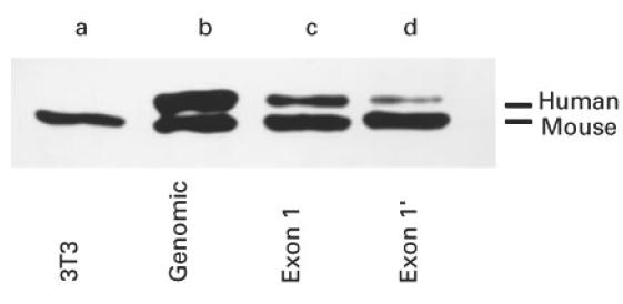
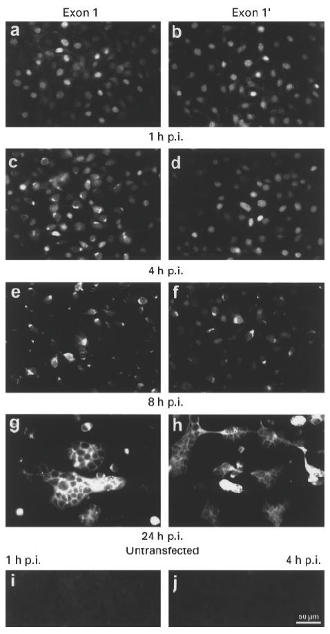
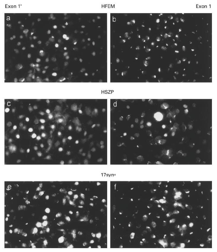
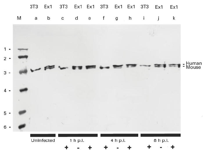
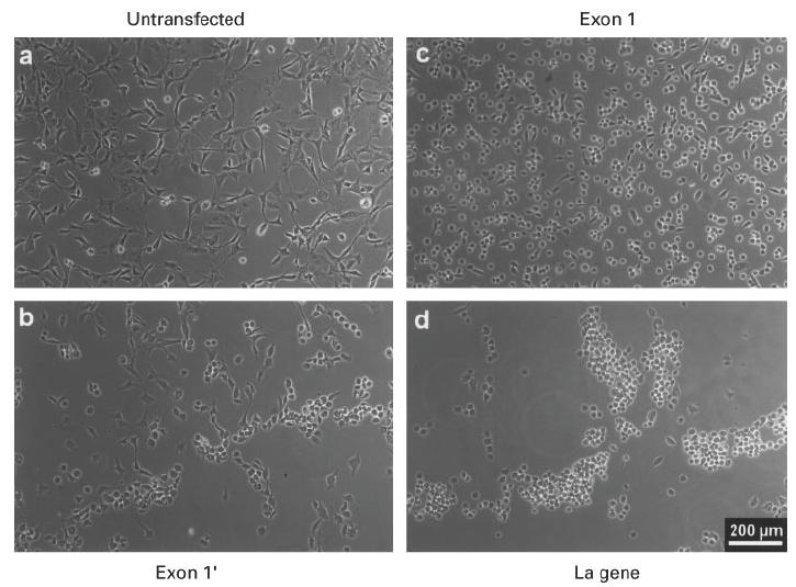
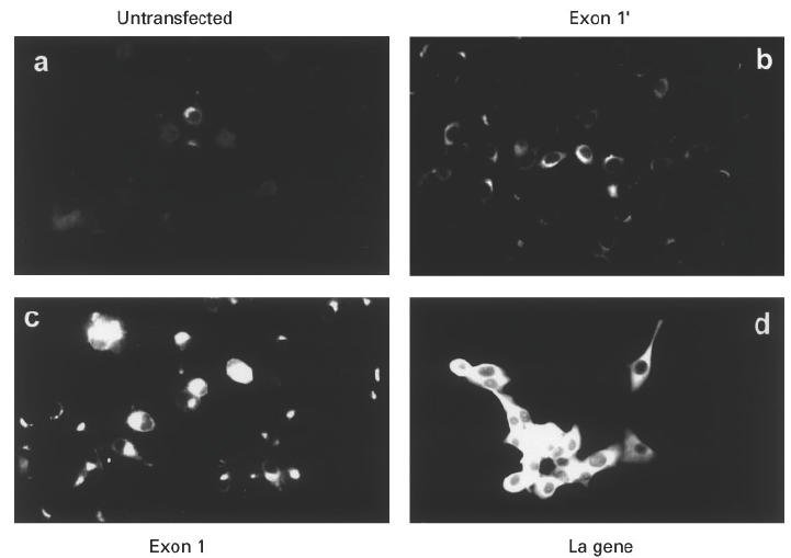
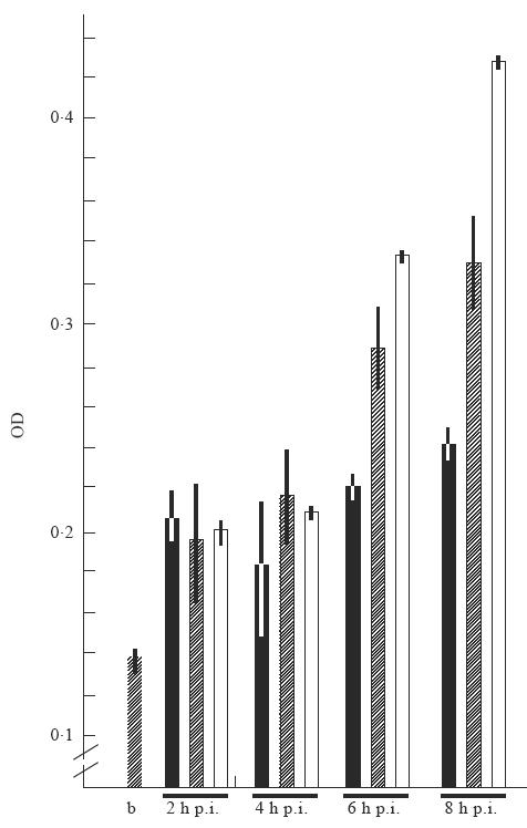
Permanently transfected mouse cell lines which expressed different levels of the human autoantigen La/SS-B were infected with different strains of herpes simplex virus type 1, including the strains ANG, HSZP, 17syn+ and HFEM. During infection the localization of the human La protein was followed using an anti-La MoAb, which recognized only the human La protein but did not cross-react with either the endogenous mouse La protein or any viral encoded protein. After infection La protein was transported from the nucleus to the cytoplasm. The time course of translocation was dependent on the amount of human La protein expressed in the respective cell line. Moreover, acceleration of viral replication was dependent on the level of expression of human La protein, suggesting that La protein is a cellular factor that facilitates virus replication.
Figures
Similar articles
-
An altered intracellular distribution of the autoantigen La/SS-B when translated from a La mRNA isoform.Exp Cell Res. 1997 Aug 1;234(2):329-35. doi: 10.1006/excr.1997.3608. Exp Cell Res. 1997. PMID: 9260901
-
Characterization of the mouse autoantigen La (SS-B). Identification of conserved RNA-binding motifs, a putative ATP binding site and reactivity of recombinant protein with poly(U) and human autoantibodies.J Immunol. 1993 Apr 1;150(7):3091-100. J Immunol. 1993. PMID: 8454877
-
Molecular analysis of the RNA and protein components recognized by anti-La(SS-B) autoantibodies.Clin Exp Immunol. 1985 Dec;62(3):685-95. Clin Exp Immunol. 1985. PMID: 2417765 Free PMC article.
-
Translocation of the nuclear autoantigen La to the cell surface of herpes simplex virus type 1 infected cells.Autoimmunity. 1992;12(1):37-45. doi: 10.3109/08916939209146128. Autoimmunity. 1992. PMID: 1617103
-
Activation of a murine autoreactive B cell by immunization with human recombinant autoantigen La/SS-B: characterization of the autoepitope.J Autoimmun. 1995 Dec;8(6):825-42. doi: 10.1016/s0896-8411(95)80020-4. J Autoimmun. 1995. PMID: 8824709
Cited by
-
A Review of the Multipronged Attack of Herpes Simplex Virus 1 on the Host Transcriptional Machinery.Viruses. 2021 Sep 14;13(9):1836. doi: 10.3390/v13091836. Viruses. 2021. PMID: 34578417 Free PMC article. Review.
-
SS-56, a novel cellular target of autoantibody responses in Sjögren syndrome and systemic lupus erythematosus.J Clin Invest. 2001 Sep;108(6):861-9. doi: 10.1172/JCI13469. J Clin Invest. 2001. PMID: 11560955 Free PMC article.
-
Neo-epitopes are required for immunogenicity of the La/SS-B nuclear antigen in the context of late apoptotic cells.Clin Exp Immunol. 2006 Feb;143(2):237-48. doi: 10.1111/j.1365-2249.2005.03001.x. Clin Exp Immunol. 2006. PMID: 16412047 Free PMC article.
References
-
- Tan EM. Antinuclear antibodies: diagnostic markers for autoimmune diseases and probes for cell biology. Adv Immunol. 1989;44:93–151. - PubMed
-
- Madore SJ, Wieben ED, Pederson T. Eukaryotic small ribonucleoproteins. Anti-La human autoantibodies react with U1 RNA–protein complexes. J Biol Chem. 1984;259:1929–33. - PubMed
-
- Pruijn GJM, Slobbe RL, van Venroooij WJ. Structure and function of La and Ro RNPs. Mol Biol Rep. 1990;14:43–48. - PubMed
Publication types
MeSH terms
Substances
LinkOut - more resources
Full Text Sources
Miscellaneous
