. 1998 Jun 29;141(7):1503-13.
doi: 10.1083/jcb.141.7.1503.

The SNARE machinery is involved in apical plasma membrane trafficking in MDCK cells

Affiliations

The SNARE machinery is involved in apical plasma membrane trafficking in MDCK cells

S H Low et al. J Cell Biol. .

Abstract

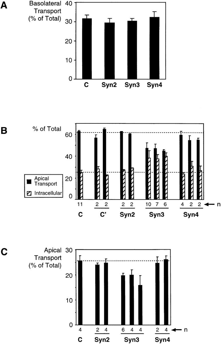
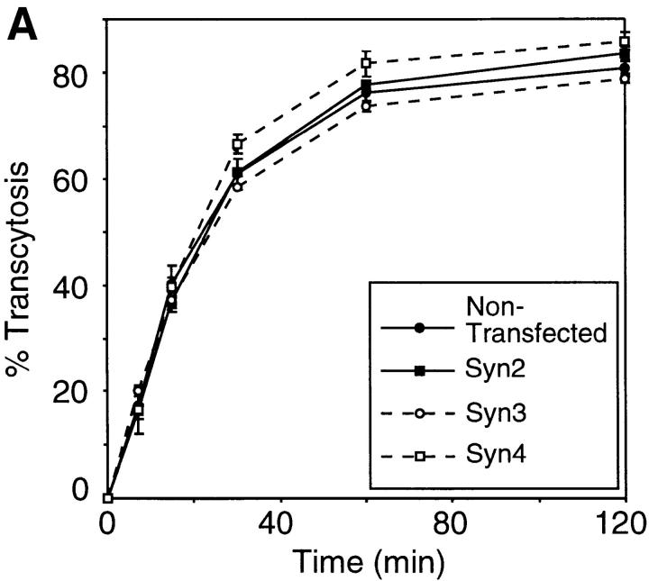
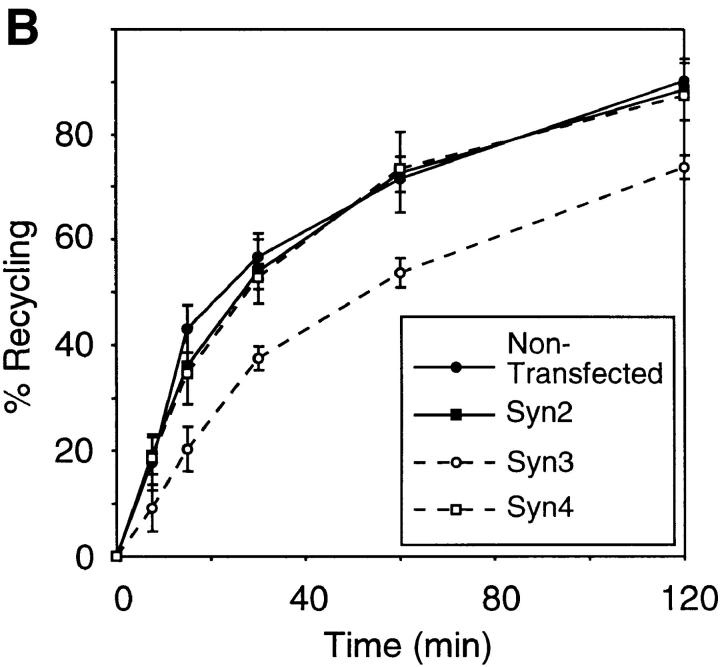
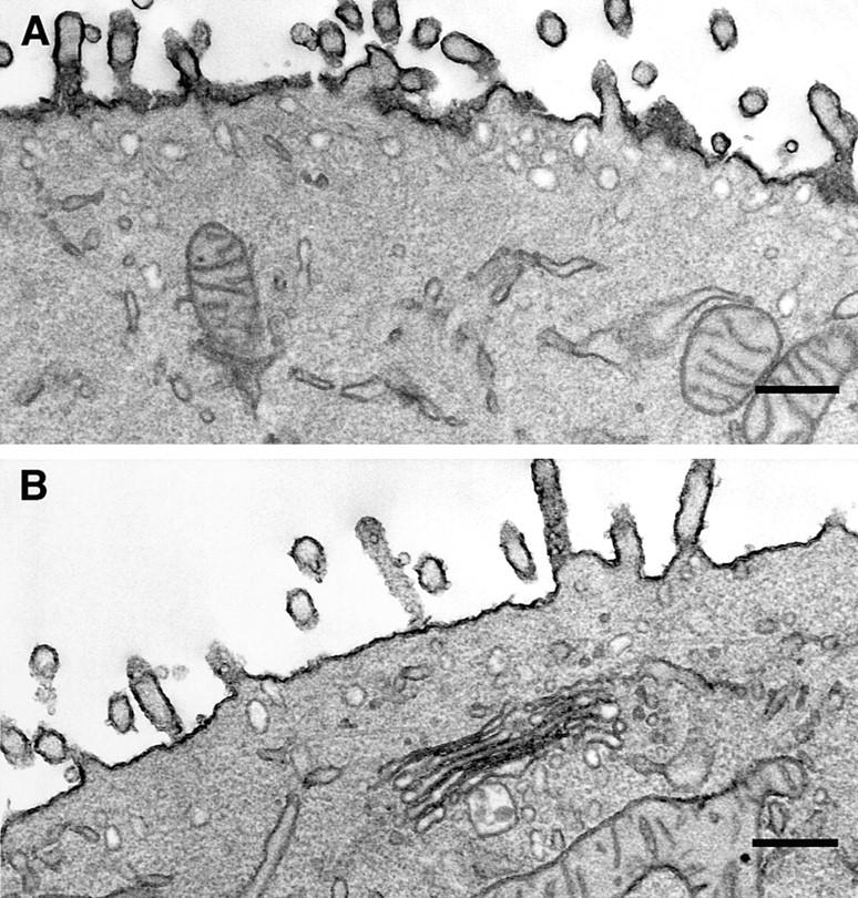
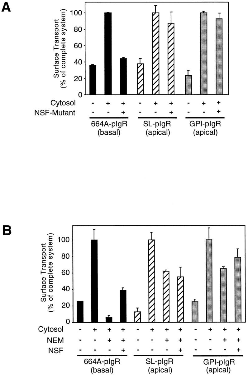
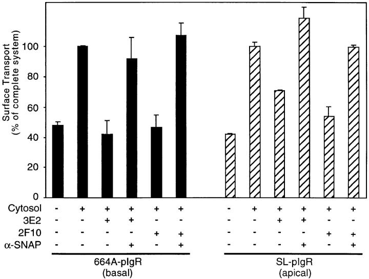
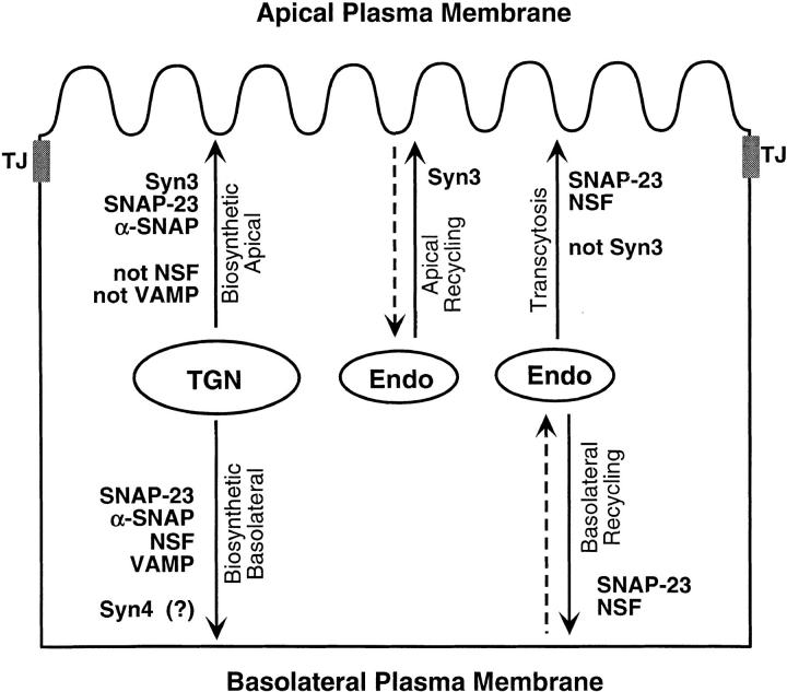
We have investigated the controversial involvement of components of the SNARE (soluble N-ethyl maleimide-sensitive factor [NSF] attachment protein [SNAP] receptor) machinery in membrane traffic to the apical plasma membrane of polarized epithelial (MDCK) cells. Overexpression of syntaxin 3, but not of syntaxins 2 or 4, caused an inhibition of TGN to apical transport and apical recycling, and leads to an accumulation of small vesicles underneath the apical plasma membrane. All other tested transport steps were unaffected by syntaxin 3 overexpression. Botulinum neurotoxin E, which cleaves SNAP-23, and antibodies against alpha-SNAP inhibit both TGN to apical and basolateral transport in a reconstituted in vitro system. In contrast, we find no evidence for an involvement of N-ethyl maleimide-sensitive factor in TGN to apical transport, whereas basolateral transport is NSF-dependent. We conclude that syntaxin 3, SNAP-23, and alpha-SNAP are involved in apical membrane fusion. These results demonstrate that vesicle fusion with the apical plasma membrane does not use a mechanism that is entirely unrelated to other cellular membrane fusion events, but uses isoforms of components of the SNARE machinery, which suggests that they play a role in providing specificity to polarized membrane traffic.


Figures

Figure 1
Figure 1
Effect of syntaxin overexpression on TGN to surface transport of WT-, SL-, or GPI-pIgR in MDCK Cells. (A) MDCK cells expressing WT-pIgR were untransfected (C) or stably transfected with syntaxin 2 (Syn2), 3 (Syn3), or 4 (Syn4). As described in Materials and Methods, newly synthesized pIgR transported to the basal surface was measured in a pulse-chase protocol and expressed as a percentage of the total labeled pIgR. The values were determined using two different clones in triplicate filters each and the means and standard deviation are plotted. (B) MDCK cells expressing SL-pIgR were transfected with either syntaxin 2, 3, or 4. Untransfected parental cells (C) or cells transfected with the syntaxin 4 plasmid but not expressing the protein (C′) served as controls. Transport of newly synthesized SL-pIgR was determined in a pulse-chase experiment and the amount transported to the apical surface (black bars) or remaining in the cell at the end of the chase (hatched bars) is expressed as a percentage of the total labeled SL-pIgR. Except for the control parental cell line, multiple clones of each cell line were examined and the values shown are the means and standard deviation of several filters (n). Dotted lines indicate the average values of the control experiments. (C) MDCK cells expressing GPI-pIgR were untransfected (C) or transfected with syntaxin 2, 3, or 4. Delivery to the surface was measured as described above. The inhibition of apical transport of SL- and GPI-pIgR by syntaxin 3 overexpression is statistically significant (Student's t-test, P < 10−10).
Figure 2
Figure 2
Transcytosis and apical recycling of IgA in syntaxin-overexpressing MDCK cells. Transcytosis and recycling pathways of IgA were analyzed in MDCK cells stably expressing WT-pIgR and syntaxins 2, 3, or 4. (A)For transcytosis, radioiodinated IgA was internalized from the basal surface of the cell for 10 min, washed, and then chased at 37°C. At indicated time intervals, the apical and basal media were collected, replaced with fresh media, and then incubated further at 37°C. Finally, the radioactivity of the collected media, and that remaining in the cells was determined. The values (mean and SD) of apically released radioiodinated IgA, expressed as percent of the total radioactivity, of two independent clones each in triplicate filters, were plotted. (B) To measure apical recycling, radioiodinated IgA was added to the apical surface of the cell and allowed to endocytose for 10 min at 37°C. Excess IgA was washed away and the cells were chased at 37°C. At indicated time intervals media were collected and the radioactivity was determined. The plot represents the percentage of IgA recycled to the apical surface (mean and SD of two independent clones each in triplicate filters).
Figure 2
Figure 2
Transcytosis and apical recycling of IgA in syntaxin-overexpressing MDCK cells. Transcytosis and recycling pathways of IgA were analyzed in MDCK cells stably expressing WT-pIgR and syntaxins 2, 3, or 4. (A)For transcytosis, radioiodinated IgA was internalized from the basal surface of the cell for 10 min, washed, and then chased at 37°C. At indicated time intervals, the apical and basal media were collected, replaced with fresh media, and then incubated further at 37°C. Finally, the radioactivity of the collected media, and that remaining in the cells was determined. The values (mean and SD) of apically released radioiodinated IgA, expressed as percent of the total radioactivity, of two independent clones each in triplicate filters, were plotted. (B) To measure apical recycling, radioiodinated IgA was added to the apical surface of the cell and allowed to endocytose for 10 min at 37°C. Excess IgA was washed away and the cells were chased at 37°C. At indicated time intervals media were collected and the radioactivity was determined. The plot represents the percentage of IgA recycled to the apical surface (mean and SD of two independent clones each in triplicate filters).
Figure 3
Figure 3
Small vesicles accumulate close to the apical plasma membrane in MDCK cells over-expressing syntaxin 3. MDCK cells stably transfected for syntaxin 3 (A) or the parental cells (B) were grown in parallel as tight monolayers on polycarbonate filters. Cells were processed for transmission electron microscopy. Parts of the apical plasma membrane with microvilli are shown. Small vesicles typically 100 nm in diameter can be seen close to the plasma membrane. The numbers of these vesicles are increased significantly in cells overexpressing syntaxin 3 (see Table I). Bar, 500 nm.
Figure 4
Figure 4
NSF is involved in transport from the TGN to the basolateral but not to the apical plasma membrane. (A) TGN to basolateral or apical plasma membrane transport was reconstituted in SLO-permeabilized MDCK cells expressing either 664A-, SL-, or GPI-pIgR as reporter molecules. The cytosol dependence of the transport reaction is shown. Addition of a recombinant, ATPase-deficient mutant of NSF (inhibits NSF-dependent fusion reactions by competition with endogenous wild-type NSF) together with the cytosol inhibits transport of 664A-pIgR to the basolateral surface strongly, while it has no effect on the apical transport of SL- or GPI-pIgR. (B) The reverse experiment was done by first inhibiting the endogenous NSF in SLO-permeabilized MDCK cells by treatment with 0.05 or 0.15 mM NEM (for basolateral or apical transport, respectively). NEM treatment inhibited basolateral transport of 664A-pIgR strongly whereas a threefold higher NEM concentration inhibited apical transport of SL- or GPI-pIgR only partially. Addition of recombinant wild-type NSF could partially restore basolateral transport of 664A-pIgR after NEM treatment but not apical transport of SL- or GPI-pIgR. The complete reactions (+ Cytosol) were set to 100%, and the values represent the mean and range of representative experiments done with duplicate filters.
Figure 5
Figure 5
Antibodies against α-SNAP inhibit transport from the TGN to the basolateral and apical plasma membrane. TGN to basolateral or apical plasma membrane transport was reconstituted in SLO-permeabilized MDCK cells expressing either 664A- or SL-pIgR as reporter molecules. Addition of 110 μg/ml of the mAbs 3E2 or 2F10 against α-SNAP inhibited both the basolateral and apical transport pathways. This inhibition was completely abolished when a fourfold molar excess of recombinant α-SNAP was added to the antibodies 10 min before addition to the permeabilized cells. Note that in these experiments, the concentration of HeLa cytosol, which contains α-SNAP, was reduced by half, which also reduces the cytosol stimulation of the transport reaction. The complete reactions (+ Cytosol) were set to 100% and the values represent the mean and range of representative experiments done with duplicate filters.
Figure 6
Figure 6
BoNT-E, which cleaves SNAP-23, inhibits transport from the TGN to the basolateral and apical plasma membranes. TGN to basolateral or apical plasma membrane transport was reconstituted in SLO-permeabilized MDCK cells expressing either 664A-, SL-, or GPI-pIgR as reporter molecules. The reactions were carried out in the absence or presence of 10 (1) or 100 (11) μg/ml of purified recombinant light chains of BoNT-E or an inactive mutant of BoNT-C1. Addition of BoNT-E caused inhibition of both basolateral and apical transport reactions. Inactivation of BoNT-E before addition by boiling prevented this inhibition. Similarly, addition of the inactive mutant of BoNT-C1, which served as a negative control for possible bacterial contaminants from the purification of the toxins, had no inhibitory effect. The complete reactions (+ Cytosol) were set to 100% and the values represent the mean and range of representative experiments done with duplicate filters. Note that in three independent experiments even the weaker inhibition of apical transport of SL-pIgR was still statistically significant by Student's t-test (P < 0.002).
Figure 7
Figure 7
Model and summary of the involvement of components of the SNARE fusion machinery in polarized membrane traffic in MDCK cells. The results of this and other studies (5, 23, 27) are summarized schematically. The following pathways to the plasma membrane are depicted: biosynthetic transport (left), apical recycling (middle), basolateral to apical transcytosis (right) and basolateral recycling (right). Evidence for the involvement of one or more SNARE components has been found for each transport step. For details refer to the Discussion. TJ, tight junction; Endo, endosome.

Similar articles

Cited by

References

    1. Aalto MK, Ronne H, Keranen S. Yeast syntaxins Sso1p and Sso2p belong to a family of related membrane proteins that function in vesicular transport. EMBO (Eur Mol Biol Organ) J. 1993;12:4095–4104. - PMC - PubMed
    1. Acharya U, Jacobs R, Peters JM, Watson N, Farquhar MG, Malhotra V. The formation of Golgi stacks from vesiculated Golgi membranes requires two distinct fusion events. Cell. 1995;82:895–904. - PubMed
    1. Apodaca G, Aroeti B, Tang K, Mostov KE. Brefeldin-A inhibits the delivery of the polymeric immunoglobulin receptor to the basolateral surface of MDCK cells. J Biol Chem. 1993;268:20380–20385. - PubMed
    1. Apodaca G, Katz KA, Mostov KE. Receptor-mediated transcytosis of IgA in MDCK cells via apical endosome. J Cell Biol. 1994;125:67–86. - PMC - PubMed
    1. Apodaca G, Cardone MH, Whiteheart SW, DasGupta BR, Mostov KE. Reconstitution of transcytosis in SLO-permeabilized MDCK cells: existence of an NSF-dependent fusion mechanism with the apical surface of MDCK cells. EMBO (Eur Mol Biol Organ) J. 1996;15:1471–1481. - PMC - PubMed

Publication types

MeSH terms