

Tyrosine phosphorylation of paxillin and focal adhesion kinase by activation of muscarinic m3 receptors is dependent on integrin engagement by the extracellular matrix
- PMID: 9636140
- PMCID: PMC22590
- DOI: 10.1073/pnas.95.13.7281
Tyrosine phosphorylation of paxillin and focal adhesion kinase by activation of muscarinic m3 receptors is dependent on integrin engagement by the extracellular matrix
Abstract
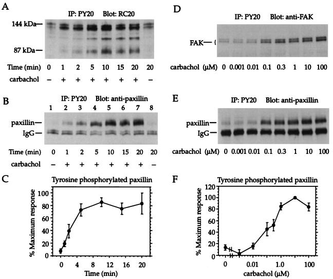
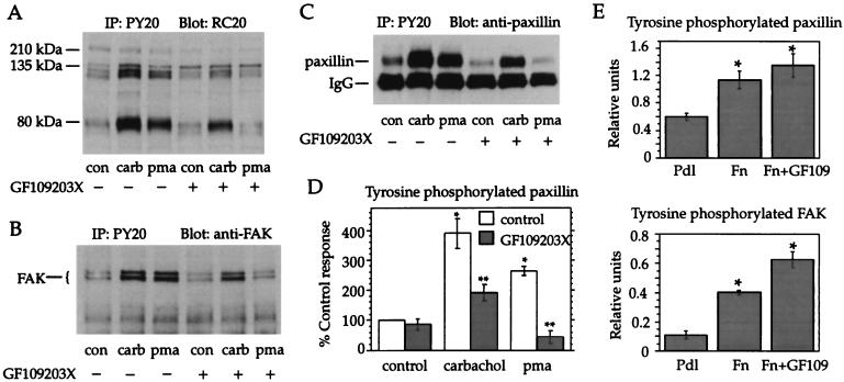
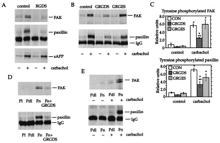
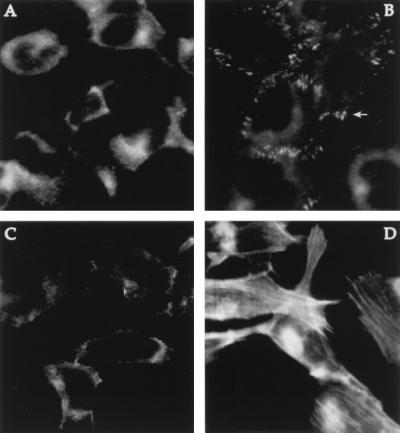
The G protein-coupled m1 and m3 muscarinic acetylcholine receptors increase tyrosine phosphorylation of several proteins, including the focal adhesion-associated proteins paxillin and focal adhesion kinase (FAK), but the mechanism is not understood. Activation of integrins during adhesion of cells to extracellular matrix, or stimulation of quiescent cell monolayers with G protein-coupled receptor ligands including bradykinin, bombesin, endothelin, vasopressin, and lysophosphatidic acid, also induces tyrosine phosphorylation of paxillin and FAK and formation of focal adhesions. These effects are generally independent of protein kinase C but are inhibited by agents that prevent cytoskeletal assembly or block activation of the small molecular weight G protein Rho. This report demonstrates that tyrosine phosphorylation of paxillin and FAK elicited by stimulation of muscarinic m3 receptors with the acetylcholine analog carbachol is inhibited by soluble peptides containing the arginine-glycine-aspartate motif (the recognition site for integrins found in adhesion proteins such as fibronectin) but is unaffected by peptides containing the inactive sequence arginine-glycine-glutamate. Tyrosine phosphorylation elicited by carbachol, but not by cell adhesion to fibronectin, is reduced by the protein kinase C inhibitor GF 109203X. The response to carbachol is dependent on the presence of fibronectin. Moreover, immunofluorescence studies show that carbachol treatment induces formation of stress fibers and focal adhesions. These results suggest that muscarinic receptor stimulation activates integrins via a protein kinase C-dependent mechanism. The activated integrins transmit a signal into the cell's interior leading to tyrosine phosphorylation of paxillin and FAK. This represents a novel mechanism for regulation of tyrosine phosphorylation by muscarinic receptors.
Figures
Similar articles
-
Activation of m3 muscarinic receptors induces rapid tyrosine phosphorylation of p125(FAK), p130(cas), and paxillin in rat pancreatic acini.Arch Biochem Biophys. 2000 May 1;377(1):85-94. doi: 10.1006/abbi.2000.1761. Arch Biochem Biophys. 2000. PMID: 10775445
-
Oxidative stress oppositely modulates protein tyrosine phosphorylation stimulated by muscarinic G protein-coupled and epidermal growth factor receptors.J Neurosci Res. 1999 Feb 1;55(3):329-40. doi: 10.1002/(SICI)1097-4547(19990201)55:3<329::AID-JNR8>3.0.CO;2-K. J Neurosci Res. 1999. PMID: 10348664
-
Tyrosine phosphorylation of p125(Fak), p130(Cas), and paxillin does not require extracellular signal-regulated kinase activation in Swiss 3T3 cells stimulated by bombesin or platelet-derived growth factor.J Cell Physiol. 2000 May;183(2):208-20. doi: 10.1002/(SICI)1097-4652(200005)183:2<208::AID-JCP7>3.0.CO;2-5. J Cell Physiol. 2000. PMID: 10737896
-
Convergent signalling in the action of integrins, neuropeptides, growth factors and oncogenes.Cancer Surv. 1995;24:81-96. Cancer Surv. 1995. PMID: 7553664 Review.
-
Paxillin interactions.J Cell Sci. 2000 Dec;113 Pt 23:4139-40. doi: 10.1242/jcs.113.23.4139. J Cell Sci. 2000. PMID: 11069756 Review.
Cited by
-
CB1 Cannabinoid Receptors Stimulate Gβγ-GRK2-Mediated FAK Phosphorylation at Tyrosine 925 to Regulate ERK Activation Involving Neuronal Focal Adhesions.Front Cell Neurosci. 2020 Jun 23;14:176. doi: 10.3389/fncel.2020.00176. eCollection 2020. Front Cell Neurosci. 2020. PMID: 32655375 Free PMC article.
-
Deletion of Mechanosensory β1-integrin From Bladder Smooth Muscle Results in Voiding Dysfunction and Tissue Remodeling.Function (Oxf). 2022 Aug 24;3(5):zqac042. doi: 10.1093/function/zqac042. eCollection 2022. Function (Oxf). 2022. PMID: 38989038 Free PMC article.
-
Orphan G protein-coupled receptor GPRC5A modulates integrin β1-mediated epithelial cell adhesion.Cell Adh Migr. 2017 Sep 3;11(5-6):434-446. doi: 10.1080/19336918.2016.1245264. Epub 2017 Feb 6. Cell Adh Migr. 2017. PMID: 27715394 Free PMC article.
-
Src family kinase involvement in muscarinic receptor-induced tyrosine phosphorylation in differentiated SH-SY5Y cells.Neurochem Res. 2001 Jul;26(7):809-16. doi: 10.1023/a:1011612118779. Neurochem Res. 2001. PMID: 11565612
-
Prediction of functional phosphorylation sites by incorporating evolutionary information.Protein Cell. 2012 Sep;3(9):675-90. doi: 10.1007/s13238-012-2048-z. Epub 2012 Jul 16. Protein Cell. 2012. PMID: 22802047 Free PMC article.
References
-
- Bonner T I, Buckley N J, Young A C, Brann M R. Science. 1987;237:527–532. - PubMed
-
- Bonner T I, Young A C, Brann M R, Buckley N J. Neuron. 1988;1:403–410. - PubMed
-
- Peralta E G, Ashkenazi A, Winslow J W, Ramachandran J, Capon D J. Nature (London) 1988;334:434–437. - PubMed
-
- Sandmann J, Peralta E G, Wurtman R J. J Biol Chem. 1991;266:6031–6034. - PubMed
MeSH terms
Substances
LinkOut - more resources
Full Text Sources
Molecular Biology Databases
Miscellaneous
