. 1998 Jul;18(7):3668-80.
doi: 10.1128/MCB.18.7.3668.

Role of histone H1 as an architectural determinant of chromatin structure and as a specific repressor of transcription on Xenopus oocyte 5S rRNA genes

Affiliations

Role of histone H1 as an architectural determinant of chromatin structure and as a specific repressor of transcription on Xenopus oocyte 5S rRNA genes

T Sera et al. Mol Cell Biol. 1998 Jul.

Abstract

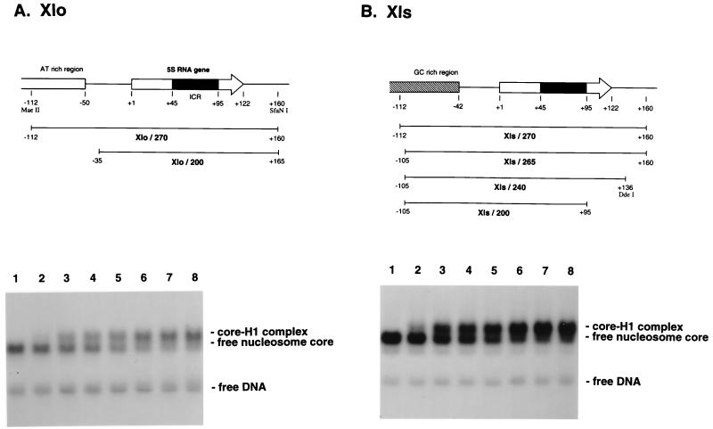
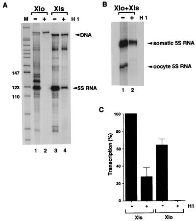
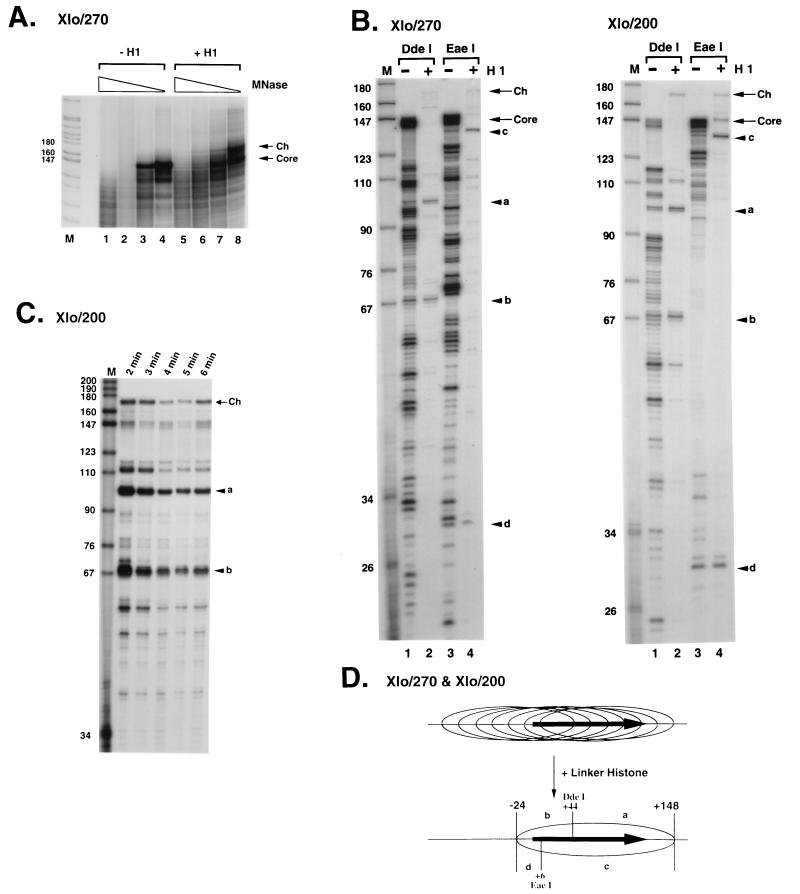
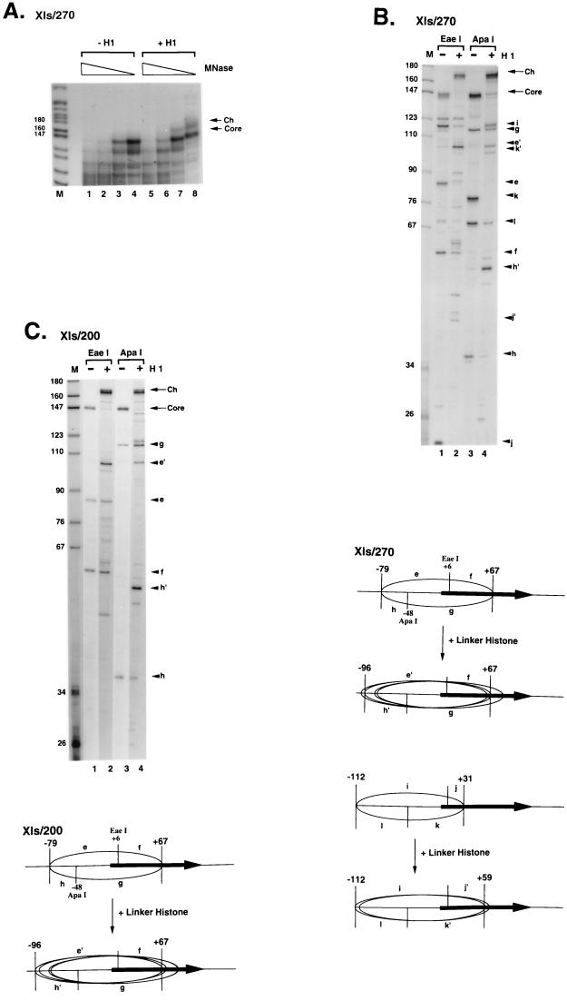
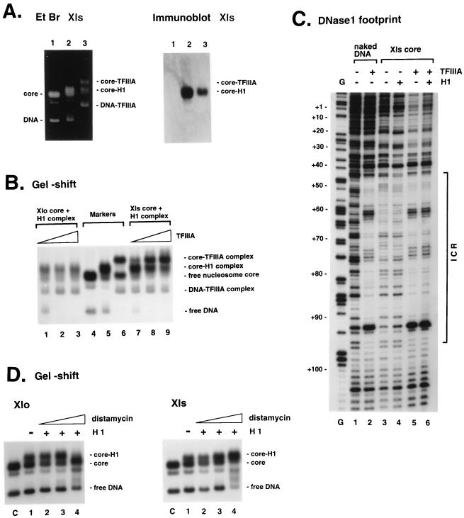
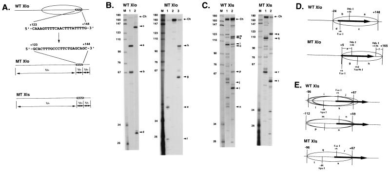
We explore the role of histone H1 as a DNA sequence-dependent architectural determinant of chromatin structure and of transcriptional activity in chromatin. The Xenopus laevis oocyte- and somatic-type 5S rRNA genes are differentially transcribed in embryonic chromosomes in vivo depending on the incorporation of somatic histone H1 into chromatin. We establish that this effect can be reconstructed at the level of a single nucleosome. H1 selectively represses oocyte-type 5S rRNA genes by directing the stable positioning of a nucleosome such that transcription factors cannot bind to the gene. This effect does not occur on the somatic-type genes. Histone H1 binds to the 5' end of the nucleosome core on the somatic 5S rRNA gene, leaving key regulatory elements in the promoter accessible, while histone H1 binds to the 3' end of the nucleosome core on the oocyte 5S rRNA genes, specifically blocking access to a key promoter element (the C box). TFIIIA can bind to the somatic 5S rRNA gene assembled into a nucleosome in the presence of H1. Because H1 binds with equivalent affinities to nucleosomes containing either gene, we establish that it is the sequence-selective assembly of a specific repressive chromatin structure on the oocyte 5S rRNA genes that accounts for differential transcriptional repression. Thus, general components of chromatin can determine the assembly of specific regulatory nucleoprotein complexes.


Figures

FIG. 1
FIG. 1
Upper panels, DNA structures of the X. laevis oocyte (A) and somatic 5S RNA genes (B) used. Arrows show the location and orientation of the 120-bp 5S RNA gene. Black boxes indicate the internal control region (ICR). Numbers represent the positions relative to the start site of transcription, +1. Lower panels, lack of significant differences in the affinities of linker histone H1 toward nucleosome cores of Xlo/200 (A) and Xls/200 (B). Reconstituted nucleosome cores were mixed with various amounts of H1 and analyzed by 0.7% agarose gel electrophoresis. Concentrations of H1 added: 0 M (lane 1), 3.5 nM (lane 2), 8.7 nM (lane 3), 17 nM (lane 4), 35 nM (lane 5), 67 nM (lane 6), 106 nM (lane 7), and 174 nM (lane 8). Small amounts of the naked DNA were included to show no affinity of H1 toward the naked DNA under the experimental conditions.
FIG. 2
FIG. 2
Specific repression of the oocyte 5S RNA gene transcription by linker histone H1. For conditions for binding of H1 and subsequent transcription by Xenopus GV extract, see Materials and Methods. (A) Transcription using Xlo/270 core alone (lane 1), Xlo/270 core-H1 complex (lane 2), Xls/265 core alone (lane 3), or Xls/265 core-H1 complex (lane 4) as the template for transcription. The transcripts were analyzed by PAGE (8% denaturing polyacrylamide gel). Sizes are indicated in nucleotides. (B) Transcription using mixtures of Xlo/270 and Xls/270 nucleosome cores in the absence (lane 1) or presence (lane 2) of H1. The oocyte and somatic 5S RNA products were analyzed by semidenaturing PAGE.
FIG. 3
FIG. 3
Nucleosome positioning on the X. laevis oocyte 5S rRNA gene. (A) Micrococcal nuclease digestion of reconstituted nucleosome cores of Xlo/270. Reconstituted nucleosome cores (50 ng of DNA) in the absence (lanes 1 to 4) or presence (lanes 5 to 8) of 1 ng of linker histone H1 were digested with 0.15, 0.075, 0.038, and 0.019 U of micrococcal nuclease (MNase; 5 min, room temperature). Products of digestion were labeled with [γ-32P]ATP and analyzed by PAGE (6% nondenaturing polyacrylamide gel). Lane M shows fragments of pBR322 digested by MspI. The positions of digestion products corresponding to the nucleosome core DNA fragments and chromatosome DNA fragment are indicated on the right as arrows labeled Core and Ch, respectively. Sizes are indicated in nucleotides. (B) Mapping of nucleosomes cores and chromatosomes by the combination of micrococcal nuclease and restriction endonuclease digestion. DNA from nucleosome cores and chromatosomes protected from micrococcal nuclease digestion (A) was recovered and digested with two kinds of restriction endonucleases to determine DNA regions contacting with histones at the nucleotide level. Although many digestion fragments were observed in the experiment with the Xlo/270 nucleosome core (lane 1 and 3), only one predominant set of digestion products was observed in each digestion using the chromatosome: fragments of 101 nt (a) and 68 nt (b) in DdeI digestion (lane 2) and fragments of 138 nt (c) and 30 nt (d) in EaeI digestion (lane 4). For the Xlo/200 nucleosome core, the mapping experiment yielded the same result as that in panel A. Lane M, MspI digestion products of pBR322 as size markers. Arrows labeled Core and Ch show DNA fragments recovered from nucleosome cores and chromatosomes, respectively. (C) Time independence of micrococcal nuclease digestion pattern of Xlo/200 cores. After micrococcal nuclease digestion, 170-bp DNA fragments corresponding to the chromatosomes were isolated and digested with DdeI to determine the positioning. Incubation times for micrococcal nuclease digestion are shown at the top. (D) Summary of mapping data shown in panel B. Positions of restriction fragments shown in panel B and restriction sites of DdeI and EaeI are indicated. Positions of nucleosome cores and the chromatosome composed of Xlo/270 and Xlo/200 are indicated by ellipsoids. The horizontal closed arrows are the 5S RNA genes.
FIG. 4
FIG. 4
Nucleosome positioning on the X. laevis somatic 5S rRNA gene. (A) Micrococcal nuclease digestion of reconstituted nucleosome cores of Xls/270. Reconstituted nucleosome cores (50 ng of DNA) in the absence (lanes 1 to 4) or presence (lanes 5 to 8) of 1 ng of linker histone H1 were digested with 0.15, 0.075, 0.038, and 0.019 U of micrococcal nuclease (MNase; 5 min, room temperature). Products of digestion were labeled with [γ-32P]ATP and analyzed by PAGE (6% nondenaturing polyacrylamide gel). Lane M shows fragments of pBR322 digested by MspI. The positions of digestion products corresponding to the nucleosome core DNA fragments and chromatosome DNA fragment are indicated on the right as arrows labeled Core and Ch, respectively. Sizes are indicated in nucleotides. (B and C) Upper panels, mapping of nucleosome cores and chromatosomes by the combination of micrococcal nuclease and restriction endonuclease digestion. DNA from nucleosome cores and chromatosomes protected from micrococcal nuclease digestion (A) was recovered and digested with two kinds of restriction endonucleases to determine DNA regions contacting histones at the nucleotide level. In the mapping of the Xls/270 nucleosome core, two sets of digestion products were observed in each digestion: fragments of 85 nt (e) and 57 nt (f) and fragments of 118 nt (i) and 21 nt (j) in EaeI digestion (lane 1); fragments of 114 nt (g) and 36 nt (h) and fragments of 79 nt (k) and 69 nt (l) in ApaI digestion (lane 3). In the same experiment using the chromatosome, two sets of major digestion products were observed: fragments e′ (104 nt) and f and fragments i and j′ (42 nt) in EaeI digestion (lane 2); fragments g and h′ (53 nt) and fragments k′ (101 nt) and l in ApaI digestion (lane 4). For Xls/200 nucleosome core, only one set of major digestion products was observed in each digestion. Nucleosome core, fragments e and f in EaeI digestion (lane 1) and fragments g and h in ApaI digestion (lane 3); chromatosome, fragments e′ and f in EaeI digestion (lane 2) and fragments g and h′ in ApaI digestion (lane 4); lane M, MspI digestion products of pBR322 as size markers. Arrows labeled Core and Ch show DNA fragments recovered from nucleosome cores and chromatosomes, respectively. Lower panels, summary of mapping data shown in the upper panels. Positions of restriction fragments shown in upper panels and restriction sites of EaeI and ApaI are indicated. In Xls/200, one nucleosome core positioning from −79 to +67 and multiple chromatosomes positioning from −96 to +67 are observed. Ellipses represent the positions of major nucleosome cores and chromatosomes. The horizontal closed arrows are the 5S RNA genes.
FIG. 5
FIG. 5
(A) Elimination of H1 bound to Xls core by TFIIIA. Nucleosome core-H1 complexes of Xls/200 were mixed with 200 ng of TFIIIA and analyzed by agarose gel electrophoresis (Materials and Methods) before ethidium bromide (Et Br) staining (left) and Western blotting with an anti-H1 antiserum (right). Lanes 1 and 2 show the nucleosome core and core-H1 complex used, respectively; lane 3 shows core complexes formed after incubation of TFIIIA with the core-H1 complex. (B) Specific binding of TFIIIA to the somatic 5S RNA gene incorporated into a nucleosome containing histone H1. Nucleosome core-H1 complexes of Xlo/200 (lanes 1 to 3) or Xls/200 (lanes 7 to 9) were mixed with various amounts of TFIIIA and analyzed by 0.7% agarose gel electrophoresis. After the reconstituted nucleosome core was incubated with linker histone H1 for 30 min at room temperature to form a complete 1:1 complex of the nucleosome core and H1 (see Materials and Methods), TFIIIA was added to the reaction mixture and the mixture was incubated for additional 30 min. Amounts of TFIIIA used were 1 ng (lanes 1 and 7), 10 ng (lanes 2 and 8), and 100 ng (lanes 3 and 9). Each reaction mixture also contained small amounts of the naked DNA as internal control for TFIIIA binding. Positions of the nucleosome core, nucleosome core-H1 complex, and nucleosome core-TFIIIA complex are shown as markers in lanes 4, 5, and 6, respectively. (C) DNase I digestion of the TFIIIA-nucleosome core complexes assembled on the somatic 5S RNA gene. Autoradiographs show digestion patterns of the coding strand: naked DNA (lane 1), DNA-TFIIIA complex (lane 2), nucleosome core (lane 3), core-H1 complex (lane 4), and core-TFIIIA complex (lane 5). Lane 6 shows the DNase I digestion pattern of the complex observed in panel B, lanes 7 to 9, corresponding to the core-TFIIIA complex. Conditions for binding of TFIIIA and H1 and subsequent DNase I digestion are described in Materials and Methods. Lane G shows positions of guanines in the sequence, cut by dimethylsulfate-piperidine. Numbers on the left correspond to positions in the sequence of the somatic 5S RNA gene. The location of the internal control region (ICR) is shown on the right. (D) Preferential inhibition of H1 binding to Xlo core by distamycin. After incubation of Xlo/200 (left) or Xls/200 (right) with distamycin (lane 1, 0 M; lane 2, 8.6 × 10⁻⁵ M; lane 3, 1.7 × 10⁻⁴ M; lane 4, 3.4 × 10⁻⁴ M), binding abilities of H1 to the cores were analyzed in a 0.7% nondenaturing agarose gel. Conditions for binding of distamycin and H1 are described in Materials and Methods.
FIG. 6
FIG. 6
Mapping of nucleosome positioning on wild-type and mutant X. laevis oocyte- and somatic-type 5S rRNA genes. (A) DNA sequence 3′ to the wild-type oocyte-type 5S rRNA (WT Xlo) gene from +123 to +144 is shown by a hatched box relative to the wild-type chromatosome position (ellipsoid). This is replaced by the sequence 3′ to the somatic-type 5S rRNA (Xls) gene from +123 to +144 to create the MT Xlo DNA sequence. Replacement of the sequence from +123 to +144 3′ to the wild-type somatic gene (WT Xls) with the oocyte sequence generates the MT Xls DNA sequence. (B and C) Mapping of chromatosome boundaries on WT Xlo, MT Xlo, WT Xls, and MT Xls sequences by using DNA fragments 270 bp in length. Procedures for WT Xlo and WT Xls were as for Fig. 3 and 4. The digestion fragments in the MT Xlo chromatosome are completely different from those observed in the WT Xlo chromosome: fragments of 41 nt (e) and 25 nt (t) in DdeI digestion (lane 1); fragments of 60 nt (g) and 98 nt (h) in Sau96I digestion (lane 3). In the mapping, Sau96I (lane 3) was additionally used to determine the chromatosome position because of no digestion by EaeI (lane 2). In the MT Xls chromatosome, the same fragments as those derived from one chromatosome position from −96 to +67 in WT Xls were observed in digestion by EaeI (lane 1) and ApaI (lane 2). (D and E) Chromatosome positions. DNA fragments are indicated as resolved in panels B and C.

Similar articles

Cited by

References

    1. Allan J, Hartman P G, Crane-Robinson C, Aviles F X. The structure of histone H1 and its location in chromatin. Nature. 1980;288:675–679. - PubMed
    1. Andrews M T, Brown D D. Transient activation of oocyte 5S RNA genes in Xenopus embryos by raising the level of the trans-acting factor TFIIIA. Cell. 1987;51:445–453. - PubMed
    1. Arents G, Burlingame R W, Wang B W, Love W E, Moudrianakis E N. The nucleosomal core histone octamer at 3.1Å resolution: a tripartite protein assembly and a left-handed superhelix. Proc Natl Acad Sci USA. 1991;88:10148–10152. - PMC - PubMed
    1. Bazett-Jones D P, LeBlanc B, Herfort M, Moss T. Short range DNA looping by the Xenopus HMG-box transcription factor, xUBF. Science. 1994;264:1134–1136. - PubMed
    1. Becker P B. The establishment of active promoters in chromatin. Bioessays. 1994;16:541–547. - PubMed

LinkOut - more resources