Review
. 1998 Jun;62(2):434-64.
doi: 10.1128/MMBR.62.2.434-464.1998.

Replication and control of circular bacterial plasmids

Affiliations
Review

Replication and control of circular bacterial plasmids

G del Solar et al. Microbiol Mol Biol Rev. 1998 Jun.

Abstract

An essential feature of bacterial plasmids is their ability to replicate as autonomous genetic elements in a controlled way within the host. Therefore, they can be used to explore the mechanisms involved in DNA replication and to analyze the different strategies that couple DNA replication to other critical events in the cell cycle. In this review, we focus on replication and its control in circular plasmids. Plasmid replication can be conveniently divided into three stages: initiation, elongation, and termination. The inability of DNA polymerases to initiate de novo replication makes necessary the independent generation of a primer. This is solved, in circular plasmids, by two main strategies: (i) opening of the strands followed by RNA priming (theta and strand displacement replication) or (ii) cleavage of one of the DNA strands to generate a 3'-OH end (rolling-circle replication). Initiation is catalyzed most frequently by one or a few plasmid-encoded initiation proteins that recognize plasmid-specific DNA sequences and determine the point from which replication starts (the origin of replication). In some cases, these proteins also participate directly in the generation of the primer. These initiators can also play the role of pilot proteins that guide the assembly of the host replisome at the plasmid origin. Elongation of plasmid replication is carried out basically by DNA polymerase III holoenzyme (and, in some cases, by DNA polymerase I at an early stage), with the participation of other host proteins that form the replisome. Termination of replication has specific requirements and implications for reinitiation, studies of which have started. The initiation stage plays an additional role: it is the stage at which mechanisms controlling replication operate. The objective of this control is to maintain a fixed concentration of plasmid molecules in a growing bacterial population (duplication of the plasmid pool paced with duplication of the bacterial population). The molecules involved directly in this control can be (i) RNA (antisense RNA), (ii) DNA sequences (iterons), or (iii) antisense RNA and proteins acting in concert. The control elements maintain an average frequency of one plasmid replication per plasmid copy per cell cycle and can "sense" and correct deviations from this average. Most of the current knowledge on plasmid replication and its control is based on the results of analyses performed with pure cultures under steady-state growth conditions. This knowledge sets important parameters needed to understand the maintenance of these genetic elements in mixed populations and under environmental conditions.


Figures
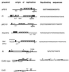
FIG. 1
FIG. 1
Origins of replication and related regions of some representative theta-replicating plasmids from gram-negative bacteria. Plasmid names (left), origins of replication (center), and Rep initiator-binding sites (right) are indicated. The symbols used are as follows: solid boxed arrows correspond to repeated Rep-binding sequences (iterons); open arrows above maps indicate inverted repeats that have partial homology to the iterons; solid arrowheads indicate repeats found in AT-rich regions (A+T); for R1, arrows indicate the imperfect palindromes initially recognized by the RepA initiator protein. Promoters are indicated as open arrowheads below the maps. Other sites of interaction are as follows: IHF-binding sites (open rectangles), dnaA boxes (solid rectangles); FIS-binding sites (hexagons), dam methylation sites (solid circles), and primase assembly sites (pas). Other sites are indicated in the figure. Maps are based on the following references: pPS10 (104, 215); pSC101 (132, 284); P1 (6); RK2 (278); R6K (277); R1 (101); ColE1 (280, 301); ColE2-type plasmids (124).
FIG. 2
FIG. 2
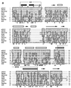
Sequence alignment and phylogenetic tree of Rep initiator proteins from theta-replicating plasmids. (a) Sequence alignment of the Rep initiator proteins of nine related plasmids of the iteron-containing class. Sequences were aligned with the program CLUSTAL W (version 1.5) by using, for pairwise alignment, gap opening and extension penalties of 10.0 and 0.1, respectively, and the protein weight matrix BLOSUM30. For multiple alignment, the delay for divergent sequences was set to 40% (294). The degree of sequence identity to the pPS10 initiator sequence for conserved residues is shown: ∗, identical residues in eight or nine of the total sequences; •, identical residues in five to seven sequences. Over the sequences is shown a secondary-structure prediction performed by the neural network algorithm PHD (260) on the output from CLUSTAL W: predicted α-helical regions (boxes) and β-strands (arrows). The two characteristic motifs found in the Rep initiators, LZ (hydrophobic heptad residues pointed to by open arrowheads) and HTH, are indicated. The EMBL database accession numbers are as follows: pPS10, X58896; pECB2, Y10829; pRO1614, L30112; pCM1, X86092; pFA3, M31727; pSC101, K00828; pCU1, Z11775; RepFIA, Y00547; R6K, M65025. (b) Phylogenetic tree for theta-type replicons from gram-negative bacteria, based on sequence alignments of their Rep proteins (such as the one shown in panel a for the pPS10 family, encircled in the tree with a dashed line). The sequences for 35 initiators were retrieved from databases, and a preliminary alignment was performed with CLUSTAL W (data not shown). Sequences that were virtually identical (pairwise scores, ≥90) were discarded, and a refined alignment was used as input data for the DISTANCES program of the Genetics Computer Group software package (95). Distance matrixes were corrected for multiple substitutions by the method of Kimura. The phylogenetic tree was built up according to the UPGMA method with the program GROWTREE (95). For further discussion, see the text.
FIG. 2
FIG. 2
Sequence alignment and phylogenetic tree of Rep initiator proteins from theta-replicating plasmids. (a) Sequence alignment of the Rep initiator proteins of nine related plasmids of the iteron-containing class. Sequences were aligned with the program CLUSTAL W (version 1.5) by using, for pairwise alignment, gap opening and extension penalties of 10.0 and 0.1, respectively, and the protein weight matrix BLOSUM30. For multiple alignment, the delay for divergent sequences was set to 40% (294). The degree of sequence identity to the pPS10 initiator sequence for conserved residues is shown: ∗, identical residues in eight or nine of the total sequences; •, identical residues in five to seven sequences. Over the sequences is shown a secondary-structure prediction performed by the neural network algorithm PHD (260) on the output from CLUSTAL W: predicted α-helical regions (boxes) and β-strands (arrows). The two characteristic motifs found in the Rep initiators, LZ (hydrophobic heptad residues pointed to by open arrowheads) and HTH, are indicated. The EMBL database accession numbers are as follows: pPS10, X58896; pECB2, Y10829; pRO1614, L30112; pCM1, X86092; pFA3, M31727; pSC101, K00828; pCU1, Z11775; RepFIA, Y00547; R6K, M65025. (b) Phylogenetic tree for theta-type replicons from gram-negative bacteria, based on sequence alignments of their Rep proteins (such as the one shown in panel a for the pPS10 family, encircled in the tree with a dashed line). The sequences for 35 initiators were retrieved from databases, and a preliminary alignment was performed with CLUSTAL W (data not shown). Sequences that were virtually identical (pairwise scores, ≥90) were discarded, and a refined alignment was used as input data for the DISTANCES program of the Genetics Computer Group software package (95). Distance matrixes were corrected for multiple substitutions by the method of Kimura. The phylogenetic tree was built up according to the UPGMA method with the program GROWTREE (95). For further discussion, see the text.
FIG. 3
FIG. 3
The theta-type replicon of the Pseudomonas plasmid pPS10. The iteron-containing origin (oriV) and motifs found in the replication initiator protein (RepA) are depicted. The minimal origin (oriV) of pPS10 plasmid is a good example for a “canonical” iteron-containing origin. It is composed of four contiguous and identical 22-bp iterons arranged as direct repeats (half arrows), flanked by a 9-bp dnaA box (hatched) and AT- and GC-rich sequences (215). The pPS10 replicon also contains the repA gene, encoding the RepA initiator protein (shadowed ovals). RepA is under a monomer-to-dimer equilibrium, which has consequences for protein activity: RepA dimers bind to an inverted repeat (with a sequence partially homologous to that of the iterons) that overlaps with the repA promoter (P), acting as self-repressor of repA expression, whereas RepA monomers bind to the iterons to form the initiation complex (94). Protein motifs found in RepA are depicted under the protein gene. The LZ motif, responsible for protein dimerization, is represented as a helical wheel projection, in which the hydrophobic spine of Leu residues and the chemical nature of the other displayed residues is indicated (103). For the HTH motif, involved in binding to DNA, the two proposed α-helices are underlined and a stretch of basic residues at the C end of the DNA recognition helix is indicated (+), whereas an arrow points to the Gly residue thought to start the turn (93).
FIG. 4
FIG. 4
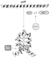
RepA-oriR complexes in initiation of R1 plasmid replication. A 100-bp region in the oriR replication origin is continuously bound by the plasmid RepA protein to form the initiation complex. There are no iterons in oriR, but two partially palindromic 10-bp sequences (sites 1 and 2) are found at the ends of that 100-bp region. They are flanked, respectively, by a consensus dnaA box and by three AT-rich sequences. These three sequences are believed to be melted to allow the DnaBC complex access to the open origin. A RepA initiator (hatched oval) dimer binds with high affinity to site 1, and then, in a second binding event, a different RepA dimer would bind with lower affinity to the distal site 2 sequence. The DNA of the oriR region could be bent to facilitate the topological proximity of sites 1 and 2, which are disposed on the same face of DNA double helix. Binding of RepA dimers to sites 1 and 2 would generate a small DNA loop, held together by protein-protein interactions. The DNA loop would be filled afterwards with more RepA molecules that are brought to the complex mainly by protein-protein interactions. This model is based on experimental gel mobility shift assays and footprinting data with both wild-type and mutant RepA proteins and oriR sequences (101). Arrowheads indicate DNase I-hypersensitive sites (the size is proportional to the intensity of cleavage), whereas arrows point to strong cleavage sites for hydroxyl radicals. The interaction of DnaA host initiator with its DNA-binding site is dependent on the previous formation of the RepA-oriR nucleoprotein complex (233). A requirement for a DnaK chaperone has been described for R1 DNA replication (105). A hypothetical role for DnaK in modulating the aggregation and activation state of RepA dimers, inspired by the findings for P1 plasmid RepA initiator (316), is also shown.
FIG. 5
FIG. 5
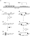
Replication of plasmid RSF1010 by the strand displacement mechanism. (a) Origin of replication and related regions. The interaction sites of RepB (inverted repeat [convergent arrows]) and of RepC (iterons [boxed arrows]) are indicated. GC- and AT-rich regions are also depicted. (b) Model for initiation of replication by the strand displacement mechanism in plasmid RSF1010 (266). Replication occurs with opposite polarities from two origins (ssiA and ssiB), which are independently used. Interactions between the plasmid-encoded proteins RepC and RepA are indicated. Priming is catalyzed by RepB′ (not shown). Thin lines indicate newly synthesized DNA, with the direction of synthesis indicated by arrowheads. See the text for details.
FIG. 6
FIG. 6
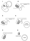
Model for RC replication. The plasmid-encoded Rep protein recognizes the dso on supercoiled DNA and introduces a site-specific nick generating a free 3′-OH end. This end is elongated by host proteins as the parental strand is being displaced. When the replication fork reaches the reconstituted dso, Rep protein catalyzes a strand transfer reaction, releasing an ssDNA intermediate and a dsDNA molecule with a parental and a newly synthesized (dotted) strand. Lagging-strand synthesis on the ssDNA molecule is initiated at the sso signal by the host RNA polymerase. This enzyme would synthesize a short primer RNA, and lagging-strand synthesis is performed by host DNA polymerases. The end products are supercoiled plasmid DNA molecules.
FIG. 7
FIG. 7
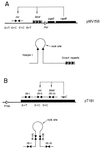
Origins of replication and related regions of plasmids replicating by the RC mode, as exemplified by plasmids pMV158 (A) and pT181 (B). The bind and nic regions of the dso are indicated. Other symbols are as in Fig. 1.
FIG. 8
FIG. 8
Model for termination of RC replication based on results from Novick’s laboratory (, , –257). Nucleophilic attacks exerted by the OH groups of the Tyr residue of Rep (Y) or by 3′-OH groups of the DNA (OH) are indicated by arrows. Solid lines, parental DNA; broken lines, newly synthesized DNA. The thick solid line indicates the nucleotides that are newly synthesized past the reconstituted dso and that will remain covalently bound to the Rep protein to generate a Rep/Rep* inactive dimer. The two subunits of the RepC dimer are differently depicted.
FIG. 9
FIG. 9
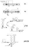
Control of plasmid DNA replication. Examples of control by Rep DNA-binding sites (plasmid P1), by antisense RNAs (plasmids R1 and pT181), and by a dual system (plasmid pMV158) are depicted. Transcripts are shown as continuous lines, with arrowheads indicating the direction of synthesis. mRNAs are shown with thicker lines than antisense RNAs; countertranscribed RNAs (thin lines with small arrowheads) are indicated. Other symbols as follows: solid ellipses, origins; rectangles, promoters; a.r.b.s., atypical ribosome-binding site; parallel vertical lines, mRNA-ctRNA interactions; plus, positive interactions; minus, and inhibitory RNA-RNA or protein-DNA interactions.

Similar articles

Cited by

References

    1. Abeles A L, Austin S J. P1 plasmid replication requires methylated DNA. EMBO J. 1987;10:3185–3189. - PMC - PubMed
    1. Abeles A L, Austin S J. P1 plasmid replication requires Escherichia coli Dam-methylated DNA. Gene. 1988;74:185–186. - PubMed
    1. Abeles A L, Brendler T, Austin S J. Evidence of two levels of control of P1 oriR and host oriC replication origins by DNA adenine methylation. J Bacteriol. 1993;175:7801–7807. - PMC - PubMed
    1. Abeles A L, Reaves L D, Austin S J. A single DnaA box is sufficient for initiation from the P1 plasmid origin. J Bacteriol. 1990;172:4386–4391. - PMC - PubMed
    1. Abeles A L, Reaves L D, Youngre-Grimes B, Austin S J. Control of P1 plasmid replication by iterons. Mol Microbiol. 1995;18:903–912. - PubMed

Publication types

LinkOut - more resources