. 1998 May 1;12(9):1356-70.
doi: 10.1101/gad.12.9.1356.

Pom1p, a fission yeast protein kinase that provides positional information for both polarized growth and cytokinesis

Affiliations

Pom1p, a fission yeast protein kinase that provides positional information for both polarized growth and cytokinesis

J Bähler et al. Genes Dev. .

Abstract

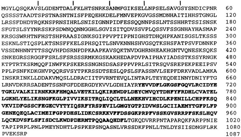
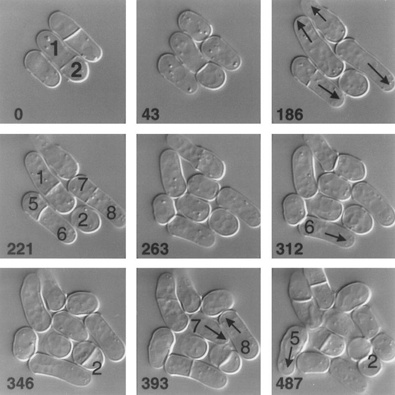
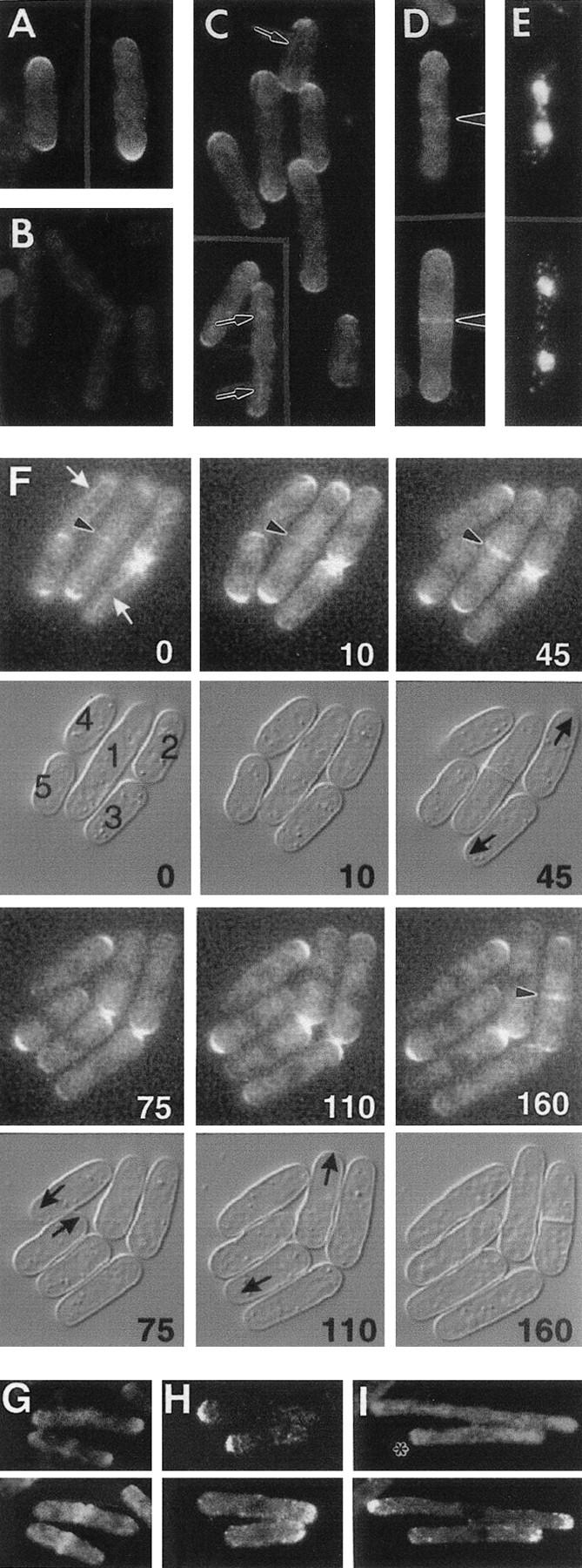
Schizosaccharomyces pombe cells have a well-defined pattern of polarized growth at the cell ends during interphase and divide symmetrically into two equal-sized daughter cells. We identified a gene, pom1, that provides positional information for both growth and division in S. pombe. pom1 mutants form functioning growth zones and division septa but show several abnormalities: (1) After division, cells initiate growth with equal frequencies from either the old or the new end; (2) most cells never switch to bipolar growth but instead grow exclusively at the randomly chosen end; (3) some cells mislocalize their growth axis altogether, leading to the formation of angled and branched cells; and (4) many cells misplace and/or misorient their septa, leading to asymmetric cell division. pom1 encodes a putative protein kinase that is concentrated at the new cell end during interphase, at both cell ends during mitosis, and at the septation site after mitosis. Small amounts of Pom1p are also found at the old cell end during interphase and associated with the actin ring during mitosis. Pom1p localization to the cell ends is independent of actin but requires microtubules and Tea1p. pom1 mutations are synthetically lethal with several other mutations that affect cytokinesis and/or the actin or microtubule cytoskeleton. Thus, Pom1p may position the growth and cytokinesis machineries by interaction with both the actin and microtubule cytoskeletons.


Figures

Figure 1
Figure 1
(A) Abnormalities in cell shape and cell division in pom1-1 mutant cells. Strain JB100 cells growing exponentially at 30°C were stained with Calcofluor and bisBenzimide to visualize septa and DNA. (Asterisks) Cells with branched or angled growth; (arrows) cells with septa in abnormal positions and/or orientations; (arrowheads) cells with longitudinal or irregular septa. (B–D) Abnormal septum placement in pom1-Δ1 mutant cells. (B) Cells of wild-type strain 972 (left panels) and pom1-Δ1 strain JB110 (right panels) growing exponentially at 36°C were stained with Calcofluor and bisBenzimide (upper panels) and with rhodamine–phalloidin to visualize F-actin (lower panels). (a, b, and c) Cells with different degrees of septum misplacement, as used for the quantitative analysis in C. (C) Septation patterns were scored in >300 cells from each culture shown in B. (Left) Septum position was scored relative to the long axis of the cell as divided into five equal compartments (see examples in B). (Right) Septum angle was scored relative to the long axis of the cell. Septa with angles of 80° to 90° were designated as orthogonal and septa with smaller angles as tilted. Septa that looked very aberrant (e.g., consisting of several sheets) or were placed longitudinally in the cell (see examples in A) were scored as irregular. (D) JB110 cells growing exponentially at 30°C were treated with FITC-conjugated lectin to stain their cell walls, then washed and grown for 45 min before staining with Calcofluor (left). The cell ends that had grown during the 45 min after labeling with lectin can be identified by their unlabeled surface (right) and are marked with asterisks (left).
Figure 2
Figure 2
Predicted sequence of Pom1p (accession no. Z50142). The kinase domain (amino acids 699–995) is shown in boldface type.
Figure 3
Figure 3
Time-lapse analysis of the growth of a pom1-1 strain. JB100 cells growing at room temperature in YE medium were observed by DIC microscopy (see Materials and Methods). The times in minutes since the beginning of observation are indicated. Individual cells are numbered for reference in the text. (Arrows) Growing cell ends.
Figure 4
Figure 4
Abnormal polarization of growth in pom1-Δ1 strains. (A) Strains 972 (wild type) and JB110 (pom1-Δ1) were grown at 36°C to 5 × 106 cells/ml, then stained with rhodamine–phalloidin to visualize F-actin organization. Strains JB20 (cdc25-22) and JB120 (pom1-Δ1 cdc25-22) were grown to the same titer at 25°C, then shifted to 36°C for 3 hr before staining. (B) Cells from the populations shown in A were scored for unipolar or bipolar actin localization, as were cells of strains SP622 (cdc10–V50) and JB121 (pom1-Δ1 cdc10–V50) (grown as described for the cdc25-22 strains in A). For strains 972 and JB110, the percentages of cells with an actin ring were also scored. In the other strains, these percentages were <5%. At least 250 cells of each strain were evaluated. (C) Strains SP622, JB121, JB20, and JB120 were grown at 25°C to 5 × 106 cells/ml, shifted to 36°C for 4 hr, and stained with Calcofluor. (D) Cells (⩾250) from each population shown in C were scored for the presence or absence of new-end growth.
Figure 5
Figure 5
Mislocalization of growth poles and of microtubule-organizing centers in pom1-Δ1 strains. (A) Cells of strains JB110 (pom1-Δ1), JB22 (cdc11-136), and JB122 (pom1-Δ1 cdc11-136) were grown at 25°C to 5 × 106 cells/ml, shifted to 36°C for 4.5 hr, and stained with Calcofluor. (B) 600 cells from each population shown in A were scored for the extent of cell branching as a function of time after the shift to 36°C. The “branched” cell population in the pom1-Δ1 single mutant consisted mainly (∼90%) of angled cells (see A, cell 1); the rest were T-shaped cells, the majority of which (∼90%) had a division scar at each end (see A, cell 2). The cdc11-136 single mutant showed <1% cell branching at all times. (C) Cells of strains 972 and JB110 were grown at 30°C to 1 × 107 cells/ml, then fixed and stained with anti-tubulin antibodies and with bisBenzimide to visualize DNA. Some cells are numbered for reference in the text. (Arrowhead and arrows) Post-anaphase microtubule-organizing centers.
Figure 6
Figure 6
Localization of Pom1p. (A–E) Immunolocalization. All strains were grown at 30°C. (A,B) Wild-type strain 972 (A) and pom1-Δ1 strain JB110 (B) were stained with antibodies to Pom1p. (C–E) The pom1–3HA strain JB111 was stained with HA-specific antibodies (C) or double-stained with HA-specific antibodies (D) and bisBenzimide to visualize the DNA (E). Arrows and arrowheads indicate structures discussed in the text. (F) Time-lapse study of Pom1p–GFP localization through the cell-cycle. Strain JB115 was grown at room temperature in EMM medium and observed by fluorescence and DIC microscopy (see Materials and Methods). The times in minutes since the beginning of observation are indicated. Individual cells are numbered for reference in the text. (White arrows) Filamentous structures; (arrowheads) central Pom1p; (black arrows) growing ends. (G–I) Comparison of Pom1p and actin localization in wild-type and mutant strains. Cells expressing Pom1p–3HA were double-stained with antibodies to HA (top) and actin (bottom). (G) Strain JB111 (wild-type except for pom1–3HA) was grown at 30°C to exponential phase before staining. (H,I) cdc10-V50 strain JB113 (H) and cdc25-22 strain JB114 (I) were grown at 25°C to 5 × 106 cells/ml, then shifted to 36°C for 3 hr before staining.
Figure 7
Figure 7
Effects of cytoskeletal inhibitors and mutations on Pom1p localization. All strains expressed Pom1p-3HA and were stained with antibodies to HA. Arrows and arrowheads indicate structures discussed in the text. (A) Strain JB111 (wild type) was grown at 30°C to 2 × 106 cells/ml, and LAT-A was added to 100 μm. After 3 hr, cells were stained separately for Pom1p and for F-actin (using rhodamine–phalloidin). (B) Strain JB111 was grown at 30°C to 5 × 106 cells/ml and TBZ was added to 100 μg/ml (right panels). A control culture (left panels) received no TBZ. After 1 hr, aliquots of cells from each culture were double-stained for Pom1p (top panels) and DNA (middle panels) or stained separately for microtubules (bottom panels). (C) Strain JB130 (nda3-KM311; left panel) was grown at 32°C to 5 × 106 cells/ml and shifted to 20°C for 1.5 hr before staining for Pom1p. Strain JB132 (ban5-3; right panel) was grown at 25°C to 5 × 106 cells/ml and shifted to 36°C for 3 hr before staining for Pom1p. (D) Strain JB131 (tea1-1) was grown at 25°C to 5 × 106 cells/ml, shifted to 36°C for 3.5 hr, and stained for Pom1p. (E) Strains 972 (wild type) and JB110 (pom1-Δ1) growing exponentially at 30°C were fixed and stained with antibodies to Tea1p.

Similar articles

Cited by

References

    1. Adachi Y, Toda T, Niwa O, Yanagida M. Differential expressions of essential and nonessential α-tubulin genes in Schizosaccharomyces pombe. Mol Cell Biol. 1986;6:2168–2178. - PMC - PubMed
    1. Alfa C, Fantes P, Hyams J, McLeod M, Warbrick E. Experiments with fission yeast. Cold Spring Harbor, NY: Cold Spring Harbor Laboratory Press; 1993.
    1. Ayscough KR, Stryker J, Pokala N, Sanders M, Crews P, Drubin DG. High rates of actin filament turnover in budding yeast and roles for actin in establishment and maintenance of cell polarity revealed using the actin inhibitor latrunculin-A. J Cell Biol. 1997;137:399–416. - PMC - PubMed
    1. Bähler, J., J.-Q. Wu, M.S. Longtine, N.G. Shah, A. McKenzie III, A.B. Steever, A. Wach, P. Philippsen, and J.R. Pringle. 1998. Heterologous modules for efficient and versatile PCR-based gene targeting in Schizosaccharomyces pombe. Yeast (in press). - PubMed
    1. Balasubramanian MK, Helfman DM, Hemmingsen SM. A new tropomyosin essential for cytokinesis in the fission yeast S. pombe. Nature. 1992;360:84–87. - PubMed

Publication types

MeSH terms