. 1998 May;18(5):2986-96.
doi: 10.1128/MCB.18.5.2986.

Virus-dependent phosphorylation of the IRF-3 transcription factor regulates nuclear translocation, transactivation potential, and proteasome-mediated degradation

Affiliations

Virus-dependent phosphorylation of the IRF-3 transcription factor regulates nuclear translocation, transactivation potential, and proteasome-mediated degradation

R Lin et al. Mol Cell Biol. 1998 May.

Abstract

The interferon regulatory factors (IRF) consist of a growing family of related transcription proteins first identified as regulators of the alpha beta interferon (IFN-alpha/beta) gene promoters, as well as the interferon-stimulated response element (ISRE) of some IFN-stimulated genes. IRF-3 was originally identified as a member of the IRF family based on homology with other IRF family members and on binding to the ISRE of the ISG15 promoter. IRF-3 is expressed constitutively in a variety of tissues, and the relative levels of IRF-3 mRNA do not change in virus-infected or IFN-treated cells. In the present study, we demonstrate that following Sendai virus infection, IRF-3 is posttranslationally modified by protein phosphorylation at multiple serine and threonine residues, which are located in the carboxy terminus of IRF-3. A combination of IRF-3 deletion and point mutations localized the inducible phosphorylation sites to the region -ISNSHPLSLTSDQ- between amino acids 395 and 407; point mutation of residues Ser-396 and Ser-398 eliminated virus-induced phosphorylation of IRF-3 protein, although residues Ser-402, Thr-404, and Ser-405 were also targets. Phosphorylation results in the cytoplasm-to-nucleus translocation of IRF-3, DNA binding, and increased transcriptional activation. Substitution of the Ser-Thr sites with the phosphomimetic Asp generated a constitutively active form of IRF-3 that functioned as a very strong activator of promoters containing PRDI-PRDIII or ISRE regulatory elements. Phosphorylation also appears to represent a signal for virus-mediated degradation, since the virus-induced turnover of IRF-3 was prevented by mutation of the IRF-3 Ser-Thr cluster or by proteasome inhibitors. Interestingly, virus infection resulted in the association of IRF-3 with the CREB binding protein (CBP) coactivator, as detected by coimmunoprecipitation with anti-CBP antibody, an interaction mediated by the C-terminal domains of both proteins. Mutation of residues Ser-396 and Ser-398 in IRF-3 abrogated its binding to CBP. These results are discussed in terms of a model in which virus-inducible, C-terminal phosphorylation of IRF-3 alters protein conformation to permit nuclear translocation, association with transcriptional partners, and primary activation of IFN- and IFN-responsive genes.


Figures

FIG. 1
FIG. 1
Sendai virus infection induces IRF-3 degradation. IRF-3 expression plasmid CMVBL-IRF3 (lanes 1 and 2) or CMVBL vector alone (lanes 3 and 4), both at 5 μg, was transiently transfected into 293 cells by the calcium phosphate method. At 24 h posttransfection, the cells were infected with Sendai virus for 16 h (lanes 2 and 4) or were left uninfected (lanes 1 and 3). Whole-cell extracts (20 μg) were prepared and analyzed by immunoblotting with anti-IRF-3 antibody.
FIG. 2
FIG. 2
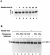
Sendai virus-induced phosphorylation and degradation of IRF-3 protein. (A) rtTA-IRF-3 cells, which were selected as described in Materials and Methods, were induced to express IRF-3 by doxycycline treatment for 24 h. At 24 h after doxycycline addition, the cells were infected with Sendai virus for 4, 8, 12, 16, 20, or 24 h (lanes 2 to 7) or were left uninfected (lane 1). IRF-3 protein was detected in whole-cell extracts (10 μg) by immunoblotting. Two forms of IRF-3, which were designated form I and form II, were detected. (B) At 24 h post-Dox induction, rtTA-IRF-3 cells were infected with Sendai virus for 16 h (lanes 4 to 8) or were left uninfected (lanes 1 to 3). Whole-cell extracts from untreated cells (20 μg) or Sendai virus-infected cells (60 μg) were incubated with 0.3 units of PPA (lanes 2, 3, 7, and 8) or 5 U of CIP (lanes 4 and 5) in the absence (lanes 1, 2, 4, 6, and 7) or presence (lanes 3, 5, and 8) of phosphatase inhibitors. Phosphorylated IRF-3 protein appears as a distinct band in immunoblots, migrating more slowly than IRF-3 forms I and II.
FIG. 3
FIG. 3
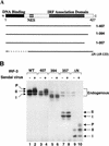
Analysis of IRF-3 deletion mutants in Sendai virus-induced phosphorylation. (A) Schematic representation of four IRF-3 deletions. Thick solid lines and thin dashed lines indicate included and excluded sequences, respectively. The N-terminal IRF homology domain, the NES, and the C-terminal IRF association domain are indicated. (B) Expression plasmids (5 μg each) encoding wild type and deletion mutants of IRF-3 (as indicated above the lanes) were transiently transfected into 293 cells; at 24 h posttransfection, cells were infected with Sendai virus for 16 h (lanes 2, 4, 6, 8, and 10) or were left uninfected (lanes 1, 3, 5, 7, and 9). Whole-cell extracts (20 μg) were prepared from infected and control cells and were analyzed by immunoblotting for IRF-3 forms I and II and for the presence of phosphorylated IRF-3 (P-IRF-3) with anti-IRF-3 antibody.
FIG. 4
FIG. 4
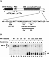
Analysis of IRF-3 point mutations in Sendai virus-induced phosphorylation. (A) Schematic representation of IRF-3 point mutations. The N-terminal IRF homology domain, the NES element, and the C-terminal IRF association domain are indicated. aa 382 to 414 and 141 to 147 are shown. The amino acids targeted for alanine or aspartic acid substitution are shown in larger letters. The point mutations are indicated below the sequence: 2A, S396A and S398A; 3A, S402A, T404A, and S405A; 5A, S396A, S398A, S402A, T404A, and S405A); 5D, S396D, S398D, S402D, T404D, and S405D; J2A, S385A and S386A; NES, S145A and S146A). (B) Expression plasmids (5 μg each) encoding wild type (WT) and point mutants of IRF-3 (as indicated above the lanes) were transiently transfected into 293 cells; at 24 h posttransfection, the cells were infected with Sendai virus for 16 h (lanes 2, 4, 6, 8, 10, 12, 14, 16, and 18) or were left uninfected (lanes 1, 3, 5, 7, 9, 11, 13, 15, and 17). Whole-cell extracts (20 μg) were prepared from infected and control cells and analyzed by immunoblotting for IRF-3 forms I and II and for the presence of phosphorylated IRF-3 (P-IRF-3) with anti-IRF-3 antibody.
FIG. 5
FIG. 5
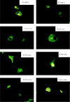
Virus-dependent cytoplasm-to-nucleus translocation of IRF-3. The subcellular localization of the GFP–IRF-3 (A and B), GFP–IRF-3(5A) (C and D), GFP–IRF-3(5D) (E and F), and GFP–IRF-3(NES) (G and H) in uninfected (A, C, E, and G) and Sendai virus-infected COS-7 cells at 8 h after infection was analyzed. GFP fluorescence in living cells was analyzed with a Leica fluorescence microscope by using a ×40 objective.
FIG. 6
FIG. 6
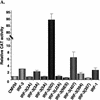
Transactivation of PRDI-PRDIII- and ISRE-containing promoters by IRF-3. 293 cells were transfected with IFN-β–CAT (A and B) or ISG15-CAT (C) reporter plasmids and the various expression plasmids as indicated below the bar graph. CAT activity was analyzed at 48 h posttransfection with 100 μg (IFN-β–CAT) or 10 μg (ISG15-CAT) of total protein extract for 1 to 2 h at 37°C. Relative CAT activity was measured as fold activation (relative to the basal level of reporter gene in the presence of CMVBI vector alone after normalization with cotransfected β-Gal activity); values are the averages of three experiments, with variability shown by the error bars. WT, wild type.
FIG. 6
FIG. 6
Transactivation of PRDI-PRDIII- and ISRE-containing promoters by IRF-3. 293 cells were transfected with IFN-β–CAT (A and B) or ISG15-CAT (C) reporter plasmids and the various expression plasmids as indicated below the bar graph. CAT activity was analyzed at 48 h posttransfection with 100 μg (IFN-β–CAT) or 10 μg (ISG15-CAT) of total protein extract for 1 to 2 h at 37°C. Relative CAT activity was measured as fold activation (relative to the basal level of reporter gene in the presence of CMVBI vector alone after normalization with cotransfected β-Gal activity); values are the averages of three experiments, with variability shown by the error bars. WT, wild type.
FIG. 7
FIG. 7
Stabilization of IRF-3 by proteasome inhibitors. IRF-3 ΔN (Δ9-133) (B) or IRF-3 ΔN2A (C) expression plasmids were transiently transfected into 293 cells; at 24 h posttransfection, cells were infected with Sendai virus and treated for 12 h with calpain inhibitor I (100 μM [lanes 2 and 5]) or MG132 proteasome inhibitor (40 μM [lanes 3 and 6]). Ethanol, the solvent for calpain inhibitor I and MG132, was added to the cells as a control (lanes 1 and 4). Endogenous (A) and transfected (B and C) IRF-3 proteins were detected in whole-cell extracts (20 μg) by immunoblotting.
FIG. 8
FIG. 8
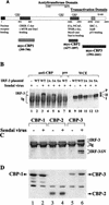
IRF-3 interacts with CBP in virus-infected cells. (A) Schematic representation of CBP, illustrating the domains involved in interaction with host or viral proteins (modified from reference 29) and the myc-tagged CBPs (CBP1, CBP2, and CBP3) used for immunoprecipitation. (B) 293 cells were transfected with wild type (WT) and point mutants of IRF-3 expression plasmid (5 μg, as indicated above the lanes) or were left untransfected (lanes 1 and 8). At 24 h after transfection, the cells were infected with Sendai virus for 16 h (lanes 1, 3 to 8, and 10 to 13) or were left uninfected (lanes 1 and 9). Whole-cell extracts (300 μg, except lane 1, which was 600 μg) were immunoprecipitated with anti-CBP antibody A22 (lanes 1 to 6) or with preimmune serum (lane 7). The immunoprecipitated complexes (lanes 1 to 7) or 30 μg of whole-cell extracts (lanes 8 to 13) was run on SDS–5% PAGE gels and subsequently probed with anti-IRF-3 antibody. (C) 293 cells were cotransfected with myc-tagged CBP expression plasmids (as indicated above the lanes) and the IRF-3 ΔN (Δ9-133) expression plasmid. At 24 h after transfection, the cells were infected with Sendai virus (lanes 2, 4, and 6) or were left uninfected (lanes 1, 3, and 5). Whole-cell extracts (300 μg) were immunoprecipitated with monoclonal anti-myc-tag antibody 9E10. The immunoprecipitated complexes were run on SDS–5% PAGE gels, and different forms of IRF-3 in the precipitates were analyzed by immunoblotting with anti-IRF-3 antibody. (D) Whole-cell extracts (30 μg) from panel C were also analyzed directly for the expression of myc-tagged CBP by immunoblotting using anti-myc antibody 9E10.

Similar articles

Cited by

References

    1. Arany Z, Sellers W R, Livingston D M, Eckner R. E1A-associated p300 and CREB-associated CBP belong to a conserved family of coactivators. Cell. 1994;77:799–800. - PubMed
    1. Au W-C, Moore P A, Lowther W, Juang Y-T, Pitha P M. Identification of a member of the interferon regulatory factor family that binds to the interferon-stimulated response element and activates expression of interferon-induced genes. Proc Natl Acad Sci USA. 1995;92:11657–11661. - PMC - PubMed
    1. Avantaggiati M L, Ogryzko V, Gardner K, Giordano A, Levine A S, Kelly K. Recruitment of p300/CBP in p53-dependent signal pathways. Cell. 1997;89:1175–1184. - PubMed
    1. Baldwin A S., Jr The NF-κB and IκB proteins: new discoveries and insights. Annu Rev Immunol. 1996;14:649–681. - PubMed
    1. Bannister A J, Kouzarides T. The CBP coactivator is a histone acetyltransferase. Nature. 1996;384:641–643. - PubMed

Publication types

MeSH terms