Role of the N-terminal zinc finger of human immunodeficiency virus type 1 nucleocapsid protein in virus structure and replication
- PMID: 9557738
- PMCID: PMC109678
- DOI: 10.1128/JVI.72.5.4442-4447.1998
Role of the N-terminal zinc finger of human immunodeficiency virus type 1 nucleocapsid protein in virus structure and replication
Abstract
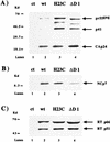
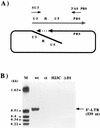
Nucleocapsid protein (NCp7) of human immunodeficiency virus type 1 is found covering the genomic RNA in the interior of the viral particle. It is a highly basic protein with two zinc fingers of the form CX2CX4HX4C which exhibit strong affinity for a zinc cation. To study the structure-function relationship of the N-terminal zinc finger of NCp7, this domain was either deleted or changed to CX2CX4CX4C. We examined virus formation and structure as well as proviral DNA synthesis. Our data show that these two NC mutations result in the formation of particles with an abnormal core morphology and impair the end of proviral DNA synthesis, leading to noninfectious viruses.
Figures


Similar articles
-
Mutations in the N-terminal domain of human immunodeficiency virus type 1 nucleocapsid protein affect virion core structure and proviral DNA synthesis.J Virol. 1997 Sep;71(9):6973-81. doi: 10.1128/JVI.71.9.6973-6981.1997. J Virol. 1997. PMID: 9261426 Free PMC article.
-
1H NMR structure and biological studies of the His23-->Cys mutant nucleocapsid protein of HIV-1 indicate that the conformation of the first zinc finger is critical for virus infectivity.Biochemistry. 1994 Oct 4;33(39):11707-16. doi: 10.1021/bi00205a006. Biochemistry. 1994. PMID: 7918387
-
The central globular domain of the nucleocapsid protein of human immunodeficiency virus type 1 is critical for virion structure and infectivity.J Virol. 1995 Mar;69(3):1778-84. doi: 10.1128/JVI.69.3.1778-1784.1995. J Virol. 1995. PMID: 7853517 Free PMC article.
-
The chaperoning and assistance roles of the HIV-1 nucleocapsid protein in proviral DNA synthesis and maintenance.Curr HIV Res. 2004 Jan;2(1):79-92. doi: 10.2174/1570162043485022. Curr HIV Res. 2004. PMID: 15053342 Review.
-
Structure, biological functions and inhibition of the HIV-1 proteins Vpr and NCp7.Biochimie. 1997 Nov;79(11):673-80. doi: 10.1016/s0300-9084(97)83501-8. Biochimie. 1997. PMID: 9479450 Review.
Cited by
-
Effect of Human Immunodeficiency Virus on Trace Elements in the Brain.Biol Trace Elem Res. 2021 Jan;199(1):41-52. doi: 10.1007/s12011-020-02129-4. Epub 2020 Apr 1. Biol Trace Elem Res. 2021. PMID: 32239375
-
Influence of HIV-1 Genomic RNA on the Formation of Gag Biomolecular Condensates.J Mol Biol. 2023 Aug 15;435(16):168190. doi: 10.1016/j.jmb.2023.168190. Epub 2023 Jun 27. J Mol Biol. 2023. PMID: 37385580 Free PMC article.
-
DNA condensation by the nucleocapsid protein of HIV-1: a mechanism ensuring DNA protection.Nucleic Acids Res. 2003 Sep 15;31(18):5425-32. doi: 10.1093/nar/gkg738. Nucleic Acids Res. 2003. PMID: 12954779 Free PMC article.
-
Pan-retroviral Nucleocapsid-Mediated Phase Separation Regulates Genomic RNA Positioning and Trafficking.Cell Rep. 2020 Apr 21;31(3):107520. doi: 10.1016/j.celrep.2020.03.084. Cell Rep. 2020. PMID: 32320662 Free PMC article.
-
A new role for HIV nucleocapsid protein in modulating the specificity of plus strand priming.Virology. 2008 Sep 1;378(2):385-96. doi: 10.1016/j.virol.2008.06.002. Epub 2008 Jul 15. Virology. 2008. PMID: 18632127 Free PMC article.
References
-
- Barat C, Schatz O, Le Grice S, Darlix J L. Analysis of the interaction of HIV-1 replication primer tRNALys,3 with nucleocapsid protein and reverse transcriptase. J Mol Biol. 1993;231:185–190. - PubMed
Publication types
MeSH terms
Substances
LinkOut - more resources
Full Text Sources
Other Literature Sources

