The class II membrane glycoprotein G of bovine respiratory syncytial virus, expressed from a synthetic open reading frame, is incorporated into virions of recombinant bovine herpesvirus 1
- PMID: 9557663
- PMCID: PMC109603
- DOI: 10.1128/JVI.72.5.3804-3811.1998
The class II membrane glycoprotein G of bovine respiratory syncytial virus, expressed from a synthetic open reading frame, is incorporated into virions of recombinant bovine herpesvirus 1
Abstract
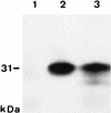
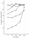
The bovine herpesvirus 1 (BHV-1) recombinants BHV-1/eG(ori) and BHV-1/eG(syn) were isolated after insertion of expression cassettes which contained either a genomic RNA-derived cDNA fragment (BHV-1/eG(ori)) or a modified, chemically synthesized open reading frame (ORF) (BHV-1/eG(syn)), which both encode the attachment glycoprotein G of bovine respiratory syncytial virus (BRSV), a class II membrane glycoprotein. Northern blot analyses and nuclear runoff transcription experiments indicated that transcripts encompassing the authentic BRSV G ORF were unstable in the nucleus of BHV-1/eG(ori)-infected cells. In contrast, high levels of BRSV G RNA were detected in BHV-1/eG(syn)-infected cells. Immunoblots showed that the BHV-1/eG(syn)-expressed BRSV G glycoprotein contains N- and O-linked carbohydrates and that it is incorporated into the membrane of infected cells and into the envelope of BHV-1/eG(syn) virions. The latter was also demonstrated by neutralization of BHV-1/eG(syn) infectivity by monoclonal antibodies or polyclonal anti-BRSV G antisera and complement. Our results show that expression of the BRSV G glycoprotein by BHV-1 was dependent on the modification of the BRSV G ORF and indicate that incorporation of class II membrane glycoproteins into BHV-1 virions does not necessarily require BHV-1-specific signals. This raises the possibility of targeting heterologous polypeptides to the viral envelope, which might enable the construction of BHV-1 recombinants with new biological properties and the development of improved BHV-1-based live and inactivated vector vaccines.
Figures


Similar articles
-
Fusion of the green fluorescent protein to amino acids 1 to 71 of bovine respiratory syncytial virus glycoprotein G directs the hybrid polypeptide as a class II membrane protein into the envelope of recombinant bovine herpesvirus-1.J Gen Virol. 2000 Apr;81(Pt 4):1051-5. doi: 10.1099/0022-1317-81-4-1051. J Gen Virol. 2000. PMID: 10725432
-
Expression of bovine viral diarrhoea virus glycoprotein E2 by bovine herpesvirus-1 from a synthetic ORF and incorporation of E2 into recombinant virions.J Gen Virol. 1999 Nov;80 ( Pt 11):2839-2848. doi: 10.1099/0022-1317-80-11-2839. J Gen Virol. 1999. PMID: 10580045
-
Resistance to bovine respiratory syncytial virus (BRSV) induced in calves by a recombinant bovine herpesvirus-1 expressing the attachment glycoprotein of BRSV.J Gen Virol. 1998 Jul;79 ( Pt 7):1759-67. doi: 10.1099/0022-1317-79-7-1759. J Gen Virol. 1998. PMID: 9680140
-
Gene contents in a 31-kb segment at the left genome end of bovine herpesvirus-1.Vet Microbiol. 1996 Nov;53(1-2):67-77. doi: 10.1016/s0378-1135(96)01235-7. Vet Microbiol. 1996. PMID: 9010999 Review.
-
A review of the biology of bovine herpesvirus type 1 (BHV-1), its role as a cofactor in the bovine respiratory disease complex and development of improved vaccines.Anim Health Res Rev. 2007 Dec;8(2):187-205. doi: 10.1017/S146625230700134X. Anim Health Res Rev. 2007. PMID: 18218160 Review.
Cited by
-
A hepadnavirus regulatory element enhances expression of a type 2 bovine viral diarrhea virus E2 protein from a bovine herpesvirus 1 vector.J Virol. 2003 Aug;77(16):8775-82. doi: 10.1128/jvi.77.16.8775-8782.2003. J Virol. 2003. PMID: 12885896 Free PMC article.
-
BHV-1: new molecular approaches to control a common and widespread infection.Mol Med. 1999 May;5(5):261-84. Mol Med. 1999. PMID: 10390543 Free PMC article. Review.
-
Recombinant bovine adenovirus-3 co-expressing bovine respiratory syncytial virus glycoprotein G and truncated glycoprotein gD of bovine herpesvirus-1 induce immune responses in cotton rats.Mol Biotechnol. 2015 Jan;57(1):58-64. doi: 10.1007/s12033-014-9801-x. Mol Biotechnol. 2015. PMID: 25173687
-
Construction and manipulation of an infectious clone of the bovine herpesvirus 1 genome maintained as a bacterial artificial chromosome.J Virol. 2002 Jul;76(13):6660-8. doi: 10.1128/jvi.76.13.6660-6668.2002. J Virol. 2002. PMID: 12050379 Free PMC article.
-
Expression of the genomic form of the bovine viral diarrhea virus E2 ORF in a bovine herpesvirus-1 vector.Virus Genes. 2003 Aug;27(1):83-91. doi: 10.1023/a:1025180604047. Virus Genes. 2003. PMID: 12913361
References
-
- Bello L J, Whitbeck C, Lawrence W C. Bovine herpesvirus 1 as a live vector for expression of foreign genes. Virology. 1992;190:666–673. - PubMed
-
- Chowdhury S. Construction and characterization of an attenuated bovine herpesvirus type 1 (BHV-1) recombinant virus. Vet Microbiol. 1996;52:13–23. - PubMed
-
- Collins P L. The molecular biology of human respiratory syncytial virus (RSV) of the genus Pneumovirus. In: Kingsburg D W, editor. The paramyxoviruses. New York, N.Y: Plenum Press; 1991. pp. 103–162.
-
- Dormitzer P R, Ho D Y, Mackow E R, Mocarsky E S, Greenberg H B. Neutralizing epitopes on herpes simplex virus-1-expressed rotavirus VP7 are dependent on coexpression of other rotavirus proteins. Virology. 1992;187:18–32. - PubMed
Publication types
MeSH terms
Substances
LinkOut - more resources
Full Text Sources
Other Literature Sources
Miscellaneous

