. 1998 Apr;72(4):2697-707.
doi: 10.1128/JVI.72.4.2697-2707.1998.

Separate DNA elements containing ATF/CREB and IE86 binding sites differentially regulate the human cytomegalovirus UL112-113 promoter at early and late times in the infection

Affiliations

Separate DNA elements containing ATF/CREB and IE86 binding sites differentially regulate the human cytomegalovirus UL112-113 promoter at early and late times in the infection

S M Rodems et al. J Virol. 1998 Apr.

Abstract

The human cytomegalovirus (HCMV) UL112-113 promoter represents a useful model for studying temporal regulation of viral gene expression. Stimulation of this promoter by the 86-kDa immediate-early protein (IE86) is controlled by sequences between nucleotides -113 and -59, which include both an ATF/CREB and an IE86 binding site. In transient assays, the ATF/CREB site is essential, and the IE86 site, although nonessential, can enhance transcription. With recombinant viruses, we have assessed the function of these promoter elements in the context of the viral genome. Transcription from the inserted UL112-113 promoter shows the same temporal pattern as the endogenous promoter, including the switch to an upstream RNA start site late in infection. Deletion of sequences containing the IE86 site results in a decrease in the level of early transcription and elimination of late transcription. In contrast, when the ATF/CREB site is deleted, early RNA synthesis is almost completely abolished, but late transcription is comparable to that of the wild type, with repositioning of the RNA start site downstream by the number of nucleotides deleted. Replacement of sequences between -108 and -95 with the HCMV cis-repression signal from the major immediate-early promoter had no effect on the level of late RNAs but resulted in the repositioning of the RNA start site 39 nucleotides upstream. These results suggest that the ATF/CREB site is functional only at early times, while sequences containing the IE86 site modulate the level of early RNAs and may be required for activating late transcription in a distance-dependent manner.


Figures

FIG. 1
FIG. 1
UL112-113 promoters inserted into recombinant viruses. (A) Sequences of the HCMV UL112-113 promoter from −113 to +1. Previously identified regulatory elements are indicated by the shaded boxes beneath the sequence. The IE86 binding site represents the sequences protected by IE86 in a DNase I footprint analysis (24), whereas the CREB/ATF and TATA sites are the respective consensus sequences. (B) Map of UL112-113 promoter mutations used in this study. The name of each recombinant virus is indicated at the right. The weak IE86 binding site, CREB/ATF site, and TATA box are indicated by shaded boxes. The black box indicates the replacement of the weak IE86 binding site and/or adjacent A-T rich sequences with the HCMV CRS from the major IE promoter. Dashed lines denote deleted sequences. (C) Sequence between −113 and −85 in v148(Δ84-58)-CAT, v148(Δ84-58)CRS-CAT, and v148(Δ84-58)IICRS-CAT. Sequences protected by IE86 in a DNase I footprint are indicated by the shaded box above the sequence. Sequences outlined in black are those replaced in v148(Δ84-58)CRS-CAT and v148(Δ84-58)IICRS-CAT. The CRS is indicated by the bracket.
FIG. 2
FIG. 2
Site of recombination in the HCMV genome and Southern blot analysis of recombinant viruses. (A) Schematic representation of the site of recombination within the HCMV genome. Shown is the HCMV Towne strain. White boxes denote the terminal repeat sequences. Black bars beneath the genome indicate the location of the endogenous UL112-113 genes and the site of insertion within the region between the US9 and US12 genes. (B) Expanded map of the region between the US9 and US12 genes in recombinant HCMV. Where indicated, the direction of the open reading frame is shown with an arrow. Sizes of fragments expected to hybridize to the CAT probe in Southern blots are shown above the map; sizes of fragments expected to hybridize to the genomic probe are shown below the map. Angled lines between the shaded bars of the genomic probe indicate that the probe is contiguous. (C) Southern blot of genomic DNA from mock- and virus-infected cells, using the CAT probe for detection. Viruses analyzed are indicated above the autoradiogram. The expected size range of the hybridized DNA fragment is indicated on the right. Positions of molecular weight markers are indicated on the left. W.T., wild type. (D) Southern blot of genomic DNA from mock- and virus-infected cells, using the genomic probe for detection. Viruses analyzed are indicated above the autoradiogram. The expected size ranges of the hybridized DNA fragments are indicated on the right. Positions of molecular weight markers are indicated on the left.
FIG. 3
FIG. 3
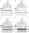
Western blot analysis of the UL112-113 proteins during infection with recombinant viruses. Western blot analysis was performed with total protein isolated from infected cells at 8 (A), 24 (B), 48 (C), and 72 (D) h after infection as described in Materials and Methods. The recombinant viruses used are indicated above each blot. Sizes of the UL112-113 proteins are indicated in kilodaltons on the right of each blot; positions of molecular weight markers are indicated in kilodaltons on the left of each blot. The band detected just above 66 kD appears to be a cross-reactive protein and has been previously observed in some experiments (36). The band near 116 kDa is detected only with recombinant viruses and may represent LacZ expressed from the human β-actin promoter. wt, wild type.
FIG. 4
FIG. 4
Analysis of CAT RNA and protein expression during infection with v358-CAT recombinant virus. (A) Map of the UL112-113 promoter and CAT gene inserted into v358-CAT. Sequences from −323 to +35 were linked to the CAT gene and inserted into a recombinant virus as described in Materials and Methods. Arrows indicate the location and direction of transcription of RNA from the early start site (+1) and the late start site (−62). Previously identified regulatory regions are indicated by the shaded boxes. (B) CAT activity detected in lysates from v358-CAT-infected cells. Protein lysates from infected cells were harvested at 8, 24, 48, and 72 h after infection with v358-CAT. CAT activity was determined as described in Materials and Methods and is represented as percent acetylation per microgram of lysate used in the reaction. Time points are indicated below the histogram. (C) Primer extension analysis of total RNA isolated from v358-CAT-infected cells. Total RNA was isolated from infected cells at 8, 24, 48, and 72 h after infection, and primer extension analysis was performed as described in Materials and Methods, using a primer which detects CAT mRNA. Extension products were separated by denaturing polyacrylamide gel electrophoresis and subjected to autoradiography. Products were electrophoresed adjacent to a sequencing ladder (lanes T, G, C, and A) generated with plasmid p148-CAT and the identical CAT primer to identify transcriptional start sites. Locations of start sites are indicated at the right. Time points are indicated below the autoradiogram. hpi, hours postinfection.
FIG. 5
FIG. 5
Analysis of CAT protein expression during infection with recombinant viruses containing UL112-113 promoter deletions. Protein lysates were prepared at 8, 24, 48, and 72 h postinfection (hpi), and CAT activity was determined as described in Materials and Methods. Activity is represented as percent acetylation per microgram of lysate used in the reaction. Solid bars represent the average of two independent infections; error bars represent the range of the two values. Promoter deletions contained in the recombinant viruses analyzed are indicated on the left.
FIG. 6
FIG. 6
Analysis of CAT RNA expression during infection with recombinant viruses containing UL112-113 promoter deletions. Primer extension analysis of total RNA isolated from cells infected with v148-CAT (A), v119-CAT (B), and v148(Δ84-58)-CAT (C) was performed as described in the legend to Fig. 4. Promoter deletions contained in the recombinant viruses analyzed are indicated adjacent to each panel. Arrows indicate the position of the transcriptional start sites detected in the assay. Products were electrophoresed adjacent to a sequencing ladder (lanes T, G, C, and A) generated with the plasmid p148-CAT and the identical CAT primer to identify transcriptional start sites. Each panel as well as Fig. 4C was derived from the same autoradiogram but was cropped for display purposes. hpi, hours postinfection.
FIG. 7
FIG. 7
Analysis of CAT protein expression during infection with v148(Δ84-58)IICRS-CAT. Protein lysates were prepared at 8, 24, 48, and 72 h after infection with either v148(Δ84-58)-CAT or v148(Δ84-58)IICRS-CAT, and CAT activity was determined as described in Materials and Methods. Activity is represented as percent acetylation per microgram of lysate used in the reaction. Solid bars represent the average of two independent infections; error bars represent the range of the two values. Promoter deletions and site-directed mutations contained in the recombinant viruses analyzed are indicated on the left. hpi, hours postinfection.
FIG. 8
FIG. 8
Analysis of CAT RNA expression during infection with v148(Δ84-58)-CAT, v148(Δ84-58)CRS-CAT, and v148(Δ84-58)IICRS-CAT. Primer extension analysis of total RNA isolated from cells infected with v148(Δ84-58)-CAT (A), v148(Δ84-58)IICRS-CAT (B), and v148(Δ84-58)CRS-CAT (C) was performed as described in the legend to Fig. 4 except that only the 72-h time point was analyzed for v148(Δ84-58)CRS-CAT. Promoter deletions contained in the recombinant viruses analyzed are indicated adjacent to each panel. Arrows indicate positions of the transcriptional start sites detected in the assay. Numbers represent the positions of the nucleotides relative to the sequence of the promoter in v148-CAT. Each panel was derived from the same autoradiogram but was cropped for display purposes. hpi, hours postinfection.
FIG. 9
FIG. 9
Summary of RNA start sites from recombinant viruses and schematic representation of promoter mutations used in this study. Names of the viruses containing the promoters are indicated at the left. Major early (E) RNA start sites are denoted by open arrowheads, whereas major late (L) RNA start sites are denoted by closed arrowheads. Minor RNA start sites are shown as dashed arrows. Sequence elements are as described in the legend to Fig. 1. Numbers represent the positions of the nucleotides relative to the sequence of the promoter in v148-CAT.

Similar articles

Cited by

References

    1. Arlt H, Lang D, Gebert S, Stamminger T. Identification of binding sites for the 86-kilodalton IE2 protein of human cytomegalovirus within an IE2-responsive viral early promoter. J Virol. 1994;68:4117–4125. - PMC - PubMed
    1. Britt W, Alford C. Cytomegalovirus. In: Fields B N, Knipe D M, Howley P M, editors. Fields virology. 3rd ed. Philadelphia, Pa: Lippincott-Raven Publishers; 1996. pp. 2493–2523.
    1. Chang C-P, Malone C L, Stinski M F. A human cytomegalovirus early gene has three inducible promoters that are regulated differentially at various times after infection. J Virol. 1989;63:281–290. - PMC - PubMed
    1. Chrivia J C, Kwok R P S, Lamb N, Hagiwara M, Montminy M R, Goodman R H. Phosphorylated CREB binds specifically to the nuclear protein CBP. Nature. 1993;365:855–859. - PubMed
    1. DeMarchi J M. Human cytomegalovirus DNA: restriction enzyme cleavage maps and map locations for immediate-early, early, and late RNAs. Virology. 1981;114:23–28. - PubMed

Publication types

MeSH terms

LinkOut - more resources