Osteoclast differentiation factor is a ligand for osteoprotegerin/osteoclastogenesis-inhibitory factor and is identical to TRANCE/RANKL
- PMID: 9520411
- PMCID: PMC19881
- DOI: 10.1073/pnas.95.7.3597
Osteoclast differentiation factor is a ligand for osteoprotegerin/osteoclastogenesis-inhibitory factor and is identical to TRANCE/RANKL
Abstract
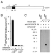
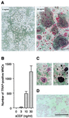
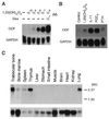
Osteoclasts, the multinucleated cells that resorb bone, develop from hematopoietic cells of monocyte/macrophage lineage. Osteoclast-like cells (OCLs) are formed by coculturing spleen cells with osteoblasts or bone marrow stromal cells in the presence of bone-resorbing factors. The cell-to-cell interaction between osteoblasts/stromal cells and osteoclast progenitors is essential for OCL formation. Recently, we purified and molecularly cloned osteoclastogenesis-inhibitory factor (OCIF), which was identical to osteoprotegerin (OPG). OPG/OCIF is a secreted member of the tumor necrosis factor receptor family and inhibits osteoclastogenesis by interrupting the cell-to-cell interaction. Here we report the expression cloning of a ligand for OPG/OCIF from a complementary DNA library of mouse stromal cells. The protein was found to be a member of the membrane-associated tumor necrosis factor ligand family and induced OCL formation from osteoclast progenitors. A genetically engineered soluble form containing the extracellular domain of the protein induced OCL formation from spleen cells in the absence of osteoblasts/stromal cells. OPG/OCIF abolished the OCL formation induced by the protein. Expression of its gene in osteoblasts/stromal cells was up-regulated by bone-resorbing factors. We conclude that the membrane-bound protein is osteoclast differentiation factor (ODF), a long-sought ligand mediating an essential signal to osteoclast progenitors for their differentiation into osteoclasts. ODF was found to be identical to TRANCE/RANKL, which enhances T-cell growth and dendritic-cell function. ODF seems to be an important regulator in not only osteoclastogenesis but also immune system.
Figures


Similar articles
-
A novel molecular mechanism modulating osteoclast differentiation and function.Bone. 1999 Jul;25(1):109-13. doi: 10.1016/s8756-3282(99)00121-0. Bone. 1999. PMID: 10423033
-
A new member of tumor necrosis factor ligand family, ODF/OPGL/TRANCE/RANKL, regulates osteoclast differentiation and function.Biochem Biophys Res Commun. 1999 Mar 24;256(3):449-55. doi: 10.1006/bbrc.1999.0252. Biochem Biophys Res Commun. 1999. PMID: 10080918 Review.
-
Osteoblasts/stromal cells stimulate osteoclast activation through expression of osteoclast differentiation factor/RANKL but not macrophage colony-stimulating factor: receptor activator of NF-kappa B ligand.Bone. 1999 Nov;25(5):517-23. doi: 10.1016/s8756-3282(99)00210-0. Bone. 1999. PMID: 10574571
-
Osteoclast differentiation factor (ODF) induces osteoclast-like cell formation in human peripheral blood mononuclear cell cultures.Biochem Biophys Res Commun. 1998 May 8;246(1):199-204. doi: 10.1006/bbrc.1998.8586. Biochem Biophys Res Commun. 1998. PMID: 9600092
-
The molecular basis of osteoclast differentiation and activation.Novartis Found Symp. 2001;232:235-47; discussion 247-50. doi: 10.1002/0470846658.ch16. Novartis Found Symp. 2001. PMID: 11277084 Review.
Cited by
-
Role of LRF/Pokemon in lineage fate decisions.Blood. 2013 Apr 11;121(15):2845-53. doi: 10.1182/blood-2012-11-292037. Epub 2013 Feb 8. Blood. 2013. PMID: 23396304 Free PMC article. Review.
-
The role of Snail in prostate cancer.Cell Adh Migr. 2012 Sep-Oct;6(5):433-41. doi: 10.4161/cam.21687. Epub 2012 Sep 1. Cell Adh Migr. 2012. PMID: 23076049 Free PMC article. Review.
-
Fas-independent T-cell apoptosis by dendritic cells controls autoimmune arthritis in MRL/lpr mice.PLoS One. 2012;7(12):e48798. doi: 10.1371/journal.pone.0048798. Epub 2012 Dec 12. PLoS One. 2012. PMID: 23300516 Free PMC article.
-
Bone-targeted pH-responsive cerium nanoparticles for anabolic therapy in osteoporosis.Bioact Mater. 2021 May 20;6(12):4697-4706. doi: 10.1016/j.bioactmat.2021.04.038. eCollection 2021 Dec. Bioact Mater. 2021. PMID: 34095626 Free PMC article.
-
Effects of eldecalcitol on cortical bone response to mechanical loading in rats.BMC Musculoskelet Disord. 2015 Jun 30;16:158. doi: 10.1186/s12891-015-0613-3. BMC Musculoskelet Disord. 2015. PMID: 26123128 Free PMC article.
References
-
- Suda T, Takahashi N, Martin T J. Endocr Rev. 1992;13:66–80. - PubMed
-
- Suda T, Takahashi N, Martin T J. In: Endocrine Review Monographs. Bikle D D, Negrovilar A, editors. Vol. 4. Bethesda: Endocrine Soc.; 1995. pp. 266–270.
-
- Suda T, Udagawa N, Nakamura I, Miyaura C, Takahashi N. Bone. 1995;17:87S–91S. - PubMed
-
- Roodman G D. Endocr Rev. 1996;17:308–332. - PubMed
-
- Takahashi N, Akatsu T, Uadagawa N, Sasaki T, Yamaguchi A, Moseley J M, Martin T J, Suda T. Endocrinology. 1988;123:2600–2602. - PubMed
MeSH terms
Substances
Associated data
- Actions
LinkOut - more resources
Full Text Sources
Other Literature Sources
Molecular Biology Databases
Research Materials
Miscellaneous

