. 1998 Mar 15;12(6):858-67.
doi: 10.1101/gad.12.6.858.

A cooperative interaction between U2AF65 and mBBP/SF1 facilitates branchpoint region recognition

Affiliations

A cooperative interaction between U2AF65 and mBBP/SF1 facilitates branchpoint region recognition

J A Berglund et al. Genes Dev. .

Abstract

During the early events of pre-mRNA splicing, intronic cis-acting sequences are recognized and interact through a network of RNA-RNA, RNA-protein, and protein-protein contacts. Recently, we identified a branchpoint sequence binding protein in yeast (BBP). The mammalian ortholog (mBBP/SF1) also binds specifically to branchpoint sequences and interacts with the well studied mammalian splicing factor U2AF65, which binds to the adjacent polypyrimidine (PY) tract. In this paper we demonstrate that the mBBP/SF1-U2AF65 interaction promotes cooperative binding to a branchpoint sequence-polypyrimidine tract-containing RNA, and we suggest that this cooperative RNA binding contributes to initial recognition of the branchpoint sequence (BPS) during pre-mRNA splicing. We also demonstrate the essential nature of the third RBD of U2AF65 for the interaction between the two proteins, both in the presence and absence of RNA.


Figures
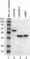
Figure 1
Figure 1
Commassie stained SDS–polyacrylamide gel of purified U2AF65, U2AF65-3, and mBBP. Three micrograms of protein was loaded in each lane. Protein markers are from GIBCO.
Figure 2
Figure 2
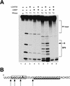
Double footprint of mBBP and U2AF65. (A) Purified mBBP, U2AF65, or both proteins together were incubated with 5′-end labeled RNA substrate in the presence of either RNase T1 (lanes 2–5) or RNase T2 (lanes 6–9), and then run in a denaturing polyacrylamide gel as described in Materials and methods. (Lanes 4,8) The concentration of mBBP used was 11 μm; (lanes 3,7) concentration of U2AF65 was 2.8 μm; (lanes 5,9) mBBP was at a concentration of 5.5 μm and U2AF65 at 1.4 μm. The PY tract and BPS are marked by brackets, and the position of the branchpoint adenosine is marked by an arrow. (B) Sequence of the 34-nucleotide RNA substrate derived from the Adenovirus major late pre-mRNA substrate. The BPS is in bold and boxed, and the branchpoint adenosine is in large text. The PY tract is in bold and underlined. Arrows represent guanosines cleaved by RNase T1 (lane 2).
Figure 3
Figure 3
The binding of a 12-nucleotide RNA to the BPS competes with mBBP binding. (A) (Lane 1) radiolabeled RNA alone; (lane 2–11) either the 12-nucleotide RNA (U2 mimic), mBBP, U2AF65, or a combination as shown above the autoradiograph. The different complexes are labeled by arrows (right). (B) Sequences of the two RNA oligoribonucleotides. The branchpoint adenosine is shown in bold and bulged out, and the PY tract is in bold and underlined.
Figure 3
Figure 3
The binding of a 12-nucleotide RNA to the BPS competes with mBBP binding. (A) (Lane 1) radiolabeled RNA alone; (lane 2–11) either the 12-nucleotide RNA (U2 mimic), mBBP, U2AF65, or a combination as shown above the autoradiograph. The different complexes are labeled by arrows (right). (B) Sequences of the two RNA oligoribonucleotides. The branchpoint adenosine is shown in bold and bulged out, and the PY tract is in bold and underlined.
Figure 4
Figure 4
Cooperative binding of mBBP and U2AF65. (A) In a gel-shift assay using a radiolabed 34-nucleotide RNA substrate derived from the Adenovirus major late pre-mRNA and purified proteins mBBP and U2AF65, we assayed for cooperative binding between mBBP and U2AF65 (Materials and Methods). (Lanes 2–6) Increasing concentrations (top) of mBBP; the complex is marked (left) by an arrow. (Lane 7) U2AF65 plus RNA; the complex is again marked by an arrow (right). (Lanes 8–12) U2AF65 at the same concentration as lane 7 plus mBBP at increasing concentrations, the same as those in lanes 2–6. The ternary complex of mBBP/U2AF65/RNA is marked by an arrow (right). (B) Graphical representation of the data shown in A (□) mBBP; (▵) mBBP + U2AF65. (C) The same experiment as in A except that U2AF65 concentration is varied (at top); the concentration of mBBP is held constant at 9 μm. The different complexes are marked at left and right. (D) Graphical representation of the data shown in C (▴) U2AF65; (▪) U2AF65 + mBBP. These experiments were repeated multiple times under multiple conditions with approximately the same 20-fold and 5-fold effects in cooperativity observed.
Figure 4
Figure 4
Cooperative binding of mBBP and U2AF65. (A) In a gel-shift assay using a radiolabed 34-nucleotide RNA substrate derived from the Adenovirus major late pre-mRNA and purified proteins mBBP and U2AF65, we assayed for cooperative binding between mBBP and U2AF65 (Materials and Methods). (Lanes 2–6) Increasing concentrations (top) of mBBP; the complex is marked (left) by an arrow. (Lane 7) U2AF65 plus RNA; the complex is again marked by an arrow (right). (Lanes 8–12) U2AF65 at the same concentration as lane 7 plus mBBP at increasing concentrations, the same as those in lanes 2–6. The ternary complex of mBBP/U2AF65/RNA is marked by an arrow (right). (B) Graphical representation of the data shown in A (□) mBBP; (▵) mBBP + U2AF65. (C) The same experiment as in A except that U2AF65 concentration is varied (at top); the concentration of mBBP is held constant at 9 μm. The different complexes are marked at left and right. (D) Graphical representation of the data shown in C (▴) U2AF65; (▪) U2AF65 + mBBP. These experiments were repeated multiple times under multiple conditions with approximately the same 20-fold and 5-fold effects in cooperativity observed.
Figure 5
Figure 5
Mutation of the branchpoint adenosine reduces formation of the ternary complex (mBBP, U2AF65, and RNA substrate). (A) A 34-nucleotide RNA substrate with the branchpoint adenosine mutated gel-shift experiments was used to perform under the same conditions as those in Fig. 4A. The mBBP/RNA complex has a different migration pattern as marked by an arrow to the side of the autoradiograph. (B) Sequence of the mutated 34-nucleotide RNA substrate used in this experiment. The branchpoint adenosine was changed to cytidine and is marked by an asterisk (*).
Figure 5
Figure 5
Mutation of the branchpoint adenosine reduces formation of the ternary complex (mBBP, U2AF65, and RNA substrate). (A) A 34-nucleotide RNA substrate with the branchpoint adenosine mutated gel-shift experiments was used to perform under the same conditions as those in Fig. 4A. The mBBP/RNA complex has a different migration pattern as marked by an arrow to the side of the autoradiograph. (B) Sequence of the mutated 34-nucleotide RNA substrate used in this experiment. The branchpoint adenosine was changed to cytidine and is marked by an asterisk (*).
Figure 6
Figure 6
Mutating the PY tract reduces cooperative formation of the ternary complex (mBBP, U2AF65, and RNA substrate). (A) This is the same experiment as in Fig. 4C except the PY tract within the 34-nucleotide RNA substrate has been mutated. The different complexes are indicated to left and right. (B) Sequence showing the double mutation of the PY tract. Two uridines in the middle were changed to guanosine, as indicated by asterisks (**).
Figure 6
Figure 6
Mutating the PY tract reduces cooperative formation of the ternary complex (mBBP, U2AF65, and RNA substrate). (A) This is the same experiment as in Fig. 4C except the PY tract within the 34-nucleotide RNA substrate has been mutated. The different complexes are indicated to left and right. (B) Sequence showing the double mutation of the PY tract. Two uridines in the middle were changed to guanosine, as indicated by asterisks (**).
Figure 7
Figure 7
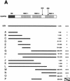
mBBP interacts with the carboxy-terminal RBD of U2AF65. (A) Yeast two-hybrid interactions. Yeast cells carrying the B42–mBBP fusion and the indicated LexA–U2AF65 fusions were obtained and tested for B-galactosidase production as described in Materials and Methods. A schematic representation of U2AF65 depicting the amino-terminal RS domain and the three RNA-binding domains RBD 1–3. The bars indicate the U2AF65 region present in each fusion and the numbers correspond to the U2AF65 amino acids fused to LexA. Plus (+) and minus (−) signs indicate β-galactosidase activity. The box around the three pluses of the P clone indicates that this fusion activates transcription independently of B42–mBBP expression. (B) LexA–U2AF65 fusions interacting with GST–mBBP. (Lane 1) Whole cell extracts were prepared from yeast cells carrying LexA alone, (lane 2) LexA–U2AF65, or (lanes 3–8) the indicated LexA–U2AF65 fusions, and 15 μl was incubated with the GST–mBBP fusion protein bound to glutathione agarose beads as described in Materials and Methods. The bound proteins were eluted in SDS sample buffer and separated in 8% polyacrylamide–SDS gels. After transfer to nitrocellulose the LexA fusion proteins were visualized with anti-LexA antibody. (Left panel) corresponds to 10 μl of the extracts directly loaded on the gel, and (right) proteins after elution from the GST–mBBP beads.
Figure 7
Figure 7
mBBP interacts with the carboxy-terminal RBD of U2AF65. (A) Yeast two-hybrid interactions. Yeast cells carrying the B42–mBBP fusion and the indicated LexA–U2AF65 fusions were obtained and tested for B-galactosidase production as described in Materials and Methods. A schematic representation of U2AF65 depicting the amino-terminal RS domain and the three RNA-binding domains RBD 1–3. The bars indicate the U2AF65 region present in each fusion and the numbers correspond to the U2AF65 amino acids fused to LexA. Plus (+) and minus (−) signs indicate β-galactosidase activity. The box around the three pluses of the P clone indicates that this fusion activates transcription independently of B42–mBBP expression. (B) LexA–U2AF65 fusions interacting with GST–mBBP. (Lane 1) Whole cell extracts were prepared from yeast cells carrying LexA alone, (lane 2) LexA–U2AF65, or (lanes 3–8) the indicated LexA–U2AF65 fusions, and 15 μl was incubated with the GST–mBBP fusion protein bound to glutathione agarose beads as described in Materials and Methods. The bound proteins were eluted in SDS sample buffer and separated in 8% polyacrylamide–SDS gels. After transfer to nitrocellulose the LexA fusion proteins were visualized with anti-LexA antibody. (Left panel) corresponds to 10 μl of the extracts directly loaded on the gel, and (right) proteins after elution from the GST–mBBP beads.
Figure 8
Figure 8
The third RBD of U2AF65 is necessary for the cooperative interaction between mBBP and U2AF65. Under the same conditions as those for the experiment shown in Fig. 4A (Materials and Methods), we looked for a cooperative interaction between mBBP and a U2AF65 protein missing the third RBD (U2AF65-3). The three different complexes are marked by arrows at left and right.
Figure 9
Figure 9
A model representing the cooperative interaction between mBBP and U2AF65 at the 3′ end of mammalian introns and the subsequent replacement of mBBP by U2 snRNP. The RNA is represented by the thick black line. The important domains within mBBP (amino terminus) and U2AF65 (third RBD) for the cooperative interaction are shown interacting with one another. The KH domain and Zn knuckles are shown binding the BPS.

Similar articles

Cited by

References

    1. Abovich N, Rosbash M. Cross-intron bridging interactions in the yeast commitment complex are conserved in mammals. Cell. 1997;89:403–412. - PubMed
    1. Abovich N, Legrain P, Rosbash M. The yeast PRP6 gene encodes a U4/U6 small nuclear ribonucleoprotein particle (snRNP) protein, and the PRP9 gene encodes a protein required for U2 snRNP binding. Mol Cell Biol. 1990;10:6417–6425. - PMC - PubMed
    1. Abovich N, Liao XC, Rosbash M. The yeast MUD2 protein: An interaction with PRP11 defines a bridge between commitment complexes and U2 snRNP addition. Genes & Dev. 1994;8:843–854. - PubMed
    1. Ackers GK, Shea MA, Johnson AD. Quantitative model for gene regulation by lamba phage repressor. Proc Natl Acad Sci. 1982;79:1129–1133. - PMC - PubMed
    1. Arning S, Gruter P, Bilbe G, Kramer A. Mammalian splicing factor SF1 is encoded by variant cDNAs and binds to RNA. RNA. 1996;2:794–810. - PMC - PubMed

Publication types

LinkOut - more resources