. 1998 Mar;72(3):2113-24.
doi: 10.1128/JVI.72.3.2113-2124.1998.

Overexpression of C/EBPbeta represses human papillomavirus type 18 upstream regulatory region activity in HeLa cells by interfering with the binding of TATA-binding protein

Affiliations

Overexpression of C/EBPbeta represses human papillomavirus type 18 upstream regulatory region activity in HeLa cells by interfering with the binding of TATA-binding protein

T Bauknecht et al. J Virol. 1998 Mar.

Abstract

The human papillomavirus type 18 (HPV-18) upstream regulatory region (URR) controls cell type-specific expression of viral oncoproteins E6 and E7. The HPV-18 URR is highly active in HeLa cells, but its activity is virtually undetectable in HepG2 cells. Previous work has shown that YY1 plays an important role in activation of the HPV-18 URR in HeLa cells, and this activating activity is dependent on its physical interaction with C/EBPbeta, which binds to the switch region adjacent to the YY1 site in the URR. Overexpression of C/EBPbeta in HepG2 cells restores C/EBPbeta-YY1 interaction, resulting in strong activation of the HPV-18 URR activity. In this report, we show that, in contrast to the effect in HepG2 cells, overexpression of C/EBPbeta represses the HPV-18 URR in HeLa cells. This C/EBPbeta-induced repression of the HPV-18 URR in HeLa cells is binding site independent. It is also promoter specific, since it activates the albumin promoter under conditions in which it represses the URR in the same cells. Biochemical analysis shows that overexpression of C/EBPbeta in HeLa cells specifically interferes with binding of TATA-binding protein to the TATA box of the HPV-18 URR, but its overexpression in HepG2 cells leads to activation of the HPV-18 URR. These results suggest that a molecular mechanism underlies the ability of C/EBPbeta to regulate transcription in a cell type-specific manner and indicate the potential of using C/EBPbeta to manipulate the activity of the HPV-18 URR in cervical carcinoma cells.


Figures

FIG. 1
FIG. 1
Schematic representation of the HPV-18 URR. The 824-bp HPV-18 URR is located within the 1,049-bp BamHI fragment of the HPV-18 genome. The URR consists of 5′ distal (positions −824 to −450), central (positions −450 to −221), and 3′ proximal (positions −221 to −1) fragments. The transcription initiation site is indicated by a crooked arrow. The relative positions of the distal YY1 site, the central overlapping AP-1 and YY1 sites, the C/EBPβ-YY1 site, the promoter-proximal AP-1 and YY1 sites, and the TBP-TATA box binding site are indicated.
FIG. 2
FIG. 2
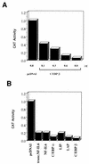
Overexpression of the C/EBP family of transcription factors represses HPV-18 URR activity in HeLa cells. (A) C/EBPβ represses HPV-18 URR activity in HeLa cells in a dose-dependent manner. HeLa cells were transfected with 3 μg of p18URR together with increasing amounts of CMV-C/EBPβ (0.1 to 0.8 μg) or 0.8 μg of pcDNAI, as indicated at the bottom of the figure, and 0.6 μg of RSV/L (18) as an internal control. Relative CAT activities were quantified relative to the activity obtained with p18URR cotransfected with 0.8 μg of pcDNAI, which was set at 1. The results are shown as bar graphs, with relative CAT activities for p18URR as follows: 0.8 μg of pcDNAI, 1.0; 0.1 μg of CMV-C/EBPβ, 0.41; 0.3 μg of CMV-C/EBPβ, 0.29; 0.6 μg of CMV-C/EBPβ, 0.12; and 0.8 μg of CMV-C/EBPβ, 0.06. (B) Different members of the C/EBP family of transcription factors repress the HPV-18 URR in HeLa cells. HeLa cells were transfected with 3 μg of p18URR together with 0.8 μg of C/EBP expression plasmids and 0.6 μg of RSV/L (18) as an internal control. CAT activities were quantified relative to the activity obtained with p18URR cotransfected with 0.8 μg of pcDNAI, which was set at 1, and the results are shown as bar graphs. The relative CAT activities for p18URR transfected with the different expression plasmids were as follows: control vector pcDNAI, 1.0; truncated NF-IL6, 0.196; NF-IL6, 0.197; C/EBPα, 0.095; LIP, 0.189; LAP, 0.086; and C/EBPβ, 0.06.
FIG. 3
FIG. 3
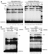
Overexpression of LIP or C/EBPβ in HeLa cells disrupts YY1-C/EBPβ complex formation on the switch region OL22. (A) Expression of LIP disrupts complex I and results in the formation of a new complex on OL22. HeLa cells were transfected with increasing amounts of CMV-LIP (5, 10, or 15 μg). 32P-labeled oligonucleotide OL22 was used in EMSAs, and anti-C/EBPβ antibodies, anti-YY1 antibodies, or preimmune serum was included in the binding reaction mixtures. Lanes: 1, standard binding reaction with extracts from nontransfected cells; 2 to 4, binding reactions with nuclear extracts from HeLa cells transfected with increasing amounts of CMV-LIP (lane 2, 5 μg; lane 3, 10 μg; lane 4, 15 μg); 5 to 7, binding reactions with nuclear extracts from cells transfected with 15 μg of CMV-LIP, incubated with anti-C/EBPβ antibodies (anti C), (lane 5), anti-YY1 antibodies (anti YY1) (lane 6), or preimmune serum (Pre) (lane 7). Specific DNA-protein complexes are indicated by numbers (I to IV). A nonspecific DNA-protein complex is indicated by an asterisk. The new complex formed on OL22 through expression of transfected CMV-LIP is indicated by an arrow. F (OL22), free probe OL22. (B) Expression of C/EBPβ leads to the formation of new complexes on OL22. Essentially the same experiments as described for panel A were carried out, but a plasmid expressing C/EBPβ was used instead of a LIP-expressing plasmid. The EMSA products shown in panels A and B were resolved on a 6.5% polyacrylamide gel.
FIG. 4
FIG. 4
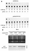
C/EBPβ represses HPV-18 activity. (A) C/EBPβ-induced repression of the HPV-18 URR is independent of the switch region OL22 in HeLa cells. CAT assays were carried out with extracts from HeLa cells transfected with a CAT plasmid containing the wild-type HPV-18 URR (p18URR) (upper panel) or with a construct containing mutations in the switch region of the HPV-18 URR (p18URR-22M1) (lower panel) (4). HeLa cells were transfected with 3 μg of p18URR or p18URR-22M1 together with increasing amounts of CMV-C/EBPβ (0.1 to 0.8 μg) and 0.6 μg RSV/L (18) as an internal control. Relative CAT activities were quantified relative to the activities obtained with p18URR and p18URR-22M1 cotransfected with 0.8 μg of pcDNAI, which were set at 1, and the results of representative CAT assays are shown. Relative CAT activities were as follows. Upper panel (p18URR): 0 μg of C/EBPβ, 1.0; 0.1 μg of C/EBPβ, 0.51; 0.2 μg of C/EBPβ, 0.36; 0.3 μg of C/EBPβ, 0.24; 0.4 μg of C/EBPβ, 0.20; 0.5 μg of C/EBPβ, 0.18; 0.6 μg of C/EBPβ, 0.12; 0.7 μg of C/EBPβ, 0.08; 0.8 μg of C/EBPβ, 0.06. Lower panel (p18URR-22M1): 0 μg of C/EBPβ, 1.0; 0.1 μg of C/EBPβ, 0.47; 0.2 μg of C/EBPβ, 0.25; 0.3 μg of C/EBPβ, 0.19; 0.4 μg of C/EBPβ, 0.15; 0.5 μg of C/EBPβ, 0.13; 0.6 μg of C/EBPβ, 0.09; 0.7 μg of C/EBPβ, 0.06; 0.8 μg of C/EBPβ, 0.05. (B) C/EBPβ represses E6-E7 mRNA expression in HeLa cells. Cells were transfected with increasing amounts of CMV-C/EBPβ or pcDNAI, as indicated in the figure, and 0.6 μg of RSV/L (18) to monitor for transfection efficiency. Cytoplasmic RNA was extracted and separated in a 1.2% agarose gel (upper panel). The filter was hybridized with a 32P-labeled HPV-18 E6-E7 DNA probe and was exposed to X-ray film for 3 days. Beside controls with transfected pcDNAI, RNA from HeLa cells transfected only with 0.6 μg of RSV/L only (18) (lane 13) and RNA from nontransfected HeLa cells (/) (lane 14) were used. The arrowheads indicate the positions of the 28S and 18S RNAs.
FIG. 5
FIG. 5
C/EBPβ, depending on its level of expression, activates or represses the albumin promoter in HeLa cells. HeLa cells were transfected with 10 μg of DNA from plasmid pALB (containing the mouse albumin promoter upstream of the CAT gene [25]) together with increasing amounts of CMV-C/EBPβ (as indicated below the bar graphs) and 0.6 μg of RSV/L (18) as an internal control. CAT activities were quantified relative to the activity obtained with pAlb cotransfected with 2.0 μg of pcDNAI, which was set at 0.1. Relative CAT activities were as follows: 0 μg of C/EBPβ, 0.1; 0.25 μg of C/EBPβ, 0.7; 0.3 μg of C/EBPβ, 0.68; 0.5 μg of C/EBPβ, 0.07; 1.0 μg of C/EBPβ, 0.03; 1.5 μg of C/EBPβ, 0.015; and 2.0 μg of C/EBPβ, 0.005.
FIG. 6
FIG. 6
Binding of TBP to the TATA box of the HPV-18 URR. (A) Formation of the specific TBP-TATA box complex I. 32P-labeled oligonucleotide 18TATA was used in EMSAs with HeLa nuclear extracts (lanes 1 to 10) or bacterial rTBP (lanes 11 to 21). Binding reactions were carried out with HeLa cell nuclear extracts in the absence (/) (lanes 1 and 10) or presence (lanes 2 to 9) of unlabeled competitor oligonucleotides. Unlabeled competitor oligonucleotides 18TATA, 18TATA-M1, 18TATAS, and 18TATAS-M1 were each used in 10- and 100-fold molar excesses, as indicated above the lanes. Binding reactions were carried out with bacterial rTBP in the absence (/) (lanes 11 and 20) or presence (lanes 12 to 19) of unlabeled competitor oligonucleotides. Unlabeled competitor oligonucleotides were used in the same manner as for the competition experiments with HeLa cell nuclear extracts. A specific antibody against bacterial rTBP (anti-rTBP) was included in the binding reaction shown in lane 21. A specific DNA-protein complex is indicated by a I (marked by an arrow); numerals II to IV indicate nonspecific DNA-protein complexes. The complex marked by an X is likely caused by binding of partially degraded TBP to the probe. F(18TATA), free probe 18TATA. EMSAs were resolved on a 6.5% polyacrylamide gel. (B) Anti-human TBP antibody specifically recognizes complex I in HeLa cells. Binding reactions with 32P-labeled 18TATA were carried out with HeLa cell nuclear extracts in the absence (/) (lanes 1 and 6) and presence (lanes 2 to 5) of unlabeled competitor. Unlabeled competitor oligonucleotides 18TATA and 18TATA-M1 were used in a 10- or 50-fold molar excess, as indicated above the lanes. A specific antibody against human TBP protein (1 μl) was included in the binding reaction shown in lane 8 (anti-TBP). In lane 7, the binding reaction mixture included 1 μl of preimmune serum (Pre). A specific DNA-protein complex is indicated by the I (marked by an arrow); numerals II to IV indicate nonspecific DNA-protein complexes. F(18TATA), free probe 18TATA. (C) Anti-human TBP antibody specifically recognizes complex I in HepG2 cells. Essentially the same binding reactions as described for panel B were carried out with nuclear extracts from HepG2 cells. A specific DNA-protein complex is indicated by the I (marked by an arrow); numerals II to V indicate nonspecific DNA-protein complexes. The complex marked by an X is likely caused by binding of partially degraded TBP to the probe. F(18TATA), free probe 18TATA.
FIG. 7
FIG. 7
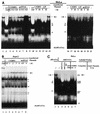
C/EBPβ interferes with TBP binding. (A) Overexpression of C/EBPβ or LIP disrupts formation of TBP-TATA complex I on oligonucleotide 18TATA in HeLa cells. HeLa cells were transfected with increasing amounts of CMV-C/EBPβ or CMV-LIP. Standard binding reactions were carried out with extracts from nontransfected cells (/) (lanes 1, 9, 15, 16, and 23) or with nuclear extracts from cells transfected with increasing amounts of CMV-C/EBPβ (lanes 3 to 5, 11, 12, 17, and 18), CMV-LIP (lanes 13, 14, 19, and 20), or pcDNAI (lanes 6 to 8, 21, and 22) as indicated above the lanes. In lanes 2 and 10, anti-human TBP antibodies were included in the binding reactions with nuclear extract from nontransfected cells. A specific DNA-protein complex is indicated by the I (marked by the arrows); numerals II to IV indicate nonspecific DNA-protein complexes. F(18TATA), free probe 18TATA. (B) Inability of C/EBPβ to disrupt TBP-TATA complex I formation in HepG2 cells. Standard binding reactions were carried out with nuclear extracts from HepG2 cells transfected with increasing amounts of CMV-C/EBPβ (lanes 2 to 6) or pcDNAI (lanes 7 and 8) as indicated above the lanes. Anti-human TBP antibodies were included in the binding reaction with nuclear extract from nontransfected HepG2 cells (lane 1). A specific DNA-protein complex is indicated by the I (marked by an arrow); numerals II to V indicate nonspecific DNA-protein complexes. The complex marked by an X is likely caused by binding of partially degraded TBP to the probe. F(18TATA), free probe 18TATA. (C) Overexpression of C/EBPβ disrupts TBP-TATA box complex formation on the albumin promoter in HeLa cells. Standard binding reactions with 32P-labeled oligonucleotide AlbTATA (encompassing the TATA box of the albumin promoter) were carried out with nuclear extracts from HeLa cells transfected (transf.) with increasing amounts of CMV-C/EBPβ as indicated above the lanes. Binding reactions with extracts from nontransfected cells were carried out in the absence (/) (lane 2) and presence (lanes 3 and 4) of unlabeled competitor oligocleotides. Unlabeled competitor oligonucleotides AlbTATA and AlbTATA-M1 were used in a 50-fold molar excess. Anti-human TBP antibodies were included in the binding reaction with nuclear extract from nontransfected HeLa cells (lane 5). In lane 1, a standard binding reaction was carried out with HeLa cell nuclear extract and 32P-labeled probe 18TATA. On the left side (for probe 18TATA), a specific DNA-protein complex is indicated by the I (marked by an arrow); numerals II to IV indicate nonspecific DNA-protein complexes. On the right side (for probe AlbTATA), a specific complex is indicated by AlbI (marked by an arrow); complexes AlbII to ALBV indicate nonspecific DNA-protein complexes. F, free probe 18TATA (lane 1) or AlbTATA (lanes 2 to 7).
FIG. 8
FIG. 8
Mutation of the TATA box strongly represses HPV-18 URR activity in HeLa cells. CAT assays were carried out with extracts prepared from HeLa cells transfected with plasmids p18URR (wild type), p18URR-22M1 (containing the mutated switch sequence OL22M1), p18URR-TATAM1 (containing the mutated TATA sequence TATA-M1), and p18URR-22M1-TATAM1 (containing the mutated switch sequence and a mutated TATA sequence). HeLa cells were transfected with 5 μg (lanes 1 to 4) or 3 μg (lanes 5 to 8) of DNA from each plasmid, as indicated in the figure, together with 0.6 μg of RSV/L (18) as an internal control. CAT activities were quantified relative to the activity obtained with p18URR (5 μg of transfected DNA) (lane 1), which was set at 1 (for 3 μg of transfected DNAs [lanes 5 to 8], the activities quantified relative to p18URR [set at 1] are shown in parentheses). The results of a representative CAT assay are shown. Relative CAT activities were as follows: p18URR (lane 1), 1.00; p18URR-TATAM1 (lane 2), 0.10; p18URR-22M1 (lane 3), 0.51; p18URR-22M1-TATAM1 (lane 4), 0.04; p18URR (lane 5), 0.447 (1); p18URR-TATAM1 (lane 6), 0.025 (0.056); p18URR-22M1 (lane 7), 0.15 (0.34); and p18URR-22M1-TATAM1 (lane 8), 0.014 (0.031).
FIG. 9
FIG. 9
Overexpression of C/EBPβ disrupts formation of the TBP-TATA complex on the HPV-16 URR in HeLa cells. 32P-labeled oligonucleotide 16TATA was used in EMSAs with HeLa cell nuclear extracts. Binding reactions were carried out with extracts from nontransfected cells in the absence (/) (lanes 2, 7, 13, and 16) and presence (lanes 3 to 6) of unlabeled competitor oligonucleotides. Unlabeled competitor oligonucleotides 16TATA and 16TATA-M1 were used in a 10- or 50-fold molar excess, as indicated above the lanes. Anti-human TBP antibodies were included in the binding reaction with nuclear extract from nontransfected HeLa cells (lane 8). Binding reactions were also carried out with nuclear extracts from HeLa cells transfected with increasing amounts of CMV-C/EBPβ (lanes 9 and 10), CMV-LIP (lanes 11 and 12), or pcDNAI (lanes 14 and 15) as indicated above the lanes. In lane 1, a standard binding reaction was carried out with HeLa cell nuclear extract and 32P-labeled probe 18TATA. On the left side (for probe 18TATA), a specific DNA-protein complex is indicated by the I (marked by an arrow); numerals II to IV indicate nonspecific DNA-protein complexes. On the right side (for probe 16TATA), a specific DNA-protein complex is indicated by 16I (marked by an arrow); 16II to 16VII indicate nonspecific DNA-protein complexes. F, free probe 18TATA (lane 1) or free probe 16TATA (lanes 2 to 16). trans., transfected.

Similar articles

Cited by

References

    1. Akira S, Isshiki H, Sugita T, Tanabe O, Kinoshita S, Nishio Y, Nakajima T, Hirano T, Kishimoto T. A nuclear factor for IL-6 expression (NF-IL6) is a member of a C/EBP family. EMBO J. 1990;9:1897–1906. - PMC - PubMed
    1. Baker C C, Phelps W C, Lindgren V, Braun M J, Gonda M A, Howley P M. Structural and transcriptional analysis of human papillomavirus type 16 sequences in cervical carcinoma cell lines. J Virol. 1987;61:962–971. - PMC - PubMed
    1. Bauknecht T, Angel P, Royer H-D, zur Hausen H. Identification of a negative regulatory domain in the human papillomavirus type 18 promoter: interaction with the transcriptional repressor YY1. EMBO J. 1992;11:4607–4617. - PMC - PubMed
    1. Bauknecht T, Jundt F, Herr I, Oehler T, Delius H, Shi Y, Angel P, zur Hausen H. A switch region determines the cell type-specific positive or negative action of YY1 on the activity of the human papillomavirus type 18 promoter. J Virol. 1995;69:1–12. - PMC - PubMed
    1. Bauknecht T, See R H, Shi Y. A novel C/EBP β-YY1 complex controls the cell-type-specific activity of the human papillomavirus type 18 upstream regulatory region. J Virol. 1996;70:7695–7705. - PMC - PubMed

Publication types

MeSH terms

LinkOut - more resources