. 1998 Feb 23;140(4):897-910.
doi: 10.1083/jcb.140.4.897.

A class VI unconventional myosin is associated with a homologue of a microtubule-binding protein, cytoplasmic linker protein-170, in neurons and at the posterior pole of Drosophila embryos

Affiliations

A class VI unconventional myosin is associated with a homologue of a microtubule-binding protein, cytoplasmic linker protein-170, in neurons and at the posterior pole of Drosophila embryos

V A Lantz et al. J Cell Biol. .

Abstract

Coordination of cellular organization requires the interaction of the cytoskeletal filament systems. Recently, several lines of investigation have suggested that transport of cellular components along both microtubules and actin filaments is important for cellular organization and function. We report here on molecules that may mediate coordination between the actin and microtubule cytoskeletons. We have identified a 195-kD protein that coimmunoprecipitates with a class VI myosin, Drosophila 95F unconventional myosin. Cloning and sequencing of the gene encoding the 195-kD protein reveals that it is the first homologue identified of cytoplasmic linker protein (CLIP)-170, a protein that links endocytic vesicles to microtubules. We have named this protein D-CLIP-190 (the predicted molecular mass is 189 kD) based on its similarity to CLIP-170 and its ability to cosediment with microtubules. The similarity between D-CLIP-190 and CLIP-170 extends throughout the length of the proteins, and they have a number of predicted sequence and structural features in common. 95F myosin and D-CLIP-190 are coexpressed in a number of tissues during embryogenesis in Drosophila. In the axonal processes of neurons, they are colocalized in the same particulate structures, which resemble vesicles. They are also colocalized at the posterior pole of the early embryo, and this localization is dependent on the actin cytoskeleton. The association of a myosin and a homologue of a microtubule-binding protein in the nervous system and at the posterior pole, where both microtubule and actin-dependent processes are known to be important, leads us to speculate that these two proteins may functionally link the actin and microtubule cytoskeletons.


Figures

Figure 1
Figure 1
95F myosin and the 195-kD protein coimmunoprecipitate. (A) A Coomassie-stained gel of proteins present in IPs from 0–3-h embryos using the following conditions: no antibody included in IPs (no Ab), 95F unconventional myosin monoclonal antibody (α95F), dorsal antibody (αdl), HRP antibody (αHRP), and affinity-purified rabbit polyclonal α6D1a antibody raised against 195-kD cloned fusion protein (α6D1a). Arrowheads indicate the proteins described in the text and are labeled 1–5. A 200-kD protein (band 1), a 195-kD protein (band 2) and its breakdown products (the predominant proteins are 190 kD [band 3] and 170 kD [band 4]; an ∼180-kD protein is sometimes present) and 95F myosin (band 5) are immunoprecipitated with α95F myosin antibody and α6D1a antibody raised against 195-kD cloned fusion protein. These proteins were not immunoprecipitated with control antibodies (αHRP, αdl). (B) Identical samples to those shown in Fig. 1 A were transferred to nitrocellulose and the resulting blot was cut in half. The upper half was probed with mouse α6D1a antiserum raised against 195-kD cloned fusion protein (α6D1a) and the lower half with α95F myosin antibody (α95F) to confirm the identity of the proteins. 0–3-h embryo extract (EE) is also shown.
Figure 2
Figure 2
The 6D1a clone isolated using the α195-kD protein antibody corresponds to the gene that encodes the 195-kD protein. Duplicate immunoblots of identical samples are shown in each panel. On each blot are IPs from 0–3-h embryos using α95F myosin antibody (IP-α95F), 0–3-h embryo extract (0–3 hr E E) and bacterial extracts containing the 6D1a-maltose–binding protein fusion protein (6D1a– MBP). (A) Antiserum from a mouse immunized with the 195-kD protein gel purified after immunoprecipitation (α195 kD). (B) Antiserum from a mouse immunized with the 6D1a–GST fusion protein (α6D1a–GST). (C) Preimmune sera from mice immunized with the 6D1a–GST fusion protein PI(α6D1a-GST). (D) Anti–95F myosin monoclonal antibody (α95F). The proteins are labeled with arrowheads and numbers as in Fig. 1.
Figure 3
Figure 3
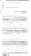
Sequence analysis of the 195-kD protein. (A) cDNAs encoding the 195-kD protein that were sequenced are depicted in schematic form. The ov 6D1a cDNA was isolated from an ovary expression library, and the partial cDNAs were isolated from an 8–12-h cDNA library. The Em 10C cDNA appears to extend to the 5′ end of the transcript whereas the Em 10A cDNA extends to the poly (A) tail. The sequence data are available from GenBank/ EMBL/DDBJ under the accession number AF041382. (B) An alignment of the entire amino acid sequence of D-CLIP-190 (uppercase) and CLIP-170 (lowercase) is shown. Identities (vertical lines), conservative substitutions (two dots), and less conservative substitutions (one dot) are determined by the Genetic Computer Group program Bestfit (Devereux et al., 1984). The first 48 amino acids of D-CLIP-190 do not align with CLIP-170. The amino terminus contains two repeats, rep1 and rep2, (black lines) that have been demonstrated to mediate binding of CLIP-170 to microtubules. The central region of both proteins is predicted to be an extended region of coiled coil (arrows indicate start and end points; Lupas et al., 1991). The predicted coiled-coil domain of D-CLIP-190 is interrupted after ∼100 amino acids by four prolines (asterisks), which are thought to disrupt α helices. The carboxy-terminal domain has two conserved sequences: a metal-binding motif (shaded) and a cysteine-rich sequence of 23 amino acids that does not conform to any previously recognized motif (dotted line).
Figure 4
Figure 4
Comparison of D-CLIP-190 and CLIP-170 predicted sequences. (A) The 195-kD protein (D-CLIP-190) is the Drosophila homologue of CLIP-170. A schematic drawing of the D-CLIP-190/CLIP-170 protein is shown. The protein can be divided into three regions: the amino-terminal domain, the coiled-coil domain, and the carboxy-terminal domain. The percent identity and similarity between D-CLIP-190 and CLIP-170 are indicated below each region. The main sequence and structural features of the protein are indicated. rep1 and rep2 are the two putative microtubule-binding motifs shared with CLIP-170. The coiled coil, the 23 amino acids–conserved motif (shaded box), and the metal-binding motif (thick black line) are also represented. (B) The MegAlign program (DNAStar), using the CLUSTAL method with the PAM250 residue weight table (Higgens et al., 1992), was used to align the putative microtubule-binding motifs from D-CLIP-190, CLIP-170 (Pierre et al., 1992), DP-150 (Holzbaur et al., 1991), Glued (Swaroop et al., 1987), BIK1 (Truehart et al., 1987), and yeast ORF YPL174c from the yeast sequence database (Genbank/EMBL/DDBJ accession number 1370367). There is a high degree of identity in this motif among the members represented. Notice that when comparing rep1 of D-CLIP-190 to that of CLIP-170 there is a higher level of conservation than when comparing rep1 to the motif from other proteins. The same is true for rep2. DP-150Glued is a component of the dynactin complex, an activator of dynein-mediated vesicle motility. DP-150Glued has one amino-terminal repeat as well as an extended region of coiled coil like CLIP-170/D-CLIP-190 (Swaroop et al., 1987; Truehart et al., 1987; Holzbaur et al., 1991). The yeast protein BIK1 is a microtubule-associated protein required for anaphase spindle movement (Berlin et al., 1990; Pellman et al., 1995). It also possesses a single repeat region and a region of coiled coil. Another yeast protein (ORF YPL174c; Genbank/EMBL/DDBJ accession number 1370367) with this motif was identified in a search of the yeast database. This protein has a putative microtubule-binding motif that contains the conserved sequence, GKN(D/S)G; however, it does not appear to have an extensive coiled-coil region (Lupas et al., 1991). (C) The metal-binding motifs from D-CLIP-190, CLIP-170 (Pierre et al., 1992), BIK1 (Truehart et al., 1987), and GAG (Berg, 1986) were aligned with the MegAlign program (DNAStar) using the CLUSTAL method with the PAM250 residue weight table (Higgens et al., 1992). The metal-binding motif consensus (MBM con) is also shown (Copeland et al., 1984; Berg, 1986). (D) A conserved motif of 23 amino acids in the carboxy terminus of D-CLIP-190 and CLIP-170 was aligned by Bestfit (Devereux et al., 1984). This highly conserved sequence, which is similar to the conserved metal-binding motif, contains several cysteines and a histidine (asterisks) whose spacing does not fit the consensus. Notably, a cysteine in CLIP-170 is not conserved in D-CLIP-190 (shaded).
Figure 5
Figure 5
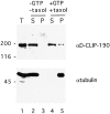
Cosedimentation of D-CLIP-190 with microtubules. Immunoblot of samples after polymerization and sedimentation of microtubules from embryo extract. Lane 1, embryo extract; lanes 2 and 4, supernatants; lanes 3 and 5, pellets without (lanes 2 and 3) or with (lanes 4 and 5) the addition of GTP and taxol. Supernatants and pellets from MT sedimentation experiments were transferred to nitrocellulose and the resulting blot was cut in half. The upper half was probed with αD-CLIP-190 antibody (mouse α6D1a) and the lower half with αtubulin antibody.
Figure 6
Figure 6
Colocalization of 95F myosin and D-CLIP-190 in the central nervous system. Confocal images of stage 14 (A–C) and stage 16 (D–L) embryos double labeled with α95F myosin (A, D, G, and J) and αD-CLIP-190 (B, E, H, and K) antibodies are shown. A lateral view is shown in A–C; ventral view is shown in D–F. High magnification views of the central nervous system in two different stage 16 embryos are shown in G–L. The embryo in G–I is oriented similarly to that shown in D–F. The embryo in J–L is oriented more laterally. An overlay of the localization of 95F myosin (red) and D-CLIP-190 (green) is shown in C, F, I, and L. Overlap in the distribution of the two proteins is yellow. Arrows indicate examples of punctate staining in which the two proteins colocalize. Bars: (F) 100 μm; (L), 10 μm.
Figure 7
Figure 7
Primary embryonic cultures stained with anti- D-CLIP-190 and 95F myosin antibodies. (A–C) A hemocyte showing colocalization of 95F myosin (A) and D-CLIP-190 (B) in large cytoplasmic organelles. The composite image in (C) shows D-CLIP-190 in green and 95F myosin in red. Regions of colocalization are yellow. (D–F) Two myoblasts and a hemocyte (well-spread cell) in which 95F myosin (D) and D-CLIP-190 (E) colocalize are shown. The composite image is shown in (F) with color scheme as in (C). Bar, 10 μm.
Figure 8
Figure 8
The posterior pole localization of D-CLIP-190 and 95F myosin is dependent on the actin cytoskeleton. Confocal images of control embryos (no drug; A–C) and embryos treated with 10 μg/ml cytochalasin D for 30 min to depolymerize actin filaments (CD; D–F) are shown. These embryos were double labeled with αD-CLIP-190 (A and D) and α95F myosin (B and E) antibodies. A composite of the individual images is shown in C and F (D-CLIP-190, green; 95F myosin, red). Overlap of the two proteins is shown in yellow. D-CLIP-190 and 95F myosin proteins are enriched at the posterior pole. D-CLIP-190 appears to be present at a higher level and present in a broader distribution than 95F myosin at the posterior pole. Levels of 95F myosin are higher than D-CLIP-190 in the cortex. The cytochalasin D–treated embryo shown still has residual protein at the posterior pole (indicated as ± in Table I). The majority of αD-CLIP-190 and α95F myosin-labeled embryos have no observable posterior pole localization (Table I).

Similar articles

Cited by

References

    1. Altschul SF, Gish W, Miller W, Myers EW, Lipman DJ. Basic local alignment search tool. J Mol Biol. 1990;215:403–410. - PubMed
    1. Amero, S.A., T.C. James, and S.C.R. Elgin. 1987. Raising antibodies to protein bands in gels. In Methods in Molecular Biology. Vol. 3. J.M. Walker, editor. Humana Press, Inc., Clifton, NJ. 355–362.
    1. Berg JM. Potential metal-binding domains in nucleic acid binding proteins. Science. 1986;232:485–487. - PubMed
    1. Berlin V, Styles CA, Fink GR. BIK1, a protein required for microtubule function during mating and mitosis in Saccharomyces cerevisiaecolocalizes with tubulin. J Cell Biol. 1990;111:2573–2586. - PMC - PubMed
    1. Bilbe G, Delabie J, Bruggen J, Richener H, Asselbergs FAM, Cerletti N, Sorg C, Odink K, Tarcsay L, Wiesendanger W, DeWolf-Peeters C, Shipman R. Restin: a novel intermediate filament-associated protein highly expressed in the Reed-Sternberg cells of Hodgkin's disease. EMBO (Eur Mol Biol Organ) J. 1992;11:2103–2113. - PMC - PubMed

Publication types

MeSH terms

Associated data