. 1998 Feb;9(2):387-402.
doi: 10.1091/mbc.9.2.387.

Involvement of ATP-dependent Pseudomonas exotoxin translocation from a late recycling compartment in lymphocyte intoxication procedure

Affiliations
Free PMC article

Involvement of ATP-dependent Pseudomonas exotoxin translocation from a late recycling compartment in lymphocyte intoxication procedure

M Alami et al. Mol Biol Cell. 1998 Feb.
Free PMC article

Abstract

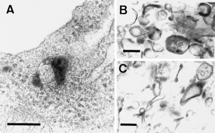
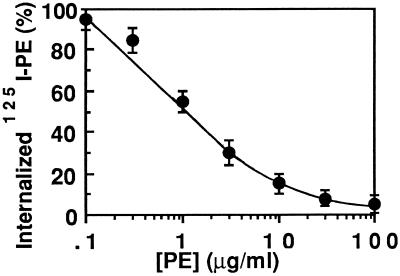
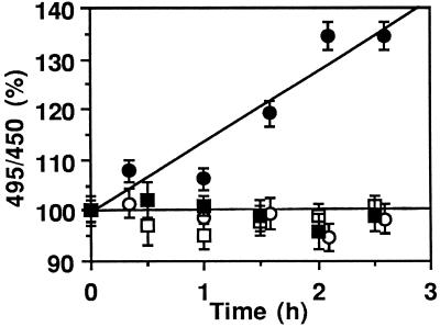
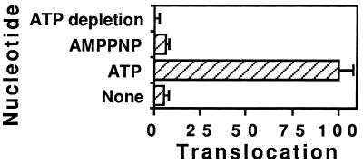
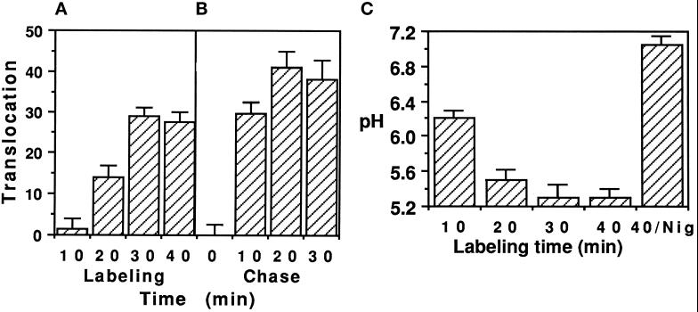
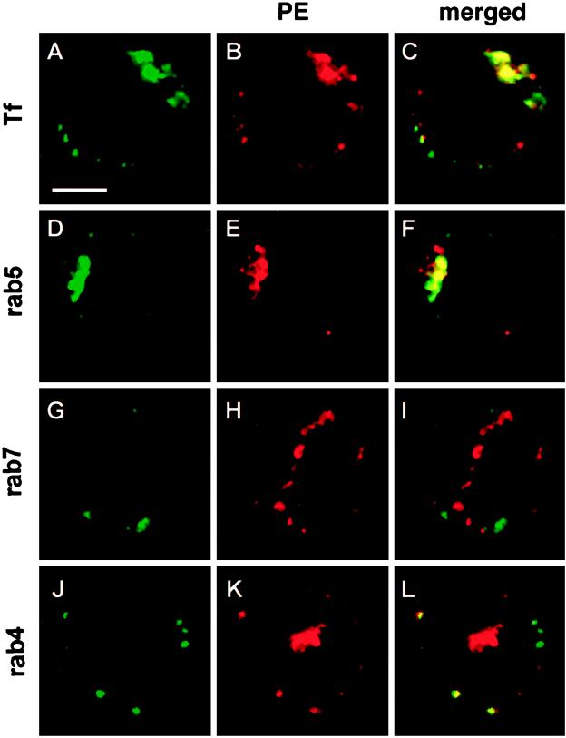
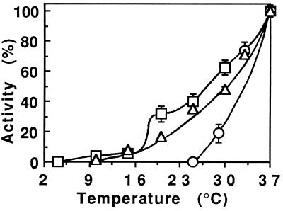
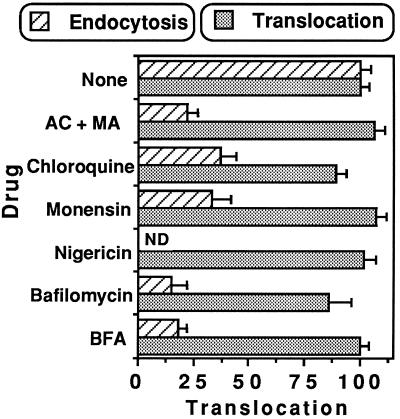
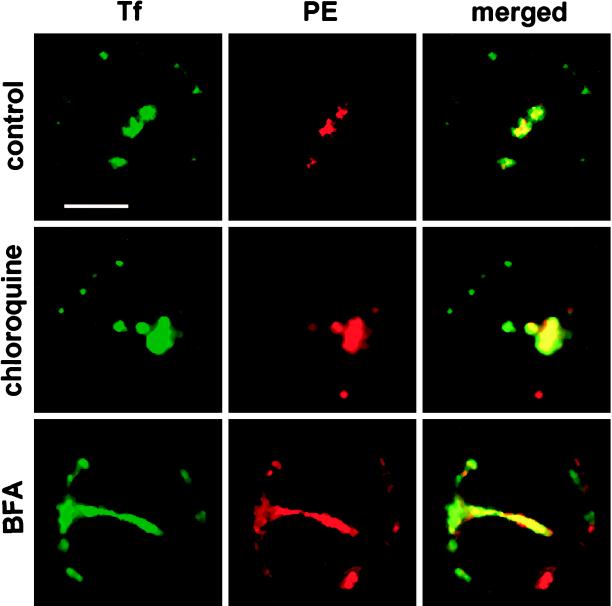
Pseudomonas exotoxin (PE) is a cytotoxin which, after endocytosis, is delivered to the cytosol where it inactivates protein synthesis. Using diaminobenzidine cytochemistry, we found over 94% of internalized PE in transferrin (Tf) -positive endosomes of lymphocytes. When PE translocation was examined in a cell-free assay using purified endocytic vesicles, more than 40% of endosomal 125I-labeled PE was transported after 2 h at 37 degrees C, whereas a toxin inactivated by point mutation in its translocation domain was not translocated. Sorting of endosomes did not allow cell-free PE translocation, whereas active PE transmembrane transport was observed after > 10 min of endocytosis when PE and fluorescent-Tf were localized by confocal immunofluorescence microscopy within a rab5-positive and rab4- and rab7-negative recycling compartment in the pericentriolar region of the cell. Accordingly, when PE delivery to this structure was inhibited using a 20 degrees C endocytosis temperature, subsequent translocation from purified endosomes was impaired. Translocation was also inhibited when endosomes were obtained from cells labeled with PE in the presence of brefeldin A, which caused fusion of translocation-competent recycling endosomes with translocation-incompetent sorting elements. No PE processing was observed in lymphocyte endosomes, the full-sized toxin was translocated and recovered in an enzymatically active form. ATP hydrolysis was found to directly provide the energy required for PE translocation. Inhibitors of endosome acidification (weak bases, protonophores, or bafilomycin A1) when added to the assay did not significantly affect 125I-labeled PE translocation, demonstrating that this transport is independent of the endosome-cytosol pH gradient. Nevertheless, when 125I-labeled PE endocytosis was performed in the presence of one of these molecules, translocation from endosomes was strongly inhibited, indicating that exposure to acidic pH is a prerequisite for PE membrane traversal. When applied during endocytosis, treatments that protect cells against PE intoxication (low temperatures, inhibitors of endosome acidification, and brefeldin A) impaired 125I-labeled PE translocation from purified endosomes. We conclude that PE translocation from a late receptor recycling compartment is implicated in the lymphocyte intoxication procedure.


Figures

Figure 1
Figure 1
Electron micrographs of BW 5147 cells and the endosome preparation after labeling with PE-gold and Tf-HRP. Cells were labeled for 30 min at 37°C with 1 μg/ml PE-gold and 20 μg/ml Tf-HRP, washed, and then either fixed directly or first fractionated to obtain endosomes. Whole cells (A) or the endosome preparation (B and C) were treated to reveal HRP activity for electron microscopy. Sections were examined after staining with uranyl acetate and lead citrate. Reaction products for Tf-HRP are contained within endocytic vesicles and PE-gold is observed within Tf-HRP-positive compartments. Bar, 0.2 μm.
Figure 2
Figure 2
Inhibition of 125I-labeled PE uptake by PE. BW cells were labeled with 1 μg/ml 125I-labeled PE for 30 min at 37°C in the presence of the indicated PE concentration. Internalization was followed as the fraction of cell-associated 125I-labeled PE resistant to Pronase treatment (0.3% in PBS) for 45 min at 2°C. Twenty to 25% of cell-associated 125I-labeled PE (5, 500 cpm/23,000 cpm) was intracellular after uptake at 37°C in the absence of competing PE. The results are expressed as a percentage of this value.
Figure 3
Figure 3
Fluorescent PE can cross the endosome membrane. BW cells were incubated for 30 min at 37°C with 2 μg/ml PE-FITC (• and ○) or PE343Q-FITC (▪ and □). Translocation was assayed as described in MATERIALS AND METHODS using isolated endosomes in the presence (closed symbols) or absence (open symbols) of 10 mM ATP + 10 mM MgCl2. Fluorescein fluorescence excitation was obtained at 450 or 495 nm and measured at 520 nm. Upon addition of 5 μM nigericin, a pH level corresponding to the buffer value was obtained for all fluorescent tracers.
Figure 4
Figure 4
Translocation of 125I-labeled PE across the endosome membrane. After labeling for 30 min at 37°C with 1 μg/ml 125I-labeled PE (▴, •, and ○) or 125I-labeled PE343Q (▪ and □), BW cells were fractionated for endosome isolation. Translocation was assayed in the presence (closed symbols) or absence (open symbols) of 10 mM ATP and was terminated by cooling on ice. (▴) An ATP-depleting system (15 mM glucose, 50 U/ml hexokinase) was added 1 h after the beginning of the assay (arrow). Ultracentrifugation for 5 min at 160,000 × g on a 17% sucrose cushion separated the translocation medium from the endosomes. The radioactivity ratio (medium/endosomes) was used to directly monitor translocation. At the beginning of the experiment, 100–250 cpm and 2000–5000 cpm were routinely present in the medium and the endosomes, respectively.
Figure 5
Figure 5
Nucleotide requirement for PE translocation. After labeling BW endosomes with 125I-labeled PE (or 125I-labeled PE343Q as control), translocation was assayed as described in the legend of Figure 4 in the presence of the indicated nucleotide.
Figure 6
Figure 6
PE translocation is restricted to mature endosomes. (A) Cells were labeled with 1 μg/ml 125I-labeled PE at 37°C for the indicated times before endosome isolation and translocation assay. (B) 125I-labeled PE endocytosis was stopped after 10 min at 37°C. Excess ligand was washed out and a chase was performed at 37°C in DMEM/BSA/LDL supplemented with PE (100 μg/ml) for the indicated period of time. (C) After labeling with 2 μg/ml PE-FITC for the indicated times at 37°C, endosomes were isolated and resuspended in translocation buffer supplemented with 2 mM ATP. Endosome pH was measured within 5 min using the 495:450 nm excitation ratio for fluorescein fluorescence. A calibration curve was plotted using PE-FITC dilutions in appropriate buffers. When indicated (Nig), 5 μM nigericin were added to the translocation buffer.
Figure 7
Figure 7
Localization of internalized PE in BW lymphocytes by confocal immunofluorescence microscopy. Cells were incubated with PE (2 μg/ml) for 30 min at 37°C and then washed, fixed, permeabilized, and processed for detection of PE using affinity-purified goat anti-PE, mouse monoclonal anti-goat and TRITC-labeled goat anti-mouse antibodies. When indicated, rab proteins were labeled using affinity-purified rabbit antibodies and FITC-labeled swine anti-rabbit antibodies. (A–C) Tf-FITC (25 μg/ml) was coendocytosed with PE. (D–F) Localization of rab5 and PE. In these sections (A–F), the label is concentrated at one pole of the cell in the pericentriolar region. (G–I) Localization of rab7 and PE. (J–L) Localization of rab4 and PE. In these cells (G–L), PE and recycling endosomes are observed within a valley between two lobes of the nucleus, whereas neither rab7 (and late endosomes) nor rab4 (and sorting endosomes) are found in this area. Representative medical optical sections recorded using the FITC and the TRITC channels, and the combination of both images are presented. Bar, 5 μm.
Figure 8
Figure 8
Translocated PE is enzymatically active. Cells were incubated with PE for 30 min at 37°C before fractionation and endosome isolation. Translocation was assayed for the indicated times in the presence of the indicated nucleotide, and translocation media were obtained by ultracentrifugation. Their ADP ribosylation activity was tested after dialysis using partially purified EF2 and 32P-labeled NAD.
Figure 9
Figure 9
Temperature dependence of 125I-labeled PE routing to the cytosol. ▵, Endocytosis was measured (after 30 min at the indicated temperature) as the fraction of cell-associated label resistant to Pronase scraping (see Figure 2). ○, Acquisition of translocation competence was deduced from translocation measurements performed at 37°C on endosomes isolated from cells labeled with 1 μg/ml 125I-labeled PE for 30 min at the indicated temperature. □, Translocation temperature dependence was observed using endosomes prepared from cells labeled with 125I-labeled PE for 30 min at 37°C. All results are expressed as percentages of the 37°C value.
Figure 10
Figure 10
When present during endocytosis, drugs protecting cells from PE intoxication impaired endosomal 125I-labeled PE translocation. The following concentrations were used: NH4Cl/methylamine (AC/MA), 20 mM each; chloroquine, 50 μM; monensin, 50 μM; nigericin, 5 μM; bafilomycin A1, 0.5 μM; BFA, 5 μg/ml. Effect on translocation: cells were labeled with 1 μg/ml 125I-labeled PE (in the absence of drug) before endosome isolation; translocation was assayed in the presence of the indicated molecules (n = 6). Effect during endocytosis, i.e., on acquisition of translocation competence: cells were preincubated for 10 min at 37°C in the presence of the indicated molecules before adding 1 μg/ml 125I-labeled PE and 150 μg/ml LDL. After 30 min at 37°C, cells were fractionated to isolate endosomes. Translocation was assayed in the absence of drug (n = 4). ND, not determined.
Figure 11
Figure 11
The morphology of the recycling compartment was markedly affected by BFA but not by chloroquine. Lymphocytes were incubated with BFA (5 μg/ml) or chloroquine (50 μM) as indicated for 10 min at 37°C before the addition of Tf-FITC (25 μg/ml) and PE (2 μg/ml). After 30 min at 37°C, cells were washed, fixed, and stained for PE using a TRITC-labeled tertiary antibody as described in the legend of Figure 7. Samples were then examined under a confocal microscope. Both medical optical sections recorded using the FITC and the TRITC channels and their overlays are presented. Bar, 5 μm.
Figure 12
Figure 12
Full-length PE translocates from purified BW endosomes. After cell labeling with 1 μg/ml 125I-labeled PE for 30 min at 37°C, endosomes were isolated and translocation was assayed for the indicated times in the presence of 10 mM ATP. Endosomal (Endos.) and translocated proteins (after TCA precipitation) were analyzed using reducing SDS/PAGE followed by autoradiography.

Similar articles

Cited by

References

    1. Allured V, Collier RJ, Caroll SF, McKay DB. Structure of exotoxin A of Pseudomonas aeruginosa at 3.0 Angstrom resolution. Proc Natl Acad Sci USA. 1986;83:1320–1324. - PMC - PubMed
    1. Bastiaens PI, Majoul IV, Verveer PJ, Söling HD, Jovin TM. Imaging the intracellular trafficking and state of the AB5 quaternary structure of cholera toxin. EMBO J. 1996;15:4246–4253. - PMC - PubMed
    1. Beaumelle B, Alami M, Hopkins CR. ATP-dependent translocation of ricin across the membrane of purified endosomes. J Biol Chem. 1993;268:23661–23669. - PubMed
    1. Beaumelle B, Bensamar L, Bienvenüe A. Selective translocation of the A chain of Diphtheria Toxin across the membrane of purified endosomes. J Biol Chem. 1992;267:11525–11531. - PubMed
    1. Beaumelle BD, Gibson A, Hopkins CR. Isolation and preliminary characterization of the major membrane boundaries of the endocytic pathway in lymphocytes. J Cell Biol. 1990;111:1811–1823. - PMC - PubMed

Publication types

MeSH terms

LinkOut - more resources