Virus attenuation after deletion of the cytomegalovirus Fc receptor gene is not due to antibody control
- PMID: 9445038
- PMCID: PMC124616
- DOI: 10.1128/JVI.72.2.1377-1382.1998
Virus attenuation after deletion of the cytomegalovirus Fc receptor gene is not due to antibody control
Abstract
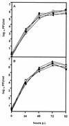
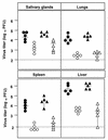
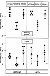
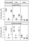
The murine cytomegalovirus (MCMV) fcr-1 gene codes for a glycoprotein located at the surface of infected cells which strongly binds the Fc fragment of murine immunoglobulin G. To determine the biological significance of the fcr-1 gene during viral infection, we constructed MCMV fcr-1 deletion mutants and revertants. The fcr-1 gene was disrupted by insertion of the Escherichia coli lacZ gene. In another mutant, the marker gene was also deleted, by recombinase cre. As expected for its hypothetical role in immunoevasion, the infection of mice with fcr-1 deletion mutants resulted in significantly restricted replication in comparison with wild-type MCMV and revertant virus. In mutant mice lacking antibodies, however, the fcr-1 deletion mutants also replicated poorly. This demonstrated that the cell surface-expressed viral glycoprotein with FcR activity strongly modulates the virus-host interaction but that this biological function is not caused by the immunoglobulin binding property.
Figures


Similar articles
-
Identification and expression of a murine cytomegalovirus early gene coding for an Fc receptor.J Virol. 1994 Dec;68(12):7757-65. doi: 10.1128/JVI.68.12.7757-7765.1994. J Virol. 1994. PMID: 7966565 Free PMC article.
-
The herpesviral Fc receptor fcr-1 down-regulates the NKG2D ligands MULT-1 and H60.J Exp Med. 2006 Aug 7;203(8):1843-50. doi: 10.1084/jem.20060514. Epub 2006 Jul 10. J Exp Med. 2006. PMID: 16831899 Free PMC article.
-
Characterization of domains of herpes simplex virus type 1 glycoprotein E involved in Fc binding activity for immunoglobulin G aggregates.J Virol. 1994 Apr;68(4):2478-85. doi: 10.1128/JVI.68.4.2478-2485.1994. J Virol. 1994. PMID: 7511171 Free PMC article.
-
The contrasting IgG-binding interactions of human and herpes simplex virus Fc receptors.Biochem Soc Trans. 2002 Aug;30(4):495-500. doi: 10.1042/bst0300495. Biochem Soc Trans. 2002. PMID: 12196122 Review.
-
Cytomegaloviral control of MHC class I function in the mouse.Immunol Rev. 1999 Apr;168:167-76. doi: 10.1111/j.1600-065x.1999.tb01291.x. Immunol Rev. 1999. PMID: 10399073 Review.
Cited by
-
Inflammatory monocytes and NK cells play a crucial role in DNAM-1-dependent control of cytomegalovirus infection.J Exp Med. 2016 Aug 22;213(9):1835-50. doi: 10.1084/jem.20151899. Epub 2016 Aug 8. J Exp Med. 2016. PMID: 27503073 Free PMC article.
-
Fast screening procedures for random transposon libraries of cloned herpesvirus genomes: mutational analysis of human cytomegalovirus envelope glycoprotein genes.J Virol. 2000 Sep;74(17):7720-9. doi: 10.1128/jvi.74.17.7720-7729.2000. J Virol. 2000. PMID: 10933677 Free PMC article.
-
Herpesvirus homologues of cellular genes.Virus Genes. 2000;21(1-2):65-75. Virus Genes. 2000. PMID: 11022790 Review.
-
The immunoevasive function encoded by the mouse cytomegalovirus gene m152 protects the virus against T cell control in vivo.J Exp Med. 1999 Nov 1;190(9):1285-96. doi: 10.1084/jem.190.9.1285. J Exp Med. 1999. PMID: 10544200 Free PMC article.
-
Modulation of innate and adaptive immunity by cytomegaloviruses.Nat Rev Immunol. 2020 Feb;20(2):113-127. doi: 10.1038/s41577-019-0225-5. Epub 2019 Oct 30. Nat Rev Immunol. 2020. PMID: 31666730 Review.
References
-
- Beersma M F C, Bijlmakers M J E, Ploegh H L. Human cytomegalovirus down-regulates HLA class I expression by reducing the stability of class I heavy chains. J Immunol. 1993;51:4455–4464. - PubMed
-
- Chou J, Kern E R, Whitley R J, Roizman B. Mapping of herpes simplex virus-1 neurovirulence to γ134.5, a gene nonessential for growth in culture. Science. 1990;250:1262–1266. - PubMed
Publication types
MeSH terms
Substances
LinkOut - more resources
Full Text Sources
Other Literature Sources
Research Materials

