Comparative Study
. 1998 Jan 1;18(1):328-38.
doi: 10.1523/JNEUROSCI.18-01-00328.1998.

Comparison of neurite outgrowth induced by intact and injured sciatic nerves: a confocal and functional analysis

Affiliations
Comparative Study

Comparison of neurite outgrowth induced by intact and injured sciatic nerves: a confocal and functional analysis

E Agius et al. J Neurosci. .

Abstract

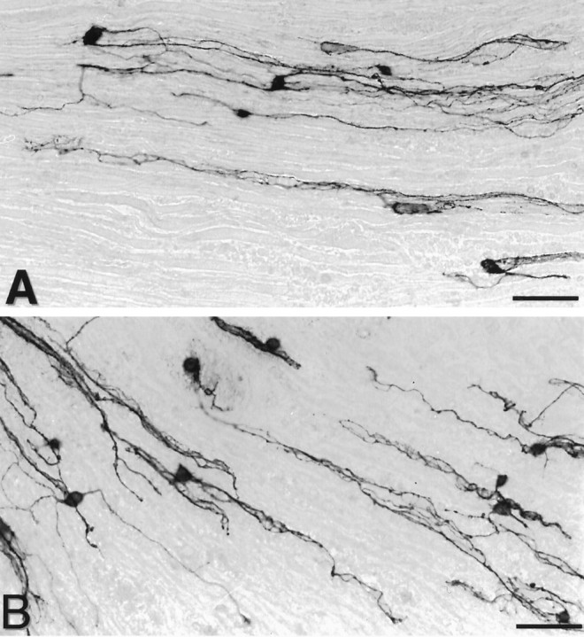
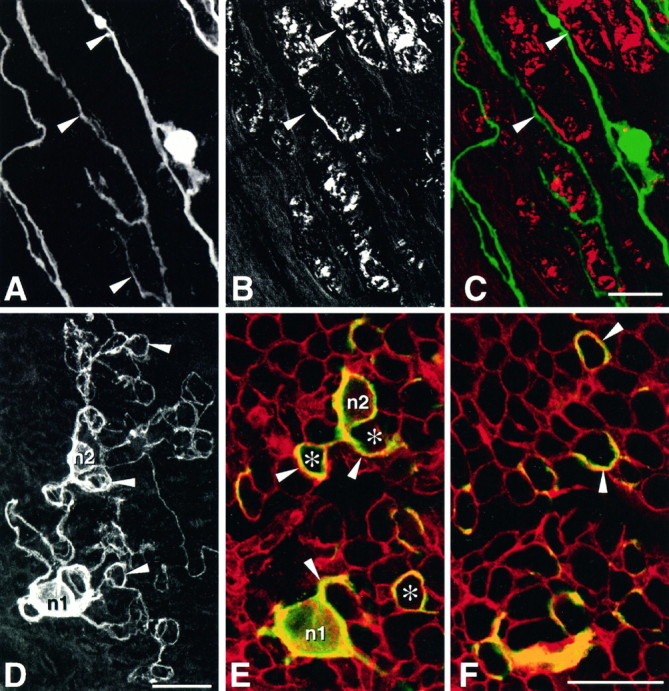
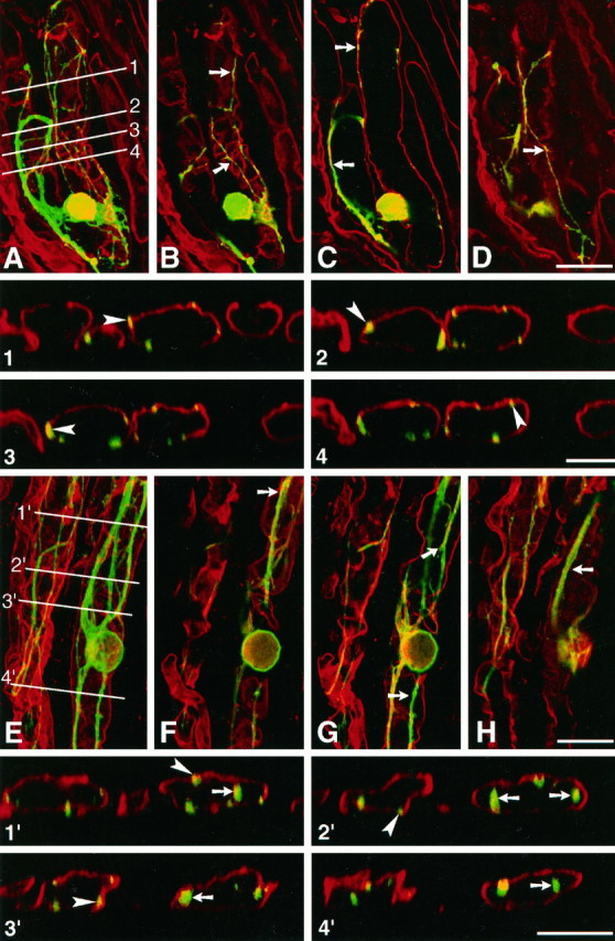
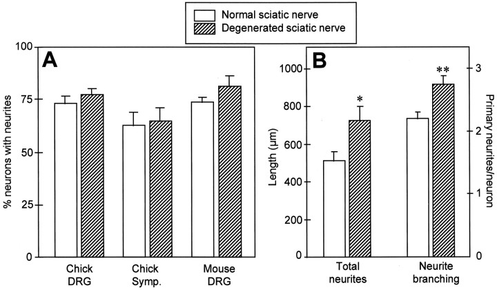
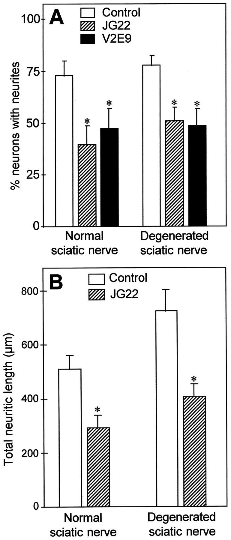
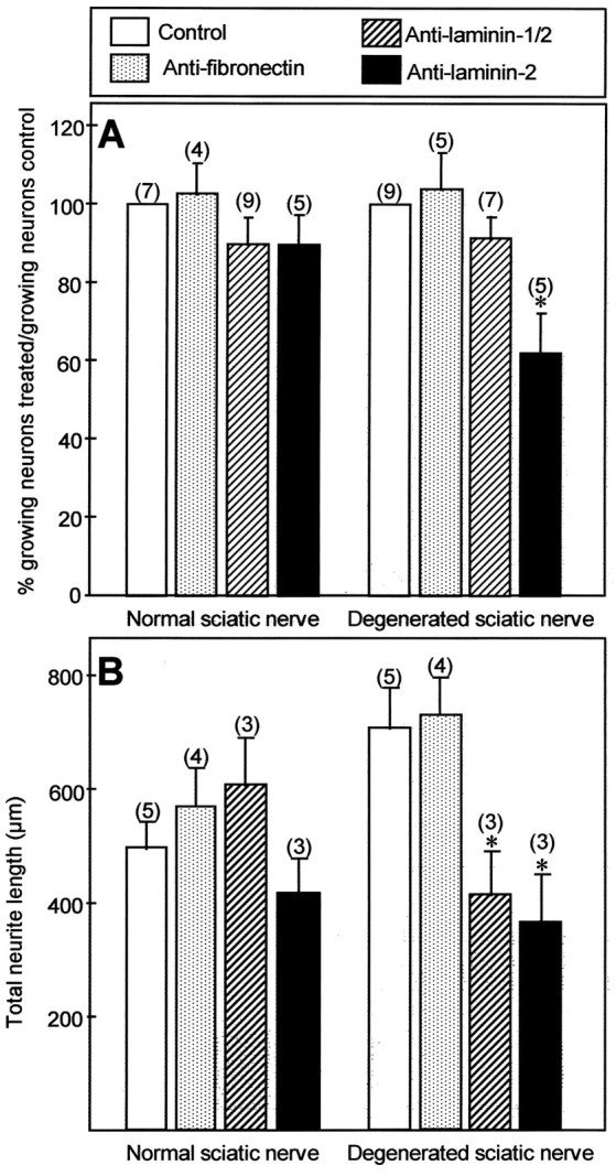
Mechanisms regulating axon growth in the peripheral nervous system have been studied by means of an in vitro bioassay, the tissue section culture, in which regenerating neurons are grown on substrata made up of tissue sections. Sections from intact and degenerated sciatic nerves proved to be different in their ability to support neurite outgrowth of embryonic chick sensory neurons from both qualitative and quantitative points of view. On denervated nerve sections, the total length of neurites elaborated per neuron was almost twice that found on intact nerve sections. In addition, confocal microscopy revealed a striking difference between intact and denervated nerve substrata: on denervated nerve sections, neurites grew inside the internal structures of endoneurial Schwann cell tubes, within the underlying tissue sections, whereas on intact nerve sections neurites extended along endoneurial basal laminae but never entered Schwann cell tubes. Perturbation experiments were used to analyze some of the molecular determinants that control neurite outgrowth in this system. Antibodies directed against the beta1-integrin subunit inhibited neurite extension on both normal and degenerated rat sciatic nerve tissue. Strikingly, however, differential inhibition was observed using antibodies directed against extracellular matrix molecules. Anti-laminin-2 (merosin) antibodies drastically reduced both the percentage of growing neurons and the total length of neurites on denervated nerve sections, but they did not modify these parameters on sections of normal nerve. Taken together, these results suggest that laminin-2/merosin promotes neurite outgrowth in peripheral nerve environments but only after Wallerian degeneration, which is when axons are allowed to extend within endoneurial tubes.


Figures

Fig. 1.
Fig. 1.
Characterization of neurite outgrowth of E13 chick sensory neurons grown for 48 hr under control conditions on longitudinal sections obtained from normal rat nerves (A) or from the distal portions of nerves transected 2 weeks before (B). Neurons were stained with anti-chicken N-CAM antibody (5e). On both types of substrata, neurons extend long processes oriented longitudinally. Scale bar, 100 μm.
Fig. 2.
Fig. 2.
Growth of E13 chick sensory neurons on longitudinal sections of denervated sciatic nerve (A–C) and on transverse sections of intact nerve (D–F). A–C, Comparison of the pattern of neurite outgrowth with respect to the localization of myelin-rich areas in the underlying substratum. A, Neurons and their neurites (arrowheads) were stained with anti-N-CAM antibodies and alkaline phosphate conjugate, observed under transmitted light, as in Figure 1, and their image was inverted.B, Myelin in the same field was observed under polarized light. C, Superimposition of both images, after color coding neurons in green and myelin inred, reveals that in most instances neurites extend in areas devoid of myelin (arrowheads).D–F, Pattern of growth of two neurons (n1 and n2) cultivated on transverse nerve sections and stained with anti-N-CAM antibodies.D, Total neuritic arborization. Neurites extend neurites that do not grow straight on the substratum but form loops (arrowheads). E, F, Confocal imaging of the same neurons. Neurons and their neurites stained with anti-N-CAM antibodies are displayed in green, whereas basal laminae stained with anti-laminin-1/2 antibodies are displayed inred. E, Horizontal optical section acquired at the surface of the substratum showing the base of the neuronal cell bodies n1 and n2 and the proximal part of their neurites (arrowheads). Note that neurites do not extend across the surface of the underlying axon units containing myelin (asterisks) but circle around, in association with laminin-stained endoneurial basal laminae.F, Horizontal optical section acquired 3 μm deeper in the preparation. Neurites (arrowheads) extend deep inside the substratum but still do not cross myelin-containing axon units. Scale bars, 25 μm.
Fig. 3.
Fig. 3.
Confocal analysis of neurite outgrowth of embryonic chicken neurons cultivated on rat sciatic nerve sections. Cultures were fixed after 48 hr and stained using an mAb directed against chicken N-CAM, revealed with FITC. Laminin-2 (merosin) in the substratum was decorated with an antiserum revealed with TRITC.A–D and 1–4, Culture on normal nerve section. A, Complete reconstruction, by extended depth of focus, of a growing neuron and underlying substratum, obtained by superimposition of consecutive images in the horizontal plane. The neuritic arborization (green) is confined to two adjacent Schwann cell tubes, defined by laminin-2 immunoreactive basal lamina scaffolds. B–D, Consecutive horizontal optical sections of the same neuron, obtained at 5 μm intervals. The plane of image C lies at midlevel of the Schwann cell tube. On normal nerve substratum, neurites extend in contact with laminin-immunoreactive basal laminae (arrows). Note inC that they do not grow inside Schwann cell tubes.Insets 1–4, Successive vertical optical sections performed perpendicular to the long axis of Schwann cell tubes, along the lines 1–4 shown in A. Such optical sections confirm that neurites grow in contact with basal laminae (arrowheads) but do not extend inside Schwann cell tubes, despite the fact that some neurites are found deep inside the tissue section. E–F and 1′–4′, Culture on degenerated nerve section. E, Complete reconstruction as in A of a growing neuron and underlying substratum. The neuritic arborization of the neuron seen on theright is confined to a single Schwann cell tube. Neurites of another neuron (cell body not visible in the micrograph) extend along a second basal lamina scaffold, on the leftof the previous neuron. F–H, Consecutive horizontal optical sections of the same field, obtained at 5 μm intervals. The plane of image G lies at midlevel of the Schwann cell tube. On denervated substrata, neurites also extend in contact with laminin-immunoreactive basal laminae (arrows). However, note in G that they grow profusely inside Schwann cell tubes. Insets 1′–4′, Successive vertical optical sections performed perpendicular to the long axis of Schwann cell tubes, along the lines 1′–4′ shown in E. Such optical sections confirm that on denervated nerve sections neurites grow not only in contact with basal laminae (arrowheads) but also extend inside Schwann cell tubes (arrows). Scale bars: A–D, E–H, 25 μm; 1–4 and 1′–4′, 10 μm.
Fig. 4.
Fig. 4.
Comparison of neurite outgrowth on normal and degenerated sciatic nerve sections. A, Quantification of the number of growing E13 chick sensory neurons (Chick DRG), E13 chick sympathetic neurons (Chick Symp.), and newborn mouse sensory neurons (Mouse DRG), cultivated for 48 hr under control conditions on normal or degenerated sciatic nerve substrata. Values are the mean and SEM of the percentage of growing neurons, from at least three independent experiments. B, Quantification of total neuritic length and neurite branching of E13 chick sensory neurons grown on normal and degenerated sciatic nerve sections. Left y-axis: mean and SEM of total neuritic length elaborated per neuron (Total neurites); right y-axis: mean and SEM of the number of primary neurites elaborated per neuron (Neurite branching). Data were calculated from at least 120 neurons. Significance compared with normal sciatic nerve: *p< 0.05; **p < 0.01. Significance was analyzed using the Student’s t test.
Fig. 5.
Fig. 5.
Inhibition of neurite outgrowth of E13 chick sensory neurons by two different antibodies, JG22 and V2E9, directed against chick β1-integrin subunit, on each type of nerve substratum. The concentrations used were 25 μg Ig/ml for both antibodies. The blocking ability of each antibody was controlled with the same neuronal preparations grown on laminin-1 or laminin-2, and the corresponding results are shown in Table 1. A, Quantification of the number of neurons extending neurites after 2 d of culture. Values represent mean and SEM of the percentage of growing neurons for five different experiments. B, Quantification of neurite length. Values represent the mean and SEM of total neuritic length elaborated per neuron, measured for at least 120 neurons.*p < 0.05. Significance was analyzed using the Student’s t test.
Fig. 6.
Fig. 6.
Effects of anti-fibronectin, anti-laminin-1/2, and anti-laminin-2 antibodies on neurite outgrowth of E13 chick sensory neurons grown for 48 hr on normal and degenerated sciatic nerve substrata. For laminin-1/2 and fibronectin, at least two different antibodies were tested, with concentrations between 25 and 50 μg Ig/ml, yielding similar results. The blocking ability of each antibody was controlled with the same neuronal preparations grown on the corresponding ECM molecule, and the corresponding results are shown in Table 2. A, Quantification of the number of neurons extending neurites after 2 d of culture. Values represent mean and SEM of the percentage of growing neurons in treated conditions normalized to the number of growing neurons in control cultures (100%) for the number of experiments indicated in parentheses.B, Quantification of neurite length. Values represent the mean and SEM of total neuritic length elaborated per neuron, measured for at least 100 neurons.*p < 0.05. Significance was analyzed using the Student’s ttest.

Similar articles

Cited by

References

    1. Agius E, Sagot Y, Duprat AM, Cochard P. Antibodies directed against the β1-integrin subunit and peptides containing the IKVAV sequence of laminin perturb neurite outgrowth of peripheral neurons on immature spinal cord substrata. Neuroscience. 1996;71:773–786. - PubMed
    1. Anton ES, Sandrock AJ, Matthew WD. Merosin promotes neurite growth and Schwann cell migration in vitro and nerve regeneration in vivo: evidence using an antibody to merosin, ARM-1. Dev Biol. 1994;164:133–146. - PubMed
    1. Arber S, Caroni P. Thrombospondin-4, an extracellular matrix protein expressed in the developing and adult nervous system, promotes neurite outgrowth. J Cell Biol. 1995;131:1083–1094. - PMC - PubMed
    1. Bastmeyer M, Beckmann M, Schwab ME, Stuermer CAO. Growth of regenerating goldfish axons is inhibited by rat oligodendrocytes and CNS myelin but not by goldfish optic nerve tract oligodendrocyte-like cells and fish myelin. J Neurosci. 1991;11:626–640. - PMC - PubMed
    1. Bedi KS, Winter J, Berry M, Cohen J. Adult rat dorsal root ganglion neurons extend neurites on predegenerated but not normal peripheral nerves in vitro. Eur J Neurosci. 1992;4:193–200. - PubMed

Publication types

MeSH terms

LinkOut - more resources