. 1997 Dec 29;139(7):1775-83.
doi: 10.1083/jcb.139.7.1775.

Cytoskeletal association is important for differential targeting of glucose transporter isoforms in Leishmania

Affiliations

Cytoskeletal association is important for differential targeting of glucose transporter isoforms in Leishmania

E L Snapp et al. J Cell Biol. .

Abstract

The major glucose transporter of the parasitic protozoan Leishmania enriettii exists in two isoforms, one of which (iso-1) localizes to the flagellar membrane, while the other (iso-2) localizes to the plasma membrane of the cell body, the pellicular membrane. These two isoforms differ only in their cytosolic NH2-terminal domains. Using immunoblots and immunofluorescence microscopy of detergent-extracted cytoskeletons, we have demonstrated that iso-2 associates with the microtubular cytoskeleton that underlies the cell body membrane, whereas the flagellar membrane isoform iso-1 does not associate with the cytoskeleton. Deletion mutants that remove the first 25 or more amino acids from iso-1 are retargeted from the flagellum to the pellicular membrane, suggesting that these deletions remove a signal required for flagellar targeting. Unlike the full-length iso-1 protein, these deletion mutants associate with the cytoskeleton. Our results suggest that cytoskeletal binding serves as an anchor to localize the iso-2 transporter within the pellicular membrane, and that the flagellar targeting signal of iso-1 diverts this transporter into the flagellar membrane and away from the pellicular microtubules.


Figures
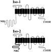
Figure 1
Figure 1
Schematic diagram showing the putative membrane topology of Pro-1 glucose transporter isoforms iso-1 and iso-2. Black boxes represent putative transmembrane domains. Loops above the boxes represent extracellular domains and the loops below the boxes represent intracellular domains. The hatched line of iso-1 and the bold black line for iso-2 indicate that the only sequence differences between the two isoforms occur within the NH2-terminal domains. Iso-1 has a 130–amino acid NH2-terminal domain, while iso-2 has a 48–amino acid NH2-terminal domain.
Figure 2
Figure 2
Immunoblots of Triton X-100 extracted L. enriettii fractions. L designates total lysate, S designates the supernatant fraction, and P designates the cytoskeletal pellet. iso-1 and iso-2 indicate the bands on the blot that correspond to each of these two glucose transporter isoforms. Samples were separated on a 10% SDS-polyacrylamide gel, blotted onto nitrocellulose, and probed with the indicated antibodies. The blots in A were probed with affinity-purified anti-P1C antibody (at a 1:100 dilution), and the blots in B were probed with anti-P1L (lane 1, 1:500), anti–α-tubulin (lane 2, 1:800), or anti–β-tubulin (lane 3, 1:1,000). Numbers at the left indicate the mobilities of molecular mass markers in kilodaltons.
Figure 2
Figure 2
Immunoblots of Triton X-100 extracted L. enriettii fractions. L designates total lysate, S designates the supernatant fraction, and P designates the cytoskeletal pellet. iso-1 and iso-2 indicate the bands on the blot that correspond to each of these two glucose transporter isoforms. Samples were separated on a 10% SDS-polyacrylamide gel, blotted onto nitrocellulose, and probed with the indicated antibodies. The blots in A were probed with affinity-purified anti-P1C antibody (at a 1:100 dilution), and the blots in B were probed with anti-P1L (lane 1, 1:500), anti–α-tubulin (lane 2, 1:800), or anti–β-tubulin (lane 3, 1:1,000). Numbers at the left indicate the mobilities of molecular mass markers in kilodaltons.
Figure 3
Figure 3
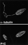
Double-label confocal laser scanning micrographs of Triton X-100–extracted L. enriettii promastigotes stained with anti-P1C and anti– α-tubulin. Cytoskeletons were fixed with methanol, stained with a 1:100 dilution of the anti-P1C antibody and a 1:500 dilution of the anti–α-tubulin antibody, and then with an FITC-conjugated anti–rabbit IgG (P1C) and a rhodamine-conjugated anti–mouse IgG (α-tubulin) secondary antibody. Cytoskeletons were examined by confocal microscopy using illumination at 488 nm to visualize the Pro-1 glucose transporter–complexed FITC antibody (P1C) or at 546 nm to visualize α-tubulin complexed with the rhodamine antibody (α-tubulin). Each micrograph represents a single 0.5-μm section through each field.
Figure 4
Figure 4
Immunoblot of pellet (P) and the supernatant (S) fractions from L. enriettii incubated with 10% DMSO (lanes 1, 2, 5, and 6) or 1 mM chlorpromazine in 10% DMSO (lanes 3, 4, 7, and 8), followed by Triton X-100 extraction. The samples were run on an 8% acrylamide gel. The blot containing lanes 1–4 was probed with the anti-P1C antibody (1:100), and the blot containing lanes 5–8 was probed with anti–α-tubulin antibody (1:1,000). The solid arrow indicates the position of iso-1, and the open arrow indicates the position of iso-2. The numbers at the left indicate the migration of molecular mass markers, with molecular masses given in kD.
Figure 5
Figure 5
Immunoblot of pellet (P) and the supernatant (S) fractions from Triton X-100–extracted L. enriettii transfected with plasmids encoding epitope tagged iso-2 (lanes 1 and 2) or epitope-tagged iso-1 (lanes 3 and 4). The blot was probed with an anti-GLUT2 antibody (1: 500). Other symbols are as indicated in Fig. 2.
Figure 6
Figure 6
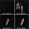
Double-label confocal laser scanning micrographs of Triton X-100–extracted L. enriettii promastigotes transfected with plasmid encoding epitope-tagged iso-1 (top) or epitope-tagged iso-2 (bottom) and stained with the rabbit anti-GLUT2 antibody directed against the epitope tag (GLUT2) and the murine anti–α-tubulin antibody (α-tubulin). Cytoskeletons were fixed with methanol, stained with 1:500 dilutions of the anti-GLUT2 antibody and anti-α-tubulin antibodies, and then with an FITC-conjugated anti–rabbit IgG (GLUT2) and a rhodamine-conjugated anti–mouse IgG (α-tubulin) secondary antibody. Cytoskeletons were examined by confocal microscopy. Each micrograph represents a single 0.5-μm section through each field.
Figure 7
Figure 7
Confocal laser microscopy of L. enriettii parasites overexpressing chimeras of the D2 hexose transporter containing the NH2-terminal domain of iso-1 (I1.D2, top) or the NH2-terminal domain of iso-2 (I2.D2, bottom). Cells were double-stained with the anti–α-tubulin antibody (α-tub, 1:400) and the anti-D2 antibody (D2, 1:200), followed by goat anti–mouse Texas red and goat anti–rabbit Bodipy Oregon green. Each micrograph represents a single 0.5-μm section through each field.
Figure 8
Figure 8
(A) Deletions of the NH2-terminal domain of iso-1. WT represents the wild-type sequence. The Δ numbers represent the number of NH2-terminal amino acids deleted in each construct. The amino acid sequence (single letter code) of the relevant portion of the NH2-terminal domain is indicated at the top with numbers designating the amino acid position starting with the initiating methionine at position 1. (B) Confocal laser scanning microscopy of L. enriettii promastigotes expressing epitope-tagged deletion constructs Δ20, Δ25, and Δ30. Stable cell lines transfected with pX63Hyg.Δ20, pX63Hyg.Δ25, or pX63Hyg.Δ30 were stained with a 1:500 dilution of the anti-GLUT2 antibody and a 1:200 dilution of FITC-conjugated secondary antibody and examined by confocal microscopy. Each micrograph represents a single 0.5-μm section through each field.
Figure 8
Figure 8
(A) Deletions of the NH2-terminal domain of iso-1. WT represents the wild-type sequence. The Δ numbers represent the number of NH2-terminal amino acids deleted in each construct. The amino acid sequence (single letter code) of the relevant portion of the NH2-terminal domain is indicated at the top with numbers designating the amino acid position starting with the initiating methionine at position 1. (B) Confocal laser scanning microscopy of L. enriettii promastigotes expressing epitope-tagged deletion constructs Δ20, Δ25, and Δ30. Stable cell lines transfected with pX63Hyg.Δ20, pX63Hyg.Δ25, or pX63Hyg.Δ30 were stained with a 1:500 dilution of the anti-GLUT2 antibody and a 1:200 dilution of FITC-conjugated secondary antibody and examined by confocal microscopy. Each micrograph represents a single 0.5-μm section through each field.
Figure 9
Figure 9
Confocal laser scanning microscopy of Triton X-100–extracted L. enriettii promastigotes expressing epitope-tagged deletion constructs Δ10, Δ20, Δ25, and Δ30. Cytoskeletons from stable cell lines transfected with pX63Hyg.Δ10, pX63Hyg.Δ20, pX63Hyg.Δ25, or pX63Hyg.Δ30 were stained with a 1:500 dilutions of the anti-GLUT2 antibody (A) and the anti–α-tubulin antibody (B) and an FITC- or a rhodamine-conjugated secondary antibody, respectively, and examined by confocal microscopy as described in Fig. 3. Each micrograph represents a single 0.5-μm section through each field.
Figure 9
Figure 9
Confocal laser scanning microscopy of Triton X-100–extracted L. enriettii promastigotes expressing epitope-tagged deletion constructs Δ10, Δ20, Δ25, and Δ30. Cytoskeletons from stable cell lines transfected with pX63Hyg.Δ10, pX63Hyg.Δ20, pX63Hyg.Δ25, or pX63Hyg.Δ30 were stained with a 1:500 dilutions of the anti-GLUT2 antibody (A) and the anti–α-tubulin antibody (B) and an FITC- or a rhodamine-conjugated secondary antibody, respectively, and examined by confocal microscopy as described in Fig. 3. Each micrograph represents a single 0.5-μm section through each field.
Figure 10
Figure 10
Immunoblots of Triton X-100 extracts from stably transfected L. donovani promastigotes overexpressing the D2 hexose transporter (lanes 1 and 2) or the MIT myo-inositol transporter (lanes 3 and 4). P designates the pellet (lanes 1 and 3) and S designates the supernatants (lanes 2 and 4) of the detergent extracts. Lanes 1 and 2 were stained with the anti-D2 antibody (1:200), and lanes 3 and 4 were stained with the anti-MIT antibody (1:3,000). Numbers to the left indicate the mobilities of protein molecular mass markers in kilodaltons.
Figure 11
Figure 11
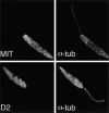
Confocal laser scanning microscopy of Triton X-100 extracted promastigotes of L. donovani overexpressing the MIT myo-inositol transporter (top) or the D2 hexose transporter (bottom). Each cytoskeleton was double-stained with either the anti-MIT antibody (MIT, 1:1,000) or the anti-D2 antibody (D2, 1:200), followed by the control α-tubulin antibody (α-tub, 1:500). Secondary FITC-conjugated or rhodamine-conjugated antibodies were used to reveal staining of the antitransporter or anti–α-tubulin antibodies, respectively. Each micrograph represents a single 0.5-μm section through each field.

Similar articles

Cited by

References

    1. Alper SL, Stuart-Tilley A, Simmons CF, Brown D, Drenckhahn D. The fodrin-ankyrin cytoskeleton of choroid plexus preferentially colocalizes with apical Na+K+-ATPase rather than with basolateral anion exchanger AE2. J Clin Invest. 1994;93:1430–1438. - PMC - PubMed
    1. Balber AE. The pellicle and the membrane of the flagellum, flagellar adhesion zone, and flagellar pocket: functionally discrete surface domains of the bloodstream form of African trypanosomes. Crit Rev Immunol. 1990;10:177–201. - PubMed
    1. Baranski TJ, Koelsch G, Hartsuck JA, Kornfeld S. Mapping and molecular modeling of a recognition domain for lysosomal enzyme targeting. J Biol Chem. 1991;266:23365–23372. - PubMed
    1. Bennett V, Stenbuck PJ. The membrane attachment protein for spectrin is associated with band 3 in human erythrocyte membranes. Nature. 1979;280:468–473. - PubMed
    1. Cairns BR, Collard MW, Landfear SM. Developmentally regulated gene from Leishmaniaencodes a putative membrane transport protein. Proc Natl Acad Sci USA. 1989;85:2130–2134. - PMC - PubMed

Publication types