. 1997 Dec 15;11(24):3327-40.
doi: 10.1101/gad.11.24.3327.

Histone acetyltransferases regulate HIV-1 enhancer activity in vitro

Affiliations

Histone acetyltransferases regulate HIV-1 enhancer activity in vitro

P L Sheridan et al. Genes Dev. .

Abstract

Specific inhibitors of histone deacetylase, such as trichostatin A (TSA) and trapoxin (TPX), are potent inducers of HIV-1 transcription in latently infected T-cell lines. Activation of the integrated HIV-1 promoter is accompanied by the loss or rearrangement of a positioned nucleosome (nuc-1) near the viral RNA start site. Here we show that TSA strongly induces HIV-1 transcription on chromatin in vitro, concomitant with an enhancer factor-assisted increase in the level of acetylated histone H4. TSA treatment, however, did not detectably alter enhancer factor binding or the positioning of nuc-1 on the majority of the chromatin templates indicating that protein acetylation and chromatin remodeling may be limiting steps that occur only on transcriptionally competent templates, or that remodeling of nuc-1 requires additional factors. To assess the number of active chromatin templates in vitro, transcription was limited to a single round with low levels of the detergent Sarkosyl. Remarkably, HIV-1 transcription on chromatin was found to arise from a small number of active templates that can each support nearly 100 rounds of transcription, and TSA increased the number of active templates in each round. In contrast, transcription on naked DNA was limited to only a few rounds and was not responsive to TSA. We conclude that HIV-1 enhancer complexes greatly facilitate transcription reinitiation on chromatin in vitro, and act at a limiting step to promote the acetylation of histones or other transcription factors required for HIV-1 enhancer activity.


Figures

Figure 1
Figure 1
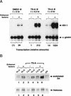
Addition of the histone deacetylase inhibitor TSA to chromatin assembly extracts strongly induces HIV-1 enhancer activity in vitro. (A) Schematic diagram of the changes in chromatin structure that accompany TSA-mediated induction of HIV-1 transcription in latently infected T cell lines (Van Lint et al. 1996a). (B) Addition of TSA at early stages in chromatin assembly enhances HIV-1 transcription on chromatin templates in vitro. HIV-1 DNA-binding proteins were added together with pHIV-1/LUC DNA during nucleosome assembly at the following concentrations: NF-κB (p65 subunit; 200 nm), LEF-1 (50 nm), Ets-1 (100 nm), TFE-3 (20 nm), Sp1 (20 nm). These experiments used a partially purified Sp1 fraction that was obtained by wheat germ agglutinin chromatography of Jurkat nuclear extracts. α-Globin gene DNA was added to the HeLa transcription extract as nonchromatin DNA. Relative HIV-1 transcription levels from duplicate reactions were quantified by phosphorImager scanning, and the specific HIV-1 and α-globin RNA transcripts are indicated with arrowheads. (C) TSA does not affect transcription from naked pHIV-1/LUC DNA. Parallel transcription reactions were carried out in HeLa nuclear extracts with or without TSA, as indicated above the panels. Reactions either lacked HIV-1 enhancer-binding proteins (lanes marked −), or contained HIV-1 enhancer-binding proteins at identical levels to those listed in A (lanes marked +) or at levels sixfold higher (lanes marked ++). The TSA stocks were prepared as suspensions in 10% DMSO and equivalent levels of DMSO were added to the control reactions lacking TSA (although this had no effect on resulting transcription levels).
Figure 1
Figure 1
Addition of the histone deacetylase inhibitor TSA to chromatin assembly extracts strongly induces HIV-1 enhancer activity in vitro. (A) Schematic diagram of the changes in chromatin structure that accompany TSA-mediated induction of HIV-1 transcription in latently infected T cell lines (Van Lint et al. 1996a). (B) Addition of TSA at early stages in chromatin assembly enhances HIV-1 transcription on chromatin templates in vitro. HIV-1 DNA-binding proteins were added together with pHIV-1/LUC DNA during nucleosome assembly at the following concentrations: NF-κB (p65 subunit; 200 nm), LEF-1 (50 nm), Ets-1 (100 nm), TFE-3 (20 nm), Sp1 (20 nm). These experiments used a partially purified Sp1 fraction that was obtained by wheat germ agglutinin chromatography of Jurkat nuclear extracts. α-Globin gene DNA was added to the HeLa transcription extract as nonchromatin DNA. Relative HIV-1 transcription levels from duplicate reactions were quantified by phosphorImager scanning, and the specific HIV-1 and α-globin RNA transcripts are indicated with arrowheads. (C) TSA does not affect transcription from naked pHIV-1/LUC DNA. Parallel transcription reactions were carried out in HeLa nuclear extracts with or without TSA, as indicated above the panels. Reactions either lacked HIV-1 enhancer-binding proteins (lanes marked −), or contained HIV-1 enhancer-binding proteins at identical levels to those listed in A (lanes marked +) or at levels sixfold higher (lanes marked ++). The TSA stocks were prepared as suspensions in 10% DMSO and equivalent levels of DMSO were added to the control reactions lacking TSA (although this had no effect on resulting transcription levels).
Figure 2
Figure 2
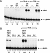
Activation of the HIV-1 enhancer in vitro is accompanied by increased levels of acetylated histone H4. (A) HIV-1 transcription reactions prepared in the presence and absence of TSA. HIV-1 enhancer-binding proteins were added at the concentrations indicated in the legend to Figure 1A, except that the concentration of Ets-1 was increased to 150 nm. (B) Western blot analysis of the level of acetylated histone H4 from the same chromatin assembly reactions shown in A, in which DMSO or TSA was added at the beginning of nucleosome assembly (t = 0 hr). Immunoblotting was carried out using antiserum specific for the acetylated form of histone H4 (Lin et al. 1989). Reaction volumes loaded on the SDS-polyacrylamide gel (18% polyacrylamide) are indicated above each lane. Total histone levels were assessed by Coomassie blue staining of aliquots from the same reactions that were analyzed by Western blot.
Figure 3
Figure 3
The two functional subregions of the HIV-1 enhancer can be induced independently by TSA in vitro. (A) NF-κB (p65 homodimer) enhancer complexes are activated by histone deacetylase inhibitors. In vitro transcription experiments were carried out with p65 (200 nm) and Sp1 (20 nm), in the presence or absence of TSA and TPX, as indicated above each lane. (B) Induction by TSA of enhancer complexes containing LEF-1 (100 nm), Ets-1 (150 nm), TFE-3 (20 nm), and Sp1 (20 nm), or equivalent levels of recombinant Sp1 or TFE-3 in the absence of any other proteins, as indicated above each lane.
Figure 4
Figure 4
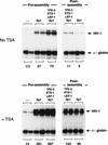
TSA activates HIV-1 transcription when the HIV-1 DNA-binding proteins are added simultaneously with the DNA at an early (preassembly) step, or when the enhancer factors are added at a late (postassembly, at t = 4.5 hr) step. Reactions were carried out in the absence (top) or presence (bottom) of TSA. These experiments used a partially purified Sp1 fraction that was obtained by wheat germ agglutinin chromatography of Jurkat nuclear extracts, which was tested alone or together with recombinant LEF-1 (100 nm), Ets-1 (150 nm), and TFE-3 (20 nm). The relative HIV-1 transcription levels are listed below each lane.
Figure 5
Figure 5
TSA treatment does not detectably alter HIV-1 enhancer factor binding or chromatin structure before transcription. (A) Analysis of nucleosome assembly by micrococcal nuclease digestion of the chromatin templates. Reactions shown in each panel were incubated with micrococcal nuclease for 3, 8, 16, and 25 min at 37°C. Blots were hybridized to a control plasmid DNA probe (located ∼1 kb upstream of the enhancer) to assess the regularity of the nucleosomal array in bulk chromatin, and with an HIV-1 primer that anneals to the TATA box to analyze the disruption in the nucleosomal array in the promoter region. DNA-binding factors present in this experiment were: NF-κB (p50; 30 nm) and Sp1 (20 nm). (B) DNase I footprint analysis of the binding of NF-κB (p50; 200 nm) and Sp1 to pHIV-1/LUC chromatin templates in the presence or absence of TSA. (C) (DNase I) This panel displays the DNase I hypersensitive sites that were generated with binding of different factors to pLTR/LUC chromatin templates, as indicated above each lane. The panel marked MNase shows the result of a micrococcal nuclease indirect end-labeling experiment to assess the positioning of nuc-1 on pLTR/LUC chromatin templates in the presence and absence of TSA. Enhancer factors were used at the following concentrations: NF-κB (p50; 60 nm), Sp1 (20 nm), IRF-1 (45 nm), and LBP-1 (150 nm). Sp1 was purified from HeLa nuclear extracts by WGA chromatography. Solid boxes are used to indicate DNase I hypersensitive sites, and the location of nuc-1 is indicated with a circle.
Figure 5
Figure 5
TSA treatment does not detectably alter HIV-1 enhancer factor binding or chromatin structure before transcription. (A) Analysis of nucleosome assembly by micrococcal nuclease digestion of the chromatin templates. Reactions shown in each panel were incubated with micrococcal nuclease for 3, 8, 16, and 25 min at 37°C. Blots were hybridized to a control plasmid DNA probe (located ∼1 kb upstream of the enhancer) to assess the regularity of the nucleosomal array in bulk chromatin, and with an HIV-1 primer that anneals to the TATA box to analyze the disruption in the nucleosomal array in the promoter region. DNA-binding factors present in this experiment were: NF-κB (p50; 30 nm) and Sp1 (20 nm). (B) DNase I footprint analysis of the binding of NF-κB (p50; 200 nm) and Sp1 to pHIV-1/LUC chromatin templates in the presence or absence of TSA. (C) (DNase I) This panel displays the DNase I hypersensitive sites that were generated with binding of different factors to pLTR/LUC chromatin templates, as indicated above each lane. The panel marked MNase shows the result of a micrococcal nuclease indirect end-labeling experiment to assess the positioning of nuc-1 on pLTR/LUC chromatin templates in the presence and absence of TSA. Enhancer factors were used at the following concentrations: NF-κB (p50; 60 nm), Sp1 (20 nm), IRF-1 (45 nm), and LBP-1 (150 nm). Sp1 was purified from HeLa nuclear extracts by WGA chromatography. Solid boxes are used to indicate DNase I hypersensitive sites, and the location of nuc-1 is indicated with a circle.
Figure 6
Figure 6
Analysis of HIV-1 chromatin structure by restriction enzyme digestion. (Top) Restriction enzyme accessibility studies carried out with HIV-1 chromatin templates. pLTR/LUC DNA was incubated with a mixture of enhancer factors at the following concentrations: NF-κB (30 nm), LEF-1 (40 nm), TFE-3 (7.5 nm), Sp1 (WGA; 20 nm), IRF-1 (45 nm), or LBP-1 (150 nm) before transcription. The chromatin templates were digested with 15 units of the various restriction enzymes indicated above each lane, and the DNA was analyzed by indirect end-labeling. The PvuII and AflII restriction enzyme digestion products were detected using the SphI primer, whereas the HindIII digests were detected using the XmnI primer. (Bottom) In vitro transcription experiments carried out with chromatin templates that were either uncut or incubated with AflII or HindIII (10 units per reaction). The location of the restriction enzyme cleavage sites relative to a downstream primer in the luciferase gene is indicated in the diagram at the bottom of the figure. Enhancer factors were present at the concentrations listed above, except that LBP-1 and IRF-1 were omitted. No TSA was added to these transcription reactions.
Figure 6
Figure 6
Analysis of HIV-1 chromatin structure by restriction enzyme digestion. (Top) Restriction enzyme accessibility studies carried out with HIV-1 chromatin templates. pLTR/LUC DNA was incubated with a mixture of enhancer factors at the following concentrations: NF-κB (30 nm), LEF-1 (40 nm), TFE-3 (7.5 nm), Sp1 (WGA; 20 nm), IRF-1 (45 nm), or LBP-1 (150 nm) before transcription. The chromatin templates were digested with 15 units of the various restriction enzymes indicated above each lane, and the DNA was analyzed by indirect end-labeling. The PvuII and AflII restriction enzyme digestion products were detected using the SphI primer, whereas the HindIII digests were detected using the XmnI primer. (Bottom) In vitro transcription experiments carried out with chromatin templates that were either uncut or incubated with AflII or HindIII (10 units per reaction). The location of the restriction enzyme cleavage sites relative to a downstream primer in the luciferase gene is indicated in the diagram at the bottom of the figure. Enhancer factors were present at the concentrations listed above, except that LBP-1 and IRF-1 were omitted. No TSA was added to these transcription reactions.
Figure 7
Figure 7
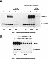
Analysis of single and multiple rounds of transcription on chromatin or nonchromatin HIV-1 DNA templates. (A) Transcription on chromatin templates in the presence and absence of TSA. Where indicated above the lanes, transcription was limited to a single round by the addition of Sarkosyl (0.1%) shortly after initiation of transcription. Assembly reactions either lacked (−) or contained (+) the following mixture of recombinant HIV-1 enhancer-binding proteins: NF-κB (p50/p65; 25 nm), LEF-1 (40 nm), and TFE-3 (7.5 nm). (B) Transcription of naked pHIV-1/LUC DNA templates in the presence of different levels of Sarkosyl, as indicated above each lane. Relative transcription levels determined by phosphorImager scanning are listed below each lane.
Figure 8
Figure 8
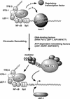
A model for the role of histone acetylation and chromatin remodeling in regulating HIV-1 enhancer activity. Possible roles for DNA-binding proteins and chromatin remodeling activities in the disruption of nuc-1 and establishing an open chromatin structure within the initiator region of the HIV-1 promoter are discussed in the text.

Similar articles

Cited by

References

    1. Ayer DE, Lawrence QA, Eisenman RN. Mad-Max transcriptional repression is mediated by ternary complex formation with mammalian homologs of yeast repressor Sin3. Cell. 1995;89:341–347. - PubMed
    1. Bannister AJ, Kouzarides T. The CBP co-activator is a histone acetyltransferase. Nature. 1996;384:641–643. - PubMed
    1. Brownell JE, Zhou J, Ranalli T, Kobayashi R, Edmonson DG, Roth SY, Allis CD. Tetrahymena histone acetyltransferase A: A homolog to yeast Gcn5p linking histone acetylation to gene activation. Cell. 1996;84:843–851. - PubMed
    1. Bulger M, Kadonaga JT. Biochemical reconstitution of chromatin with physiological nucleosome spacing. Methods Mol Genet. 1994;5:241–262.
    1. Candau R, Zhou JX, Allis CD, Berger SL. Histone acetyltransferase activity and interaction with ADA2 are critical for GCN5 function in vivo. EMBO J. 1997;16:555–565. - PMC - PubMed

Publication types

MeSH terms

Substances

LinkOut - more resources