Hepatitis B virus X protein and p53 tumor suppressor interactions in the modulation of apoptosis
- PMID: 9405677
- PMCID: PMC25100
- DOI: 10.1073/pnas.94.26.14707
Hepatitis B virus X protein and p53 tumor suppressor interactions in the modulation of apoptosis
Abstract
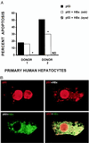
We have reported previously that the hepatitis B virus oncoprotein, HBx, can bind to the C terminus of p53 and inhibit several critical p53-mediated cellular processes, including DNA sequence-specific binding, transcriptional transactivation, and apoptosis. Recognizing the importance of p53-mediated apoptosis for maintaining homeostasis and preventing neoplastic transformation, here we further examine the physical interaction between HBx and p53 as well as the functional consequences of this association. In vitro binding studies indicate that the ayw and adr viral subtypes of HBx bind similar amounts of glutathione S-transferase-p53 with the distal C terminus of HBx (from residues 111 to 154) being critical for this interaction. Using a microinjection technique, we show that this same C-terminal region of HBx is necessary for sequestering p53 in the cytoplasm and abrogating p53-mediated apoptosis. The transcriptional transactivation domain of HBx also maps to its C terminus; however, a comparison of the ability of full-length and truncated HBx protein to abrogate p53-induced apoptosis versus transactivate simian virus 40- or human nitric oxide synthase-2 promoter-driven reporter constructs indicates that these two functional properties are distinct and thus may contribute to hepatocarcinogenesis differently. Collectively, our data indicate that the distal C-terminal domain of HBx, independent of its transactivation activity, complexes with p53 in the cytoplasm, partially preventing its nuclear entry and ability to induce apoptosis. These pathobiological effects of HBx may contribute to the early stages of hepatocellular carcinogenesis.
Figures


Similar articles
-
Hepatitis B virus X mutants derived from human hepatocellular carcinoma retain the ability to abrogate p53-induced apoptosis.Oncogene. 2001 Jun 21;20(28):3620-8. doi: 10.1038/sj.onc.1204495. Oncogene. 2001. PMID: 11439325
-
Abrogation of p53-induced apoptosis by the hepatitis B virus X gene.Cancer Res. 1995 Dec 15;55(24):6012-6. Cancer Res. 1995. PMID: 8521383
-
The transactivation and p53-interacting functions of hepatitis B virus X protein are mutually interfering but distinct.Cancer Res. 1997 Nov 15;57(22):5137-42. Cancer Res. 1997. PMID: 9371515
-
Hepatitis B virus x protein in the pathogenesis of hepatitis B virus-induced hepatocellular carcinoma.J Gastroenterol Hepatol. 2011 Jan;26 Suppl 1:144-52. doi: 10.1111/j.1440-1746.2010.06546.x. J Gastroenterol Hepatol. 2011. PMID: 21199526 Review.
-
Hepatitis B virus X protein sensitizes UV-induced apoptosis by transcriptional transactivation of Fas ligand gene expression.IUBMB Life. 2005 Sep;57(9):651-8. doi: 10.1080/15216540500239697. IUBMB Life. 2005. PMID: 16203685 Review.
Cited by
-
Compartmentalisation of Hepatitis B virus X gene evolution in hepatocellular carcinoma microenvironment and the genotype-phenotype correlation of tumorigenicity in HBV-related patients with hepatocellular carcinoma.Emerg Microbes Infect. 2022 Dec;11(1):2486-2501. doi: 10.1080/22221751.2022.2125344. Emerg Microbes Infect. 2022. PMID: 36102940 Free PMC article.
-
Occult Hepatitis B Virus Infection in Hepatic Diseases and Its Significance for the WHO's Elimination Plan of Viral Hepatitis.Pathogens. 2024 Aug 6;13(8):662. doi: 10.3390/pathogens13080662. Pathogens. 2024. PMID: 39204261 Free PMC article. Review.
-
Pathogenicity and virulence of Hepatitis B virus.Virulence. 2022 Dec;13(1):258-296. doi: 10.1080/21505594.2022.2028483. Virulence. 2022. PMID: 35100095 Free PMC article. Review.
-
Hepatitis B Virus Core Promoter A1762T/G1764A (TA)/T1753A/T1768A Mutations Contribute to Hepatocarcinogenesis by Deregulating Skp2 and P53.Dig Dis Sci. 2015 May;60(5):1315-24. doi: 10.1007/s10620-014-3492-9. Epub 2015 Jan 8. Dig Dis Sci. 2015. PMID: 25567052
-
The effect of miR-338-3p on HBx deletion-mutant (HBx-d382) mediated liver-cell proliferation through CyclinD1 regulation.PLoS One. 2012;7(8):e43204. doi: 10.1371/journal.pone.0043204. Epub 2012 Aug 17. PLoS One. 2012. Retraction in: PLoS One. 2013 Mar 8;8(3). doi: 10.1371/annotation/21379809-1376-4250-b4c2-bf51eac58a98 PMID: 22912826 Free PMC article. Retracted.
References
MeSH terms
Substances
LinkOut - more resources
Full Text Sources
Research Materials
Miscellaneous

