. 1997 Sep 22;138(6):1255-64.
doi: 10.1083/jcb.138.6.1255.

Vinculin proteolysis unmasks an ActA homolog for actin-based Shigella motility

Affiliations

Vinculin proteolysis unmasks an ActA homolog for actin-based Shigella motility

R O Laine et al. J Cell Biol. .

Abstract

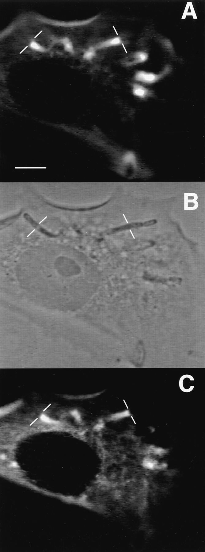
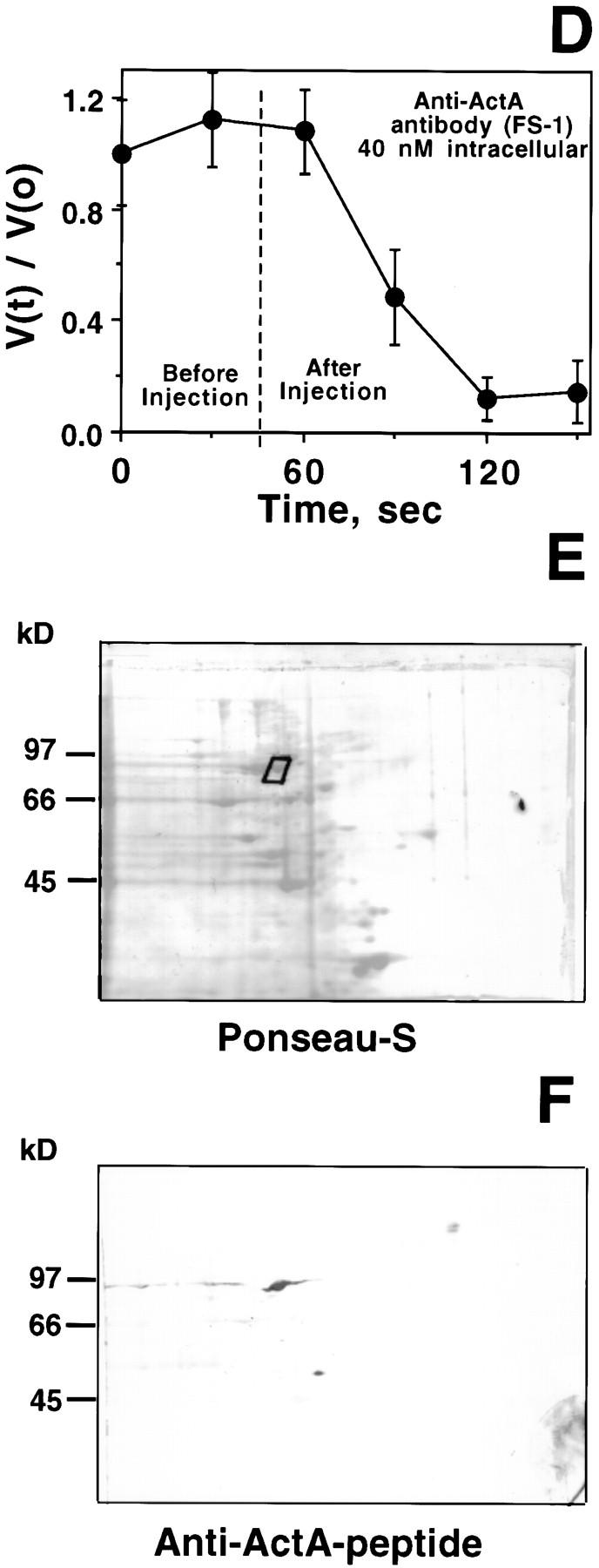
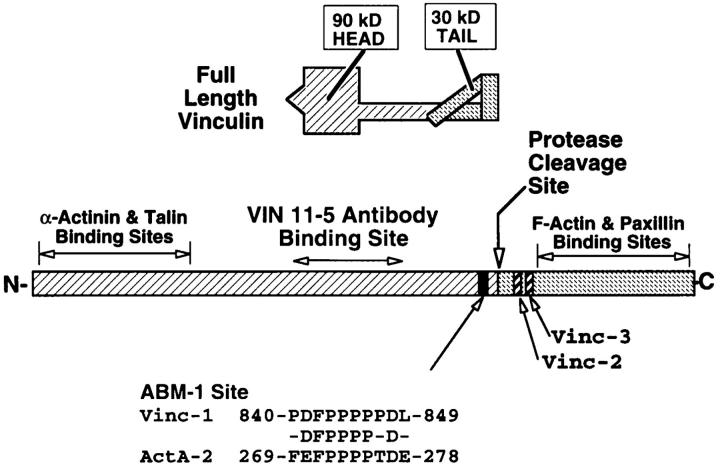
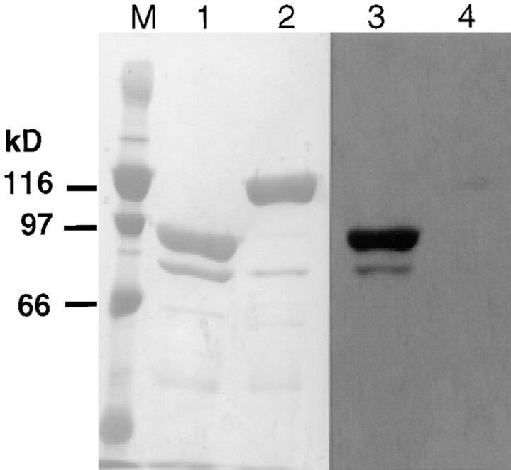
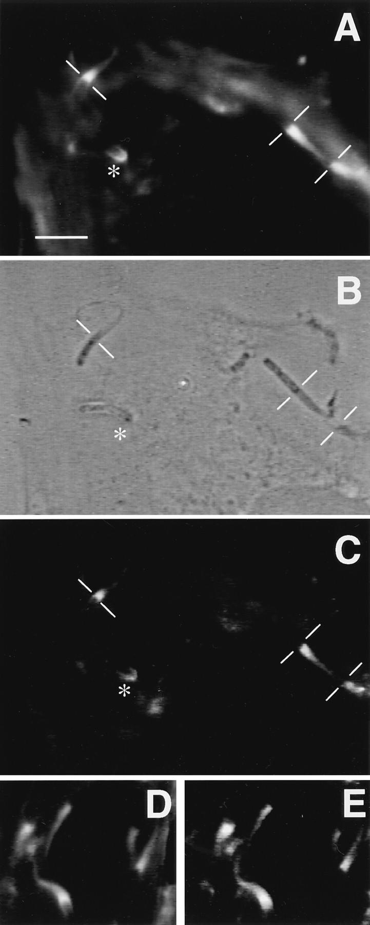
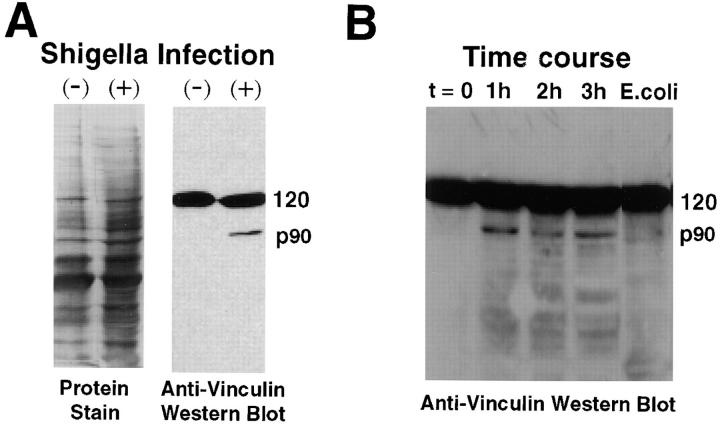
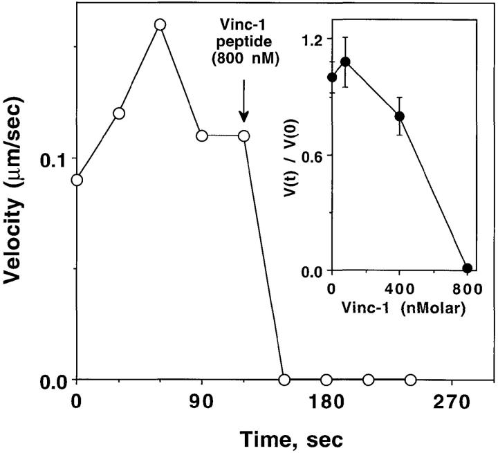
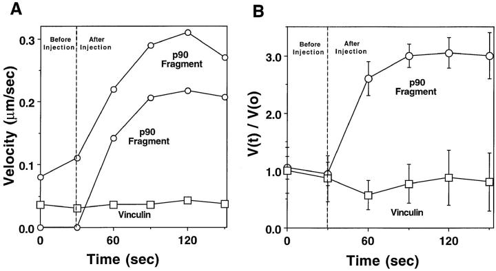
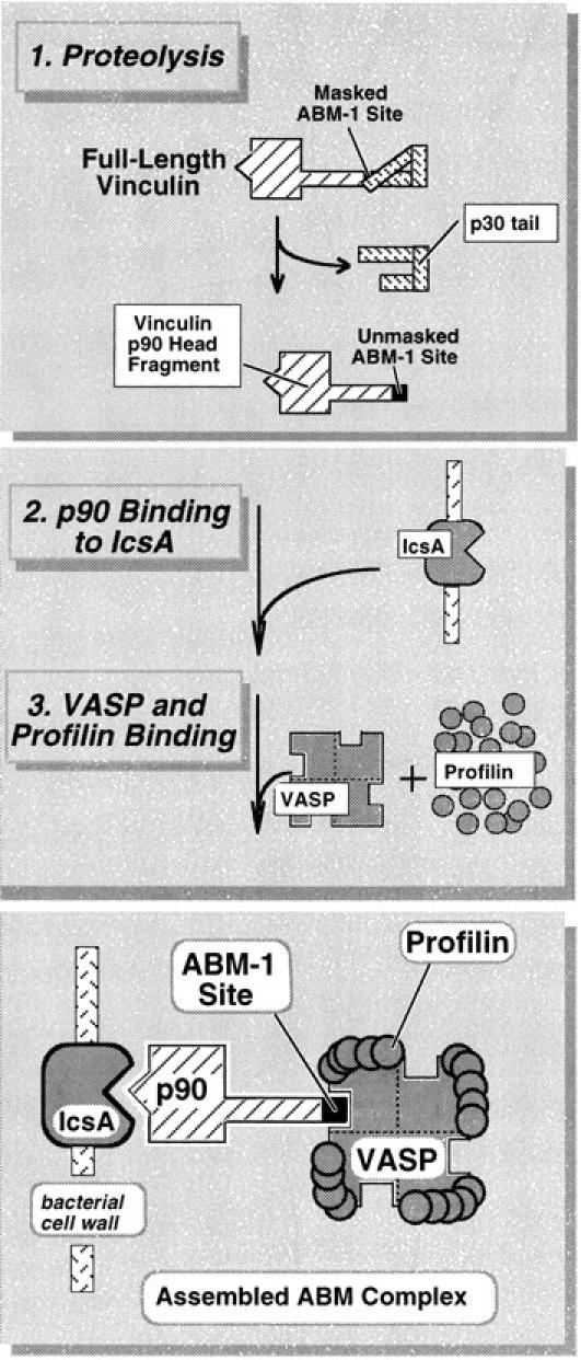
To generate the forces needed for motility, the plasma membranes of nonmuscle cells adopt an activated state that dynamically reorganizes the actin cytoskeleton. By usurping components from focal contacts and the actin cytoskeleton, the intracellular pathogens Shigella flexneri and Listeria monocytogenes use molecular mimicry to create their own actin-based motors. We raised an antibody (designated FS-1) against the FEFPPPPTDE sequence of Listeria ActA, and this antibody: (a) localized at the trailing end of motile intracellular Shigella, (b) inhibited intracellular locomotion upon microinjection of Shigella-infected cells, and (c) cross-reacted with the proteolytically derived 90-kD human vinculin head fragment that contains the Vinc-1 oligoproline sequence, PDFPPPPPDL. Antibody FS-1 reacted only weakly with full-length vinculin, suggesting that the Vinc-1 sequence in full-length vinculin may be masked by its tail region and that this sequence is unmasked by proteolysis. Immunofluoresence staining with a monoclonal antibody against the head region of vinculin (Vin 11-5) localized to the back of motile bacteria (an identical staining pattern observed with the anti-ActA FS-1 antibody), indicating that motile bacteria attract a form of vinculin containing an unmasked Vinc-1 oligoproline sequence. Microinjection of submicromolar concentrations of a synthetic Vinc-1 peptide arrested Shigella intracellular motility, underscoring the functional importance of this sequence. Western blots revealed that Shigella infection induces vinculin proteolysis in PtK2 cells and generates p90 head fragment over the same 1-3 h time frame when intracellular bacteria move within the host cell cytoplasm. We also discovered that microinjected p90, but not full-length vinculin, accelerates rates of pathogen motility by a factor of 3 +/- 0.4 in Shigella-infected PtK2 cells. These experiments suggest that vinculin p90 is a rate-limiting component in actin-based Shigella motility, and that supplementing cells with p90 stimulates rocket tail growth. Earlier findings demonstrated that vinculin p90 binds to IcsA (Suzuki, T.A., S. Saga, and C. Sasakawa. 1996. J. Biol. Chem. 271:21878-21885) and to vasodilator-stimulated phosphoprotein (VASP) (Brindle, N.P.J., M. R. Hold, J.E. Davies, C.J. Price, and D.R. Critchley. 1996. Biochem. J. 318:753-757). We now offer a working model in which proteolysis unmasks vinculin's ActA-like oligoproline sequence. Unmasking of this site serves as a molecular switch that initiates assembly of an actin-based motility complex containing VASP and profilin.


Figures

Figure 1
Figure 1
Characteristics of anti–ActA-peptide antibody: immunolocalization, inhibition of motility, and identification of human platelet p90 polypeptide. (A) Fluorescence image of Shigella-infected PtK2 using bodipy-phalloidin to label polymerized actin. The thin white bars demarcate the interface between the bacterium and the trailing actin rocket tail. (B) Phase-contrast image of the same field shown in A. (C) Indirect immunofluorescence image of the same cells using the FS-1 antibody raised against the FEFPPPPTDE sequence in Listeria ActA protein. (D) Speed measurements of Shigella in PtK2 cells before and after microinjection of FS-1 anti– ActA-peptide antibody. The dashed line indicates the time of microinjection of antibody (40 nM calculated intracellular concentration; needle concentration 0.4 μM). Bars represent the SEM of 13 different bacteria at each time point. To compare different bacteria, values were graphed as the ratio V(t)/V(0), where V(t) is the velocity at each time point and V(0) is the initial velocity at t = 0 s. Comparisons of actual pre- and postinjection speeds also demonstrated a highly significant inhibition of Shigella motility after introduction of the FS-1 antibody (mean preinjection speed: 0.11 ± 0.01 μm/s SEM n = 46, vs. postinjection: 0.02 ± 0.01 μm/s n = 48, P < 0.0001). This same concentration of antibody also significantly inhibited Listeria intracellular motility (mean preinjection velocity 0.11 ± 0.01 μm/s n = 14, vs. 0.02 ± 0.01 μm/s n = 18, P < 0.0001). (E) Ponseau S staining of an electroblot of a two-dimensional IEF/SDS electrophoresis gel. The boxed area shows the major spot identified by FS-1 antibody (raised against ActA peptide). (F) Same electroblot stained with the FS-1 antibody. Two major cross-reactive polypeptides are identified: the 90-kD polypeptide selected for microsequencing, and a 53-kD polypeptide. Bar, 10 μm.
Figure 1
Figure 1
Characteristics of anti–ActA-peptide antibody: immunolocalization, inhibition of motility, and identification of human platelet p90 polypeptide. (A) Fluorescence image of Shigella-infected PtK2 using bodipy-phalloidin to label polymerized actin. The thin white bars demarcate the interface between the bacterium and the trailing actin rocket tail. (B) Phase-contrast image of the same field shown in A. (C) Indirect immunofluorescence image of the same cells using the FS-1 antibody raised against the FEFPPPPTDE sequence in Listeria ActA protein. (D) Speed measurements of Shigella in PtK2 cells before and after microinjection of FS-1 anti– ActA-peptide antibody. The dashed line indicates the time of microinjection of antibody (40 nM calculated intracellular concentration; needle concentration 0.4 μM). Bars represent the SEM of 13 different bacteria at each time point. To compare different bacteria, values were graphed as the ratio V(t)/V(0), where V(t) is the velocity at each time point and V(0) is the initial velocity at t = 0 s. Comparisons of actual pre- and postinjection speeds also demonstrated a highly significant inhibition of Shigella motility after introduction of the FS-1 antibody (mean preinjection speed: 0.11 ± 0.01 μm/s SEM n = 46, vs. postinjection: 0.02 ± 0.01 μm/s n = 48, P < 0.0001). This same concentration of antibody also significantly inhibited Listeria intracellular motility (mean preinjection velocity 0.11 ± 0.01 μm/s n = 14, vs. 0.02 ± 0.01 μm/s n = 18, P < 0.0001). (E) Ponseau S staining of an electroblot of a two-dimensional IEF/SDS electrophoresis gel. The boxed area shows the major spot identified by FS-1 antibody (raised against ActA peptide). (F) Same electroblot stained with the FS-1 antibody. Two major cross-reactive polypeptides are identified: the 90-kD polypeptide selected for microsequencing, and a 53-kD polypeptide. Bar, 10 μm.
Figure 2
Figure 2
Structural organization of human vinculin. Full-length vinculin has a rigid 90-kD head region and a 30-kD tail region defined by a proteolytic cleavage site. As shown, the tail region is folded to indicate the latent binding site for F-actin as proposed by Johnson and Craig (1995). Below is a linearized representation of human vinculin indicating the reported binding regions and the proteolytic cleavage site that generates the p90 head and p30 tail fragments. The head fragment binds monoclonal anti-vinculin 11-5 antibody (Kilic and Ball, 1991), talin (Price et al., 1989), and α-actinin (Wachsstock et al., 1987). The location of the IcsA binding site on vinculin's head fragment has not been determined. The tail fragment contains the sites for binding F-actin (Johnson and Craig, 1995) and paxillin (Turner et al., 1990). Human vinculin also has an ABM-1 sequence (residues 840–849) named Vinc-1 located at the COOH terminus of the p90 fragment generated from vinculin by limited digestion with thermolysin, and included for comparison is the second ABM-1 repeat of Listeria ActA (residues 269–278). Also shown are the residues (including the conservative substitution of D for E) shared by the two sequences. The p30 tail contains two other oligoproline sequences (Vinc-2 and Vinc-3) that fail to fulfill the consensus features of ABM-1 homology sequences.
Figure 3
Figure 3
Anti–ActA-peptide identification of the p90 fragment from digested human vinculin. Full-length human vinculin (lanes 2 and 4) and thermolysin- digested (lanes 1 and 3) were separated by SDS-PAGE and transferred to PVDF membrane. Lanes 1 and 2 are the Coomassie blue stain of the membrane, whereas lanes 3 and 4 are a set of duplicate samples reacted against the FS-1 anti– ActA-peptide antibody. Note the p90 fragment of the digested human vinculin (lane 3) is strongly recognized by the antibody, whereas, the full-length molecule shows very faint recognition (lane 4). Lane M contains molecular weight markers.
Figure 4
Figure 4
Immunofluorescence microscopy of Shigella-infected PtK2 cells using anti-vinculin antibody. (A) Fluorescence image of Shigella-infected PtK2 cells using bodipy-phalloidin to label polymerized actin. The thin white lines demarcate the junction between the bacterium and the actin rocket tail. The asterisk identifies a bacterium that has a small focal cluster of F-actin. (B) Phase-contrast image of the same field as shown in A. (C) Indirect immunofluorescence micrograph of the same cells using the anti-vinculin 11-5 antibody (monoclonal antibody directed against the head fragment). Note that this anti-vinculin antibody localizes to the F-actin tails and to the focal F-actin cluster at one end of the bacterium identified by the asterisk. (D) An additional fluorescence image of Shigella-infected PtK2 cells using bodipy-phalloidin. (E) Same cell visualized by indirect immunofluorescence using the vin11-5 antibody. Bar, 10 μm.
Figure 6
Figure 6
Shigella infection induces the production of the 90-kD vinculin head fragment. (A) Western blots of PtK2 extracts from uninfected and infected cells using anti-vinculin clone 11-5. Left lanes, Coomassie blue–stained samples; right lanes, ECL-developed Western blots. A 120-kD cross-reactive polypeptide (full-length vinculin) is evident in both extracts. However the 90-kD head fragment is detected only in the infected cell extract. Cells were infected for 3 h before generation of the extract. (B) Time course of p90 formation after Shigella infection. Note the appearance of the p90 band within 1 h of infection. Far right lane shows extract from PtK2 cells infected for 3 h with a similar number of E. coli.
Figure 5
Figure 5
Effect of Vinc-1 peptide on Shigella speed. Representative experiment showing Shigella flexneri speed measurements before and after introduction of the Vinc-1 peptide. Arrow indicates time of microinjection of 800 nM Vinc-1 peptide, calculated as the intracellular concentration. This experiment is representative of multiple determinations (mean preinjection velocities: 0.11 ± 0.01, SEM, n = 44, vs. postinjection: 0.00 ± 0.01 n = 44, P < 0.0001). Inset demonstrates the concentration dependence of Vinc-1 peptide inhibition. The relative values V(t)/V(0) were determined as described in Fig. 1 D. Bars represent the (SEM) for 24–44 observations.
Figure 7
Figure 7
Effects of the p90 head fragment and intact vinculin on speed of Shigella motility in PtK2 cells. (A) Individual Shigella bacteria migrating in PtK2 cells before and after microinjection of the p90 head fragment or intact vinculin (estimated intracellular concentrations 0.12 μM, needle concentrations 1.2 μM). Dashed line represents the time of microinjection. Two examples of bacteria exposed to the p90 fragment are shown. In one instance the bacterium was stationary before microinjection (lower ○). After introduction of p90, the bacterium began rapidly accelerating to a speed of 0.2 μm/sec. (B) Relative velocity values before and after microinjection of the same concentrations of the p90 and vinculin depicted in A. V(t)/V(0) values were determined as described in the Fig. 1 D. Bars represent the standard error of n = 12 bacteria. All motile bacteria in each microinjected cell were analyzed before and after microinjection.
Figure 8
Figure 8
A working model for vinculin proteolysis and the assembly of the Shigella actin-based motility complex.

Similar articles

Cited by

References

    1. Beckerle MC. Identification of a new protein localized at sites of cell– substrate adhesion. J Cell Biol. 1986;103:1679–1687. - PMC - PubMed
    1. Bernardini ML, Mounier J, d'Hauteville H, Coquis-Rondon M, Sansonetti PJ. Identification of IcsA, a plasmid locus of Shigella flexnerithat governs bacterial intra- and intercellular spread through interaction with F-actin. Proc Natl Acad Sci USA. 1989;86:3867–3871. - PMC - PubMed
    1. Brindle NPJ, Hold MR, Davies JE, Price CJ, Critchley DR. The focal-adhesion vasodilator-stimulated phosphoprotein (VASP) binds to the proline-rich domain in vinculin. Biochem J. 1996;318:753–757. - PMC - PubMed
    1. Chakraborty T, Ebel F, Domann E, Niebuhr K, Gerstel B, Pistor S, Temm-Grove J, Jockusch BM, Reinhard M, Walter U, et al. A focal adhesion factor directly linking intracellularly motile Listeria monocytogenes and Listeria ivanoviito the actin-based cytoskeleton of mammalian cells. EMBO (Eur Mol Biol Organ) J. 1995;14:1314–1321. - PMC - PubMed
    1. Cudmore S, Cossart P, Griffiths G, Way M. Actin-based motility of vaccinia virus. Nature (Lond) 1995;378:636–638. - PubMed

Publication types

MeSH terms